
Lisfrank injury
Reading File
Now let me search the medical library and PubMed in parallel for Lisfranc injury content.
Finding Sources
Searching PubMed
Lisfranc injury
Excellent content found. Now let me read the most comprehensive sources in parallel - Campbell's Operative Orthopaedics (main surgical reference), Tintinalli's Emergency Medicine, and Rosen's Emergency Medicine.
Reading File
Reading File
Reading File
Now let me get more detail from Campbell's on treatment and read the full Rosen's section, plus fetch the key images:
Reading File
Reading File
Reading File
Now let me get the treatment section from Campbell's and the Tintinalli section:
Reading File
Reading File
Let me get the full treatment/management section and fetch key images:
Reading File
Loading Image
Loading Image
I now have comprehensive information to write a detailed answer. Let me compile everything.
Lisfranc Injury (Tarsometatarsal Joint Injury)
Anatomy & Biomechanics
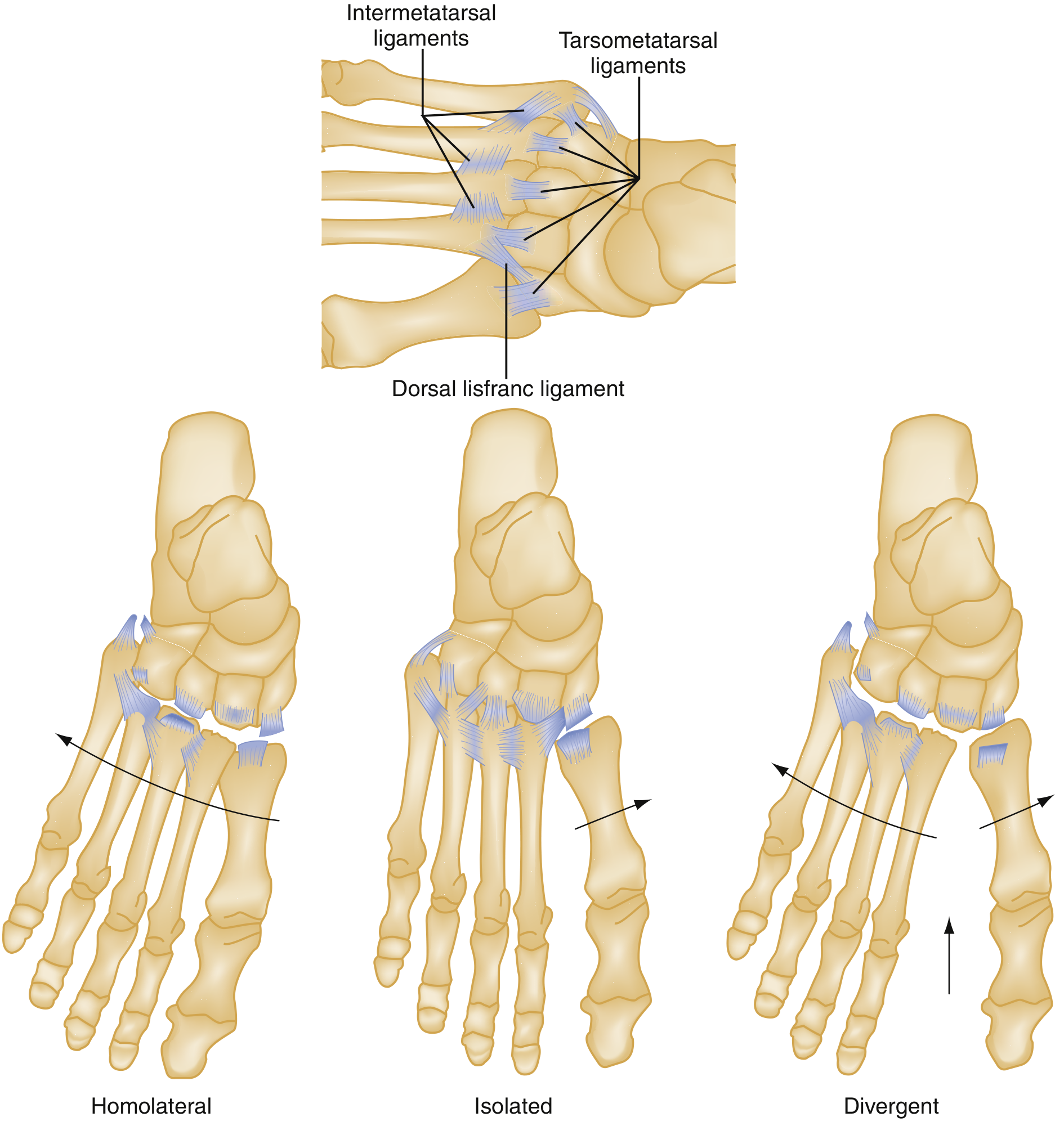
The Lisfranc joint complex refers to the tarsometatarsal (TMT) articulations - the bases of the first three metatarsals with their respective cuneiforms, and the fourth and fifth metatarsal bases with the cuboid.
Key anatomical points:
- The second metatarsal is the keystone, recessed between the medial and lateral cuneiforms - this makes it the most vulnerable to fracture
- Transverse stability is provided by the wedge-shaped metatarsal bases and cuneoform-cuboid articulations
- Longitudinal stability depends almost entirely on ligamentous support
- Strong intermetatarsal ligaments connect the bases of metatarsals 2-5, but there is no ligament between the 1st and 2nd metatarsal bases
- The Lisfranc ligament (the key stabilizer) runs from the medial cuneiform to the base of the second metatarsal
Three columns:
- Medial: 1st metatarsal, medial cuneiform, navicular facet
- Middle: 2nd and 3rd metatarsals with their cuneiforms
- Lateral: 4th and 5th metatarsals with the cuboid
Ligamentous anatomy and classification diagram (Rosen's Emergency Medicine):
Epidemiology & Mechanisms
- Historically described in equestrian riders whose foot caught in the stirrup during a fall from a horse
- High-energy mechanisms: Motor vehicle crashes, industrial crush injuries - typically produce fracture-dislocations
- Low-energy mechanisms: Sports (football, basketball), axial load on a plantarflexed foot (e.g., stepping off a curb, jumping) - typically produce ligamentous injuries
- Recent evidence suggests >50% of injuries result from low-energy mechanisms (Rosen's)
- Diagnosis is missed on initial presentation in up to 30% of cases, especially with low-energy mechanisms
Classification
Myerson Classification (modification of Quénu-Küss / Hardcastle)
| Type | Description |
|---|---|
| A (Total incongruity / Homolateral) | All 5 metatarsals displaced together, usually lateral or dorsolateral |
| B1 (Partial - medial) | Medial displacement of the 1st metatarsal (isolated) |
| B2 (Partial - lateral) | Lateral displacement of some or all lateral metatarsals (2-5) |
| C (Divergent) | Metatarsals splayed outward in opposite directions (medial and lateral) |
Nunley Classification (low-energy/ligamentous injuries)
| Stage | Description |
|---|---|
| I | Nondisplaced, stable (sprain) |
| II | 2-5 mm diastasis between 1st cuneiform and 2nd metatarsal base |
| III | >5 mm diastasis with loss of arch height |
Clinical Features
-
Midfoot pain, swelling, and inability to bear weight (especially on toes)
-
Plantar ecchymosis (bruising under the midfoot arch) - highly specific sign of Lisfranc injury
-
Tenderness along the TMT joint line
-
Pain on passive pronation/abduction of the forefoot (stress testing)
-
"Piano key" test: dorsal-plantar stress on individual metatarsals elicits pain
-
Campbell's: Compartment syndrome, though rare, can occur with high-energy injuries - presents with severe swelling and must be promptly decompressed.
Imaging
Plain Radiographs (first line)
- AP, lateral, and 30-degree oblique views of both feet
- Weight-bearing films are preferred but often impossible acutely
Radiographic findings on AP view:
| Finding | Significance |
|---|---|
| Fleck sign | Small bony avulsion between medial cuneiform and 2nd metatarsal - pathognomonic |
| Diastasis >2 mm between medial cuneiform and 2nd MT base | Unstable injury |
| Diastasis >1 mm between 1st and 2nd metatarsal bases | Unstable |
| Lateral displacement of 2nd MT off the middle cuneiform | Key alignment loss |
| Medial shaft of 2nd MT not aligned with medial aspect of middle cuneiform | Diagnostic sign |
Plain radiograph showing Lisfranc injury (Grainger & Allison's Diagnostic Radiology):
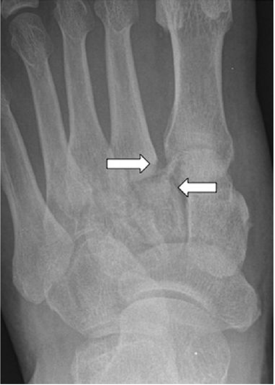
Arrows point to lateral displacement of the 2nd metatarsal off the intermediate cuneiform.
CT Scan
- Recommended when X-rays are equivocal
- Better delineation of bony structures, detects occult fractures and subluxations
- Weight-bearing CT (where available) is very useful for subtle instability
- Limitation: still fails to detect many isolated ligamentous injuries
MRI
- Most sensitive and specific for ligamentous injury
- Strong correlation with intraoperative instability
- Best reserved for suspected purely ligamentous injury with normal CT
Stress Fluoroscopy
- Can be used under anesthesia by orthopedics for definitive assessment of instability
Ultrasound
- Can evaluate the dorsal Lisfranc ligament; utility for detecting instability is unproven
Emergency Department Management
- Apply Ottawa Ankle Rules - can safely exclude midfoot fractures if no bony tenderness at the navicular or 5th metatarsal base, and patient can bear weight
- Splint, elevate, and apply ice in the ED
- Orthopedic consultation for any displaced injury or suspected compartment syndrome
- Compartment syndrome is an emergency - requires fasciotomy (medial incision for deep/abductor hallucis compartments + two dorsal incisions between metatarsals)
Definitive Treatment
Non-operative (Nunley Stage I / nondisplaced injuries)
- Non-weight-bearing short leg cast for 6 weeks
- Then weight-bearing cast for an additional 4-6 weeks
- Serial radiographs to confirm maintained alignment
- Requires strict criteria: displacement <2 mm, ligamentous stability confirmed
Operative (displaced injuries)
Indications: diastasis >2 mm, fracture-dislocation, failed closed reduction
Open Reduction and Internal Fixation (ORIF):
- Preferred over closed reduction for most displaced injuries
- Closed reduction (with finger traps, countertraction, percutaneous bone clamps) acceptable if displacement is not severe
- K-wires: used for the lateral two joints but may lose reduction in the medial and middle columns
- 4-mm cannulated or standard partially-threaded cancellous screws: excellent fixation for medial and middle columns, inserted under image control
- Dorsal/medial plating: avoids articular surface damage; fixation strength comparable to transarticular screws
Primary Arthrodesis:
- Reserved for severely comminuted fractures or significant articular damage
- Growing evidence (2024 meta-analyses) that primary arthrodesis vs. ORIF shows no significant difference in return to sport or complications for purely ligamentous injuries - PMID 40020966 and PMID 39680239
Complications
| Complication | Notes |
|---|---|
| Post-traumatic arthritis | Most common long-term complication; may require fusion |
| Compartment syndrome | Rare, high-energy injuries; requires urgent fasciotomy |
| Midfoot instability and deformity | From missed or undertreated injuries |
| Chronic pain | Significant morbidity even after treatment |
| Charcot foot | Especially in diabetics with untreated/missed injuries |
| Missed diagnosis | Up to 30% missed on initial presentation |
Key Points Summary
- The Lisfranc ligament (medial cuneiform to 2nd metatarsal base) is the primary stabilizer; there is no intermetatarsal ligament between metatarsals 1 and 2
- The fleck sign on X-ray is pathognomonic
- Plantar ecchymosis is highly specific clinically
- Missed in ~30% of cases on initial ED visit
- Any diastasis >2 mm is considered unstable and surgical
- Recent meta-analyses (2024-2025) show no significant difference between ORIF and primary arthrodesis in outcomes for purely ligamentous Lisfranc injuries
Sources: Campbell's Operative Orthopaedics 15th Ed 2026, Rosen's Emergency Medicine, Tintinalli's Emergency Medicine, Grainger & Allison's Diagnostic Radiology, Bailey and Love's Surgery 28th Ed. Recent systematic reviews: PMID 37278226, PMID 39680239, PMID 40020966, PMID 38012651.
This is a shared conversation. Sign in to Orris to start your own chat.