
Treatment of Corn on foot
corn callus foot treatment
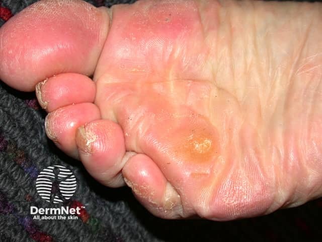
Clinical photography of a plantar hard corn (clavus) on the sole. The image shows a small, well-circumscribed hyperkeratotic lesion with a central keratotic core surrounded by a footprint of diffuse thickening (callus) on a weight-bearing area of the plantar forefoot. The surrounding skin is mildly erythematous and desquamated with fissuring at the periphery. The keratin layer appears compact, yellowish-tan, and slightly translucent at the center. There is no visible ulceration or vesiculation. The lesion is consistent with a classic hard corn caused by repetitive pressure and friction, typically over bony prominences or tight footwear. Distinguishing features from plantar warts include absence of punctate black dots, peripheral erythema, and a well-defined, conical core. Clinical significance includes guidance for conservative management: reduce friction, footwear adjustment, callus-removing keratolytics (salicylic acid), and protective padding; monitor for infection or rapid growth suggesting alternative pathology. Imaging modality supports dermatologic diagnosis; no radiologic or histopathologic data provided. This image is relevant for education on differentiating corns from calluses and plantar warts, verification of treatment response, and patient counseling on preventive foot care. Additionally, record lesion dimensions and laterality if known, document prior therapies, specify follow-up intervals, and note red flags such as increasing pain, drainage, or complications.
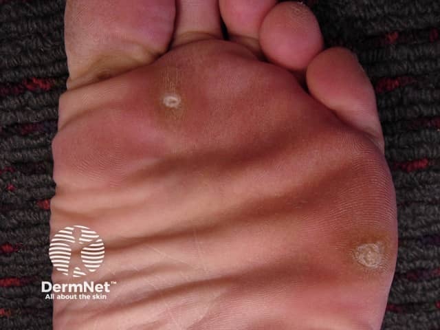
Clinical photography of the plantar surface of the foot demonstrating a classic hard corn (clavus) due to repetitive pressure. The lesion is a small, well‑circumscribed, hyperkeratotic papule with a central keratin plug, surrounded by peripheral epidermal thickening (callus) on the weight‑bearing sole. The surrounding skin is erythematous to slightly inflamed under pressure. The core appears pale yellow to white and slightly translucent; the lesion projects above the adjacent skin, producing a painful sensation with direct compression. This appearance distinguishes clavus from a plantar wart or simple callus by the presence of a dense keratinized core and a more pointed cone shape toward the dermis. The lesion is typically located at pressure points near the metatarsal heads or along the distal toe margins, often due to ill‑fitting footwear or abnormal gait. Etiology is chronic friction and pressure; pathophysiology involves localized epidermal hyperplasia and keratinocyte hypertrophy. Management focuses on debulking the hyperkeratosis (paring), application of keratolytics (salicylic acid), protective footwear, and orthotic correction; addressing footwear fit reduces recurrence risk. Differential diagnoses include plantar callus, plantar wart (verruca plantaris), and foreign body granuloma. Clinically correlates with pain on palpation and history of footwear‑related irritation. This image is educational for dermatology, podiatry, primary care, and medical education in skin pathology.
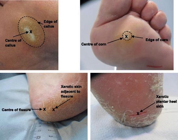
A composite of four clinical photographs demonstrating common hyperkeratotic skin lesions of the human foot and specific measurement landmarks for dermatological study. Top-left: A plantar callus on the metatarsal region, showing a broad area of yellowish, thickened stratum corneum with a dotted line marking the edge and an 'X' at the center. Top-right: A plantar corn (heloma), appearing as a smaller, more circumscribed, and deeply nucleated hyperkeratotic lesion compared to the callus. Bottom-left and Bottom-right: Clinical manifestations of xerosis and heel fissures. The skin shows significant dryness, scaling, and deep linear cracks (fissures). Annotations include 'X' marks at the center of fissures and adjacent xerotic skin to indicate measurement sites for biophysical parameters like hydration, elasticity, and texture. These images illustrate the distinction between localized pressure-induced lesions (calluses/corns) and generalized epidermal barrier dysfunction (xerosis/fissures) in podiatric medicine.
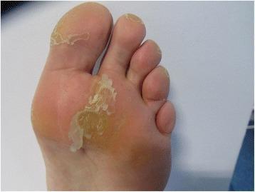
This clinical photograph displays the plantar surface of a human foot undergoing treatment for a hyperkeratotic lesion, specifically a plantar callus, following seven days of Trichloroacetic acid (TCA) application. The image focuses on the forefoot, including the metatarsal region and the hallux. The primary finding is the presence of loosely adhered, white-to-yellowish sheets of the stratum corneum (SC) exhibiting significant desquamation and scaling. This flaky appearance indicates the keratolytic effect of the TCA, which causes the thickened skin to separate and peel from the underlying epidermal layers. The central callus appears as a well-defined, slightly yellowish, thickened plaque, now surrounded by these translucent, peeling margins. In contrast, the adjacent skin and non-weight-bearing areas of the arch appear normal in texture and color. This visual representation is characteristic of the intermediate phase of chemical debridement in podiatric care, demonstrating the progression of lesion reduction through pharmaceutical intervention.
"corn" AND "foot" AND treatment
heloma clavus treatment podiatry
corn on foot treatment guidelines 2024 podiatry salicylic acid surgery
Treatment of Corn (Clavus) on the Foot
Types of Corns
| Type | Location | Characteristics |
|---|---|---|
| Hard corn (Heloma durum) | Dorsum of toes, plantar metatarsal heads | Firm, dry, well-defined central core |
| Soft corn (Heloma molle) | Between toes (4th web space most common) | Macerated, whitish, painful; caused by moisture + pressure |
| Seed corn (Heloma milliare) | Plantar surface | Tiny, multiple, non-weight-bearing |
| Subungual/periungual corn | Under or beside nail | Associated with nail deformity |
| Vascular corn | Plantar | Contains blood vessels; bleeds on paring |
1. Conservative (First-Line) Treatment
A. Footwear Modification
- The single most important intervention - addressing the root cause
- Well-fitted shoes with adequate toe box width and depth
- Avoid tight, narrow, or high-heeled footwear
- Extra-depth shoes for underlying deformities (hammer toe, bunion)
B. Padding and Offloading
- Donut-shaped foam or felt pads placed around (not over) the corn to redistribute pressure
- Silicone toe sleeves or interdigital spacers for soft corns
- Prefabricated or custom orthotics to offload pressure from metatarsal heads
- Orthopaedic insoles with metatarsal domes or bars
C. Soaking and Mechanical Debridement (Home Care)
- Soak foot in warm water 10-15 minutes to soften the corn
- Gently file with a pumice stone or foot file - reduces thickness over time
- Never use a blade at home, especially in diabetic patients
2. Keratolytic Therapy
Salicylic Acid (most widely used)
- Available as plasters, gels, liquids, and paints
- OTC concentration: 17% - adequate for most hard corns
- Medicated plasters: 40% - UK trial (J Foot Ankle Res) showed 40% salicylic acid plasters were as effective as scalpel enucleation with favourable cost outcomes
- Higher concentrations (up to 60%): used under medical supervision
- How to use: Apply only to the corn surface, protect surrounding skin with petroleum jelly, leave for 24-48 hours, remove, file gently, repeat
- Contraindicated in: diabetics, patients with peripheral neuropathy or poor circulation, broken skin
Urea-based Creams
- 15-40% urea creams - soften and hydrate hyperkeratotic skin
- Suitable for regular maintenance use
Lactic Acid
- Combined with urea in creams - mild keratolytic
- Safer for long-term use than salicylic acid
Trichloroacetic Acid (TCA)
- Applied by a clinician; causes chemical debridement and peeling
- Useful for resistant cases
3. Professional / Podiatric Treatment
Scalpel Paring / Enucleation
- The standard podiatry procedure for rapid relief
- Using a scalpel (#15 blade), the clinician shaves down the hyperkeratosis layer by layer until the central nucleus is reached and removed (enucleated)
- Provides immediate pain relief
- Must be repeated periodically as corns recur without addressing causative factors
- Caution: Do NOT perform in diabetic, immunocompromised, or vascularly compromised patients without proper assessment
Silver Nitrate Application
- Applied to the enucleated base with silver nitrate sticks
- Cauterises the base, slows regrowth
- Turns the treated area black temporarily
Intralesional Corticosteroid Injection
- For highly painful corns with associated bursitis
- Triamcinolone acetonide injected at the base
- Reduces inflammation and discomfort
4. Surgical Treatment (for Refractory Cases)
- Condylectomy / Exostectomy: Removal of the underlying bony prominence (exostosis, condyle) that creates the pressure point
- Digital osteotomy / arthroplasty: For hammer toe or claw toe deformities
- Metatarsal osteotomy: For plantar corns under metatarsal heads due to metatarsal head elevation or malposition
- Syndactyly procedure: For intractable soft corns between toes (removes part of the proximal phalanx to create space)
5. Special Situations
Diabetic Patients
- Avoid salicylic acid, blade debridement at home, and aggressive keratolytics
- Regular podiatry review is mandatory
- Even minor corn-related trauma can lead to ulceration and infection
- Custom diabetic footwear and total contact casting if ulceration develops
Soft Corns (Interdigital)
- Keep the web space dry - absorbent cotton or lamb's wool between toes
- Silicone interdigital spacers
- Antifungal powder if maceration is prominent
- Surgical removal of underlying condyle if recurrent
Children
- High salicylic acid concentrations are not recommended - risk of systemic salicylism from percutaneous absorption
- Focus on footwear, padding, and gentle mechanical debridement
6. Prevention
- Properly fitted footwear (re-measure feet periodically, especially after age 40)
- Moisture-wicking socks
- Regular use of emollient creams
- Early use of protective padding at pressure points
- Custom orthotics for structural foot deformities
Clinical Images
Quick Treatment Summary
| Scenario | Recommended Treatment |
|---|---|
| Mild, first presentation | Footwear modification + padding + pumice |
| Moderate, no diabetes | Salicylic acid 17-40% plasters + footwear review |
| Painful, requires rapid relief | Podiatry scalpel paring + offloading orthotic |
| Recurrent, underlying deformity | Surgical correction (osteotomy, condylectomy) |
| Diabetic patient | Podiatry debridement ONLY + diabetic footwear; no salicylic acid |
| Soft corn (interdigital) | Interdigital spacer + dry web space + surgical condylectomy if recurrent |
Key principle: Treating the corn without addressing the underlying mechanical cause (footwear, deformity, gait) guarantees recurrence. Structural correction is required for a lasting cure.
Can Ciprofloxacin eyedrop be given in a 3 year old child
ciprofloxacin ophthalmic drops children safety age
ciprofloxacin eye drops age limit children 1 year FDA approved ophthalmic pediatric
Can Ciprofloxacin Eye Drops Be Given to a 3-Year-Old Child?
Short Answer: Yes - it is safe and approved.
Regulatory Approval Status
"Safety and effectiveness of Ciprofloxacin Ophthalmic Solution have been established in all ages. Use of ciprofloxacin is supported by evidence from adequate and well-controlled studies of ciprofloxacin in adults, children, and neonates."
- Originally approved December 1990
- In March 2017, the FDA expanded the pediatric indication to include children younger than 1 year following a formal review under the Best Pharmaceuticals for Children Act (BPCA)
- An FDA pharmacovigilance review covering 1990-2023 found no new safety signals in pediatric patients under 17 years, and no deaths attributable to the drug
The Key Concern: Fluoroquinolone Arthropathy
| Factor | Detail |
|---|---|
| Route of administration | Topical ophthalmic - not systemic |
| Systemic absorption | Extremely minimal from eye drops |
| Animal studies | Topical ocular ciprofloxacin did NOT cause arthropathy in immature animals |
| Clinical evidence | No joint-related adverse events reported in pediatric ophthalmic use |
Indications in Children
- Bacterial conjunctivitis (pink eye) - most common use
- Bacterial corneal ulcers
- Prophylaxis after ocular trauma or foreign body
Dosing in a 3-Year-Old (Ciprofloxacin 0.3% Solution)
Bacterial Conjunctivitis
- Day 1-2: 1-2 drops in affected eye(s) every 2 hours while awake
- Day 3-7: 1-2 drops every 4 hours while awake
- Duration: 5-7 days total
Corneal Ulcer (under specialist supervision)
- More frequent dosing - every 15 min initially, tapering over days
One Important Distinction: Drops vs. Ointment
| Formulation | Age restriction |
|---|---|
| Ciprofloxacin 0.3% eye drops (solution) | Safe in all ages, including neonates |
| Ciprofloxacin 0.3% eye ointment | Not recommended in children under 2 years (per Mayo Clinic / product labeling) |
Precautions in Children
- Avoid contact with soft contact lenses (contains benzalkonium chloride preservative)
- Apply gentle nasolacrimal duct pressure after instillation to minimize systemic absorption (though this is negligible anyway)
- Supervise application - do not allow child to touch dropper tip to eye
- If no improvement after 7 days, refer to ophthalmology
- Use with caution if the child has a known hypersensitivity to any fluoroquinolone
Summary
A 3-year-old can safely receive ciprofloxacin 0.3% eye drops. The arthropathy risk associated with systemic fluoroquinolones does not apply to topical ophthalmic use. The FDA has established safety and effectiveness for all ages, including neonates, based on clinical trial data and post-market surveillance covering over 30 years of use.
Everything about febrile seizures
"febrile seizures" children management
PMID: 38653182
febrile seizures 2024 AAP guidelines classification management epilepsy risk
Febrile Seizures - Complete Overview
1. Definition
-
No intracranial infection (meningitis, encephalitis)
-
No toxic encephalopathy
-
No prior afebrile (non-febrile) seizure history
-
No acute metabolic cause
-
Tintinalli's Emergency Medicine, Rosen's Emergency Medicine, Textbook of Family Medicine 9e
2. Epidemiology
| Parameter | Data |
|---|---|
| Prevalence | 2-5% of all children in the U.S. |
| Age range | 6 months to 5-6 years |
| Peak age | 14-18 months (second year of life) |
| Sex | Boys slightly more affected than girls |
| Most common convulsive event | In children <60 months of age |
3. Classification
Simple Febrile Seizure (most common, ~70-75%)
- Generalized (tonic-clonic, not focal)
- Duration <15 minutes
- Occurs only once within a 24-hour period
- Age 6 months to 5-6 years
- No postictal neurologic deficit (other than brief drowsiness)
Complex (Complicated) Febrile Seizure (~25-30%)
-
Duration >15 minutes (febrile status epilepticus if >30 min)
-
Focal at onset or during the seizure
-
Recurs within 24 hours
-
Occurs in a child <6 months or >6 years
-
Followed by Todd's paralysis (postictal focal weakness)
-
Rosen's Emergency Medicine, Tintinalli's Emergency Medicine
4. Pathophysiology
- The exact mechanism is incompletely understood
- Fever lowers the seizure threshold in a developmentally susceptible brain
- It is thought that the rapid rise of temperature (or defervescence) - not absolute fever height - triggers the seizure, though data are not entirely consistent on this
- The immature brain (6 months to 5 years) has increased excitability and reduced inhibitory capacity
- Genetic susceptibility plays a significant role: autosomal dominant inheritance patterns identified; mutations in SCN1A (sodium channel) and GABRG2 (GABA receptor) genes found in some families with Generalized Epilepsy with Febrile Seizures Plus (GEFS+)
5. Causes / Precipitating Illnesses
-
Roseola (HHV-6) - classically causes abrupt high fever followed by febrile seizure as temperature rises
-
Otitis media
-
Upper respiratory tract infections (viral)
-
Urinary tract infections
-
Gastroenteritis
-
Influenza
-
Post-vaccination fever (MMR most commonly; also MMRV, combined DTaP+PCV13)
- MMR: risk window 8-14 days post-vaccination
- DTaP: risk window within 48 hours
-
Goodman & Gilman's, Textbook of Family Medicine 9e
6. Clinical Features
- Most seizures are generalized tonic-clonic
- Typically last under 6 minutes; fewer than 8% last >15 minutes
- Child often postictal (drowsy, confused) briefly after the event
- Most parents do not bring the child to medical attention until after the seizure has ended
- Temperature is usually >38.9°C (102°F) at time of seizure
7. Evaluation and Investigations
History
- Detailed seizure description (focal? duration? recurrence within 24h?)
- Symptoms of infection, medication use, toxic ingestions
- Developmental history, prenatal/birth history
- Family history of febrile seizures or epilepsy
- Vaccination history and recent vaccinations
Physical Examination
- Meningeal signs: neck stiffness, Kernig's sign, Brudzinski's sign
- Bulging/tense fontanelle
- Petechial rash (raises concern for meningococcemia)
- Level of consciousness and return to baseline neurologic status
- Focal neurologic deficits, Todd's paralysis
Investigations (AAP Guidelines)
| Investigation | Recommendation |
|---|---|
| Routine blood tests (CBC, electrolytes, glucose) | NOT indicated for simple febrile seizure - only if needed to evaluate fever source |
| Blood culture | Not routine |
| Urinalysis / urine culture | Consider for fever source evaluation |
| Lumbar puncture (LP) | Perform if clinical signs of meningitis are present (neck stiffness, Kernig's/Brudzinski's signs). Consider in children <12 months, unimmunized against Hib/S. pneumoniae, or on prior antibiotics. Not routine after a simple febrile seizure in fully immunized child >12 months with normal exam |
| EEG | NOT indicated for simple febrile seizures |
| Neuroimaging (CT/MRI) | NOT indicated for simple febrile seizures |
- Harriet Lane Handbook 23rd ed., Tintinalli's, Rosen's, AAP 2024 updated guidelines
8. Treatment
During the Acute Seizure
- Usually self-limited - supportive care only
- Position child safely (lateral position to prevent aspiration)
- Do not restrain
- Clear airway
| Step | Drug | Dose / Route |
|---|---|---|
| First-line | IV/IM Lorazepam | 0.1 mg/kg IV or 0.1 mg/kg IM |
| First-line alternative | Rectal Diazepam | 0.2-0.5 mg/kg rectally |
| First-line alternative | Intranasal Midazolam | 0.2 mg/kg intranasal |
| Second-line (if persists) | IV Fosphenytoin | 20 PE/kg IV |
| Second-line alternative | IV Phenobarbital | 20 mg/kg IV |
| Second-line alternative | IV Valproate | 20-40 mg/kg IV |
| Second-line alternative | IV Levetiracetam | 60 mg/kg IV |
| Third-line (refractory) | Propofol/Midazolam infusion (ICU) | Continuous infusion with intubation |
- Merck Manual 2025, Rosen's Emergency Medicine
After the Seizure
- Identify and treat the source of fever (antipyretics: paracetamol/ibuprofen)
- Note: Antipyretics do NOT prevent febrile seizure recurrence - they just provide comfort
- Observe in ED for several hours until neurologically back to baseline
- Discharge home if: alert and returning to baseline, fever source identified and treated, close follow-up available
Long-term / Prophylactic Treatment
| Scenario | Recommendation |
|---|---|
| Simple febrile seizure | No chronic AED prophylaxis - risk vs benefit unfavorable |
| Recurrent simple febrile seizures | Generally still no chronic prophylaxis |
| Complex febrile seizures / high-risk | Rectal diazepam (0.5 mg/kg) at time of fever illness may reduce recurrence - intermittent use only |
| Phenobarbital prophylaxis | Not recommended - causes cognitive impairment (IQ reduction) with unproven efficacy in preventing epilepsy |
| Valproate prophylaxis | Effective but hepatotoxicity risk makes it unsuitable for routine use in young children |
- Goodman & Gilman's, Textbook of Family Medicine 9e, Corsello et al. 2024 systematic review (PMID 38653182)
9. Recurrence
- Overall recurrence rate: ~33% (1 in 3 children)
- Children <12 months: 50% recurrence risk
- Children >12 months: 30% recurrence risk
- Most recurrences happen within 1 year of the first event
Risk Factors for Recurrence
- Age <1 year at first seizure (strongest predictor)
- Family history of febrile seizures in first-degree relatives
- Lower temperature at time of first seizure (<40°C)
- Short duration between fever onset and seizure
- Daycare attendance (higher infection exposure)
- Developmental delay
- Tintinalli's, Textbook of Family Medicine 9e, Rosen's
10. Risk of Developing Epilepsy
| Group | Epilepsy risk |
|---|---|
| General population | 0.5-1% |
| After simple febrile seizure | ~1-2% (roughly same as general population) |
| After complex febrile seizure | ~4-7% |
| If ALL risk factors present | Up to 10% |
Risk Factors for Later Epilepsy
- Pre-existing neurological disorder or developmental delay
- Family history of epilepsy (not just febrile seizures)
- Complex febrile seizure (prolonged, focal, or recurrent within 24h)
- First febrile seizure before 12 months of age
- Todd's paralysis after the seizure
- Abnormal EEG (if obtained)
11. Febrile Status Epilepticus
- Febrile seizure lasting >30 minutes (or repeated seizures totalling >30 min without recovery)
- Higher risk of subsequent hippocampal sclerosis and temporal lobe epilepsy (FEBSTAT study, 2024)
- Higher risk of meningitis vs simple febrile seizure (15-18% incidence of bacterial meningitis in some series)
- Requires LP, ICU-level management, and neurology consultation
- Managed with the full SE protocol (benzodiazepines → fosphenytoin → phenobarbital → anesthesia)
12. Parental Counseling (Key Points)
- Febrile seizures do not cause brain damage in virtually all cases
- The risk of developing epilepsy is very low (similar to general population after a simple seizure)
- ~1 in 3 children will have another febrile seizure - this is not dangerous
- Give fever-reducing medications for comfort, not to prevent seizures (antipyretics don't prevent recurrence)
- If a seizure occurs: lay child on their side, clear airway, time the seizure, do not restrain or put anything in mouth
- Call emergency services if: seizure lasts >5 minutes, child doesn't recover within 30 minutes, signs of neck stiffness, rash, child is very ill-looking
- Educate on when to use rescue diazepam if prescribed for high-risk children
- Rosen's Emergency Medicine, Tintinalli's
13. Differential Diagnosis
- Bacterial meningitis / viral encephalitis (most important to exclude)
- Sepsis with seizure
- Hyponatremia / hypoglycemia / hypocalcemia
- Toxic ingestion
- Epilepsy (first afebrile seizure with concurrent febrile illness)
- Dravet syndrome (SCN1A mutation - presents like febrile seizures initially but has worse prognosis and treatment implications)
- Intracranial hemorrhage
Dravet syndrome is an important mimic - these children have repeated febrile convulsions starting in the first year of life that appear identical to febrile seizures, but eventually develop afebrile seizures. SCN1A genetic testing should be considered in children with repeated prolonged or hemiclonic febrile seizures beginning before 12 months.
14. Summary Algorithm
Child 6 months - 6 years with Seizure + Fever
              ↓
    Signs of meningitis?
    YES → LP, admit, treat empirically
    NO  ↓
    Simple or Complex?
    SIMPLE (<15 min, generalized, once in 24h)
      → No LP, no EEG, no imaging, no AEDs
      → Treat fever source, observe, discharge
      → Parental education and reassurance
    COMPLEX (>15 min, focal, recurrent in 24h)
      → Consider LP if <12 months or unimmunized
      → Consider EEG, neurology referral
      → No routine neuroimaging unless focal neuro deficit
      → Treat active seizure >5 min with benzodiazepine
      → Evaluate for Dravet syndrome if recurrent + hemiclonic