
Teach me hemolytic anemia in simple way for pathology University examin very simple language so I can score more
Reading File
Now let me search the medical library for hemolytic anemia content in parallel.
Finding Sources
Finding Sources
Robbins Pathology is the best source for a pathology exam. Let me read the key sections in parallel.
Reading File
Reading File
Finding Sources
Excellent content from Robbins. Let me now read the detailed individual diseases sections.
Reading File
Finding Sources
Finding Sources
I have excellent content. Let me also read the sickle cell, G6PD, thalassemia, and autoimmune sections from the same Robbins block.
Reading File
I now have rich, detailed content from Robbins Pathology covering all the major hemolytic anemias. Let me also quickly fetch the key images for visual learning.
Loading Image
Loading Image
I have all the information needed for a comprehensive, exam-focused answer. Let me now compose a well-structured, easy-to-understand guide.
🩸 Hemolytic Anemia - Simple Pathology Guide for Exam
Based on Robbins & Kumar Basic Pathology (Robbins Pathology)
What is Hemolytic Anemia? (The Big Idea)
Think of it this way: Normal RBCs live for 120 days. In hemolytic anemia, they die early - sometimes in just 20 days.
When RBCs break down faster than the bone marrow can make them, you get anemia. The body fights back by:
- Making more RBCs (reticulocytosis)
- Bone marrow expands (erythroid hyperplasia)
- In severe cases, liver + spleen start making blood cells (extramedullary hematopoiesis)
Classification - The Most Important Table to Remember
| Basis | Types |
|---|---|
| Where the defect is | Intrinsic (inside the RBC) vs. Extrinsic (outside the RBC) |
| Where RBC breaks | Extravascular (in spleen) vs. Intravascular (inside blood vessels) |
Extravascular vs. Intravascular Hemolysis
This is a high-yield comparison question in exams!
| Feature | Extravascular | Intravascular |
|---|---|---|
| Where it happens | Spleen (macrophages eat damaged RBCs) | Inside blood vessels (RBCs burst) |
| Cause | RBC can't pass through spleen (less flexible) | Severe membrane damage (complement, toxins, trauma) |
| Jaundice | Yes (bilirubin from macrophage breakdown) | Less prominent |
| Splenomegaly | Yes (spleen works overtime) | Less prominent |
| Hemoglobinuria | No | Yes (Hb spills into urine - urine turns dark) |
| Hemosiderinuria | No | Yes |
| Iron deficiency | No (iron recycled by macrophages) | Possible (iron lost in urine) |
| Gallstones (pigment) | Yes (long-standing cases) | No |
| Haptoglobin | Low (seen in BOTH types) | Low |
Exam tip: Low haptoglobin is a feature of BOTH types of hemolysis.
Major Hemolytic Anemias - One by One
1. Hereditary Spherocytosis (HS)
Genetics: Autosomal Dominant (mostly)
The Problem (Simple explanation):
The RBC membrane has a "scaffold" made of proteins - Spectrin, Ankyrin, Band 3, Band 4.1, Band 4.2. If any of these are defective, the membrane becomes unstable. The RBC sheds small pieces of membrane, shrinks, and becomes a sphere (spherocyte) instead of a biconcave disc.
Spherocytes are rigid and can't squeeze through the spleen's narrow passages, so they get eaten by macrophages there.
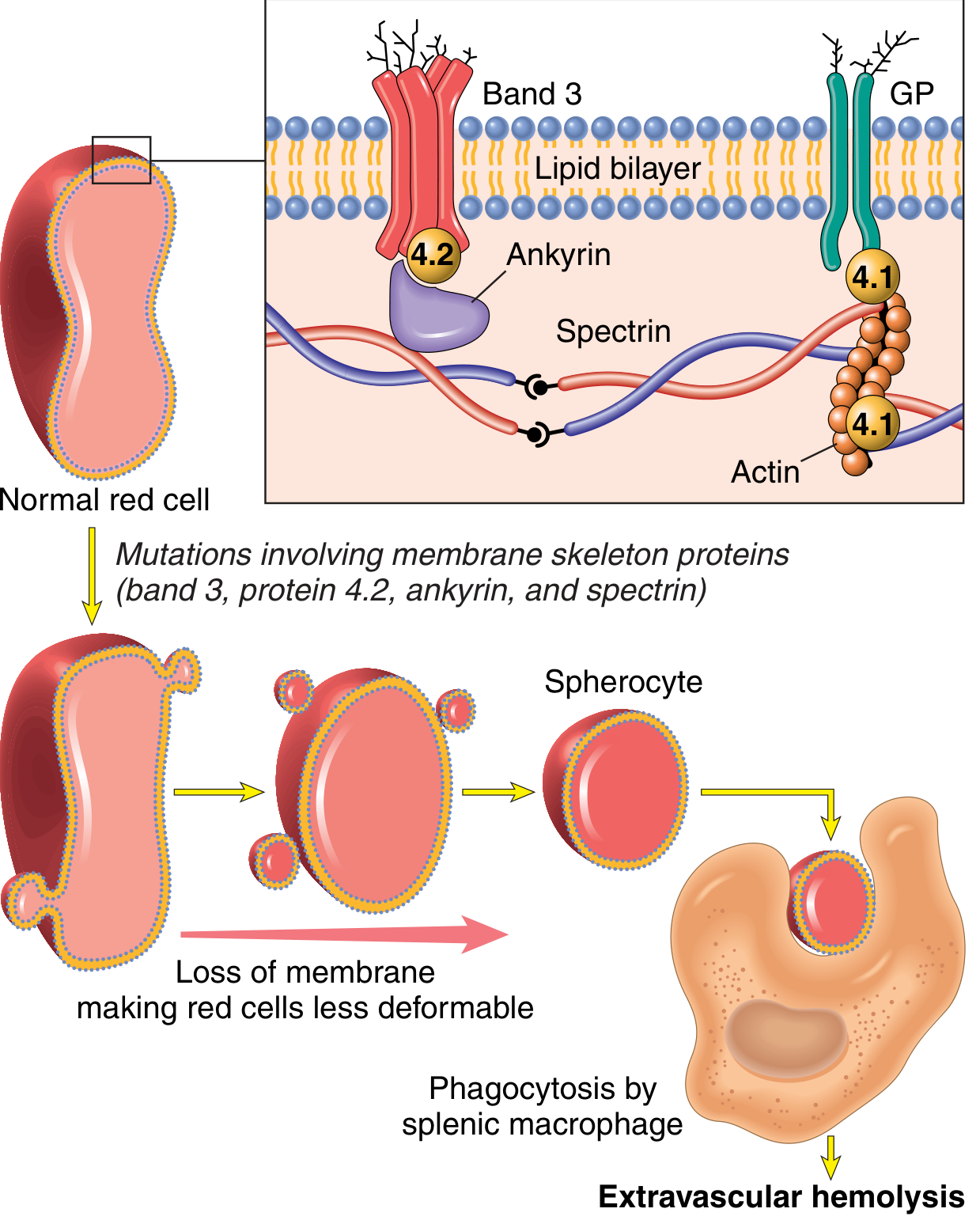
Key proteins mutated: Spectrin, Ankyrin, Band 3, Band 4.2
Type of hemolysis: Extravascular (in spleen)
Clinical features (the triad):
- Anemia
- Splenomegaly (spleen weight 500-1000g; normal is 150-200g)
- Jaundice + Gallstones (pigment stones in 40-50%)
Blood smear: Spherocytes = dark red cells with NO central pallor
Lab findings: Reticulocytosis, elevated indirect bilirubin, low haptoglobin, positive osmotic fragility test
Treatment: Splenectomy - fixes the anemia even though spherocytes remain!
2. Sickle Cell Anemia
Genetics: Autosomal Recessive
The Problem (Simple explanation):
One amino acid swap - Glutamic acid → Valine at position 6 of beta-globin → creates HbS.
When oxygen is low (deoxygenated), HbS molecules stick together and form long polymer rods that distort the RBC into a sickle shape.
Factors that make sickling WORSE:
- Low oxygen
- Dehydration (concentrates HbS)
- Acidosis
- Infection/inflammation (slows blood flow)
- HbF reduces sickling (that's why newborns are protected initially)
Two major consequences:
- Hemolytic anemia - mean RBC life span only 20 days (1/6 of normal)
- Vaso-occlusion - sickled cells block small vessels → pain crises + tissue infarcts
Organs affected:
- Spleen: repeated infarcts → "autosplenectomy" (spleen shrinks and becomes fibrotic) → patient vulnerable to bacterial infections (especially encapsulated bacteria like Pneumococcus)
- Bones: marrow hyperplasia → "crew-cut" appearance on skull X-ray; osteonecrosis of femoral head
- Kidney: papillary necrosis, hematuria
- Brain: stroke
- Eyes: retinopathy
- Lungs: acute chest syndrome
Blood smear: Sickle cells, target cells, anisocytosis, poikilocytosis
Lab: Low Hb, high reticulocytes, elevated bilirubin, Hb electrophoresis shows HbS
3. Thalassemia
Genetics: Autosomal Codominant
The Problem (Simple explanation):
Normal hemoglobin (HbA) = 2 alpha + 2 beta chains. In thalassemia, one type of chain is produced in reduced amounts. The imbalance is the problem.
Beta-Thalassemia Major (Most severe - exam favourite!)
- Both beta-globin genes are mutated → almost no beta chains made
- Alpha chains accumulate → precipitate inside RBC → damage membrane → ineffective erythropoiesis + hemolysis
- Compensatory massive bone marrow expansion → bone deformities
- "Chipmunk face" (maxillary expansion), "hair-on-end" skull X-ray
- Requires regular blood transfusions → iron overload → heart failure, liver cirrhosis, organ damage
- Splenomegaly is massive
Alpha-Thalassemia
- Loss of 1-2 alpha genes: silent carrier to mild anemia
- Loss of 3 alpha genes: HbH disease
- Loss of all 4 alpha genes: Hb Barts → hydrops fetalis (fatal in utero)
Blood smear: Microcytic, hypochromic RBCs, target cells
4. G6PD Deficiency
Genetics: X-linked Recessive (males affected more)
The Problem (Simple explanation):
G6PD enzyme protects RBCs from oxidative damage by making NADPH. Without G6PD, RBCs can't fight oxidative stress → hemoglobin gets oxidized → forms Heinz bodies (denatured Hb clumps) → RBCs destroyed.
Triggers (remember these for exams!):
- Drugs: Primaquine, Dapsone, Nitrofurantoin, Sulfonamides
- Infection
- Fava beans ("Favism")
Type of hemolysis: Episodic (only during triggers, not constant)
Blood smear during crisis: Heinz bodies (on special stain), bite cells (macrophages bite out Heinz body pieces)
Key point: Between attacks, blood smear is NORMAL.
5. Autoimmune Hemolytic Anemia (AIHA)
The Problem: Body makes antibodies against its own RBCs.
| Feature | Warm AIHA | Cold AIHA |
|---|---|---|
| Antibody type | IgG | IgM |
| Temperature | Binds at 37°C (body temp) | Binds at cold temps |
| Hemolysis type | Extravascular (spleen) | Intravascular |
| Causes | SLE, CLL, drugs | Mycoplasma infection, EBV (mono) |
| Treatment | Steroids | Avoid cold |
Diagnosis: Direct Coombs test (DAT) - positive. RBCs are coated with antibody.
6. Microangiopathic Hemolytic Anemia (MAHA) - Bonus!
The Problem: RBCs get shredded as they pass through damaged or partially blocked small vessels (mechanical fragmentation).
Causes: TTP, HUS, DIC, malignant hypertension, artificial heart valves
Blood smear: Schistocytes (helmet cells, fragmented RBCs) - this is pathognomonic!
High-Yield Lab Pattern for ALL Hemolytic Anemias
| Test | Result | Why |
|---|---|---|
| Hemoglobin | Low | RBCs destroyed |
| Reticulocytes | HIGH | Bone marrow compensating |
| Indirect (unconjugated) bilirubin | HIGH | Hb breakdown product |
| Haptoglobin | LOW | Binds free Hb, then removed from circulation |
| LDH | HIGH | Released from broken RBCs |
| Peripheral smear | Abnormal RBC shapes | Key to diagnosis |
Quick Memory Summary
| Disease | Key Defect | Key Finding |
|---|---|---|
| Hereditary Spherocytosis | Spectrin/Ankyrin mutation | Spherocytes, no central pallor |
| Sickle Cell | HbS (Glu→Val) | Sickle cells, vaso-occlusion |
| Thalassemia | Reduced globin chain synthesis | Microcytic hypochromic, target cells |
| G6PD Deficiency | Oxidant sensitivity | Heinz bodies, bite cells, triggered by drugs |
| Warm AIHA | IgG antibody | + Coombs test, responds to steroids |
| Cold AIHA | IgM antibody | + Coombs, triggered by cold, mycoplasma |
| MAHA | Mechanical fragmentation | Schistocytes |
Common Exam Questions & Answers
Q: What is the hallmark of ALL hemolytic anemias?
A: Reticulocytosis (bone marrow compensation) + low haptoglobin.
Q: Why does splenectomy cure hereditary spherocytosis?
A: The spleen is the site of RBC destruction. Remove the spleen → no destruction → anemia corrects. Spherocytes still exist but are no longer destroyed.
Q: What is the most common cause of death in thalassemia major?
A: Cardiac failure from iron overload (hemosiderosis from repeated transfusions).
Q: What blood smear finding is pathognomonic of MAHA?
A: Schistocytes (fragmented RBCs).
Q: Sickle cell patients get infections with which organisms?
A: Encapsulated bacteria - Streptococcus pneumoniae, H. influenzae, Salmonella (especially for osteomyelitis).
Source: Robbins & Kumar Basic Pathology (Robbins Pathology), Chapter 10
This is a shared conversation. Sign in to Orris to start your own chat.