
CVS physiology Neet mds notes
Reading File
Now let me search the library for CVS physiology content relevant to NEET MDS.
Finding Sources
Finding Sources
Finding Sources
Finding Sources
Finding Sources
Finding Sources
Now let me read the main CVS physiology block from Ganong's and Guyton simultaneously:
Reading File
Finding Sources
Excellent! Now let me read several key sections in parallel:
Reading File
Reading File
Reading File
Reading File
Reading File
Reading File
I now have comprehensive material to compile detailed NEET MDS notes. Let me also fetch the key diagram images and read the arterial pressure/blood pressure section:
Reading File
Loading Image
I now have enough comprehensive material. Let me compile the complete NEET MDS CVS Physiology notes:
CVS Physiology - NEET MDS Notes
Based on Ganong's Review of Medical Physiology, 26th Edition
1. CONDUCTING SYSTEM OF THE HEART
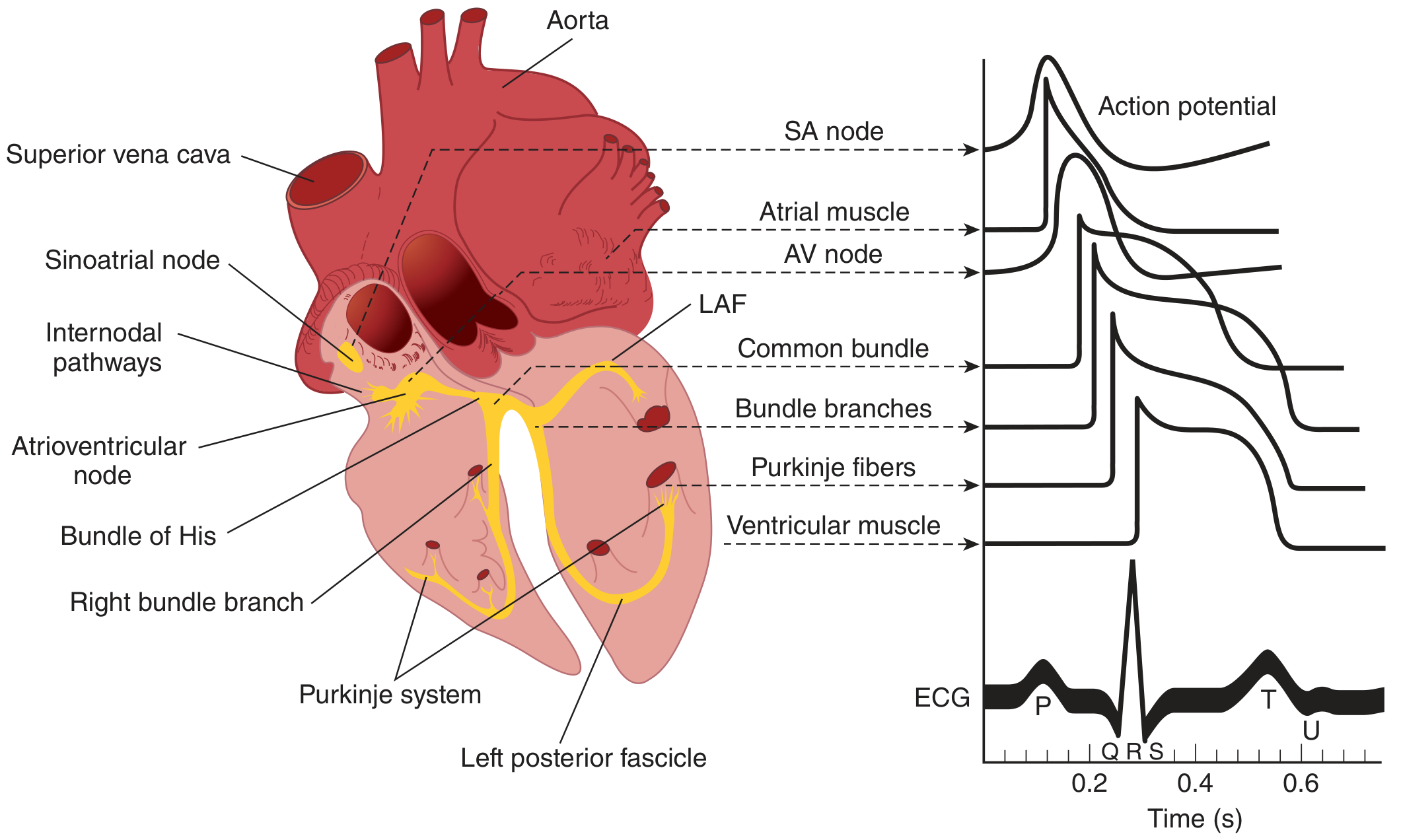
Components & Anatomy
| Structure | Location | Rate of Discharge |
|---|---|---|
| SA Node | Junction of SVC + Right atrium | 60-100/min (primary pacemaker) |
| AV Node | Right posterior interatrial septum | 40-60/min |
| Bundle of His | Top of interventricular septum | 40-60/min |
| Bundle Branches (L & R) | Subendocardially down IVS | - |
| Purkinje Fibers | Spread to all ventricular myocardium | 20-40/min |
Key points for NEET MDS:
- SA node = normal pacemaker (discharges fastest)
- Right vagus -> mainly SA node; Left vagus -> mainly AV node
- Right sympathetics -> SA node; Left sympathetics -> AV node
- Internodal tracts: Anterior (Bachmann bundle to left atrium), Middle (tract of Wenckebach), Posterior (tract of Thorel)
- Atrial fibers are separated from ventricular fibers by a fibrous ring; the ONLY conduction pathway between atria and ventricles is the Bundle of His
- Purkinje fibers: largest cells, fewest mitochondria and striations, specialized for rapid conduction
- SA node cells: smallest, sparsely striated, highest internal resistance
2. CARDIAC ACTION POTENTIALS
A. Ventricular Muscle Action Potential (Resting Membrane Potential = -90 mV)
| Phase | Name | Ion Movement |
|---|---|---|
| Phase 0 | Rapid depolarization | Na+ influx (fast voltage-gated Na+ channels, INa) |
| Phase 1 | Initial rapid repolarization | Inactivation of Na+ channels |
| Phase 2 | Plateau | Ca2+ influx via slow L-type Ca2+ channels (ICa) |
| Phase 3 | Slow repolarization | K+ efflux through multiple K+ channels |
| Phase 4 | Resting potential | -90 mV; stable in working myocardium |
B. Pacemaker (SA Node) Action Potential - Prepotential / Pacemaker Potential
- After each impulse, IK declines
- Channel permeable to both Na+ and K+ activates - called "h" channel (funny/hyperpolarization-activated) - produces If (funny current / Ih)
- This Ih causes initial slow depolarization (first part of prepotential)
- T-type (transient) Ca2+ channels open - complete the prepotential
- L-type (long-lasting) Ca2+ channels produce the actual impulse
- No stable resting potential - spontaneous depolarization is the key feature
NEET Mnemonics:
- "0-1-2-3-4": Depolarize, Notch, Plateau, Repolarize, Rest
- Plateau (phase 2) = due to Ca2+ (important for cardiac contractility)
- Pacemaker has "funny current" (If) - activated by HYPERpolarization
3. ECG (Electrocardiogram)
Normal ECG Waves
| Wave/Interval | Represents | Normal Duration |
|---|---|---|
| P wave | Atrial depolarization | 0.08-0.1 s |
| PR interval | AV conduction time | 0.12-0.20 s |
| QRS complex | Ventricular depolarization | <0.12 s |
| ST segment | Ventricular plateau (all cells depolarized) | - |
| T wave | Ventricular repolarization | - |
| U wave | Repolarization of Purkinje fibers | - |
| QT interval | Ventricular systole (electrical) | 0.35-0.44 s |
Normal Cardiac Rate
- Normal: 60-100 beats/min
- Tachycardia: >100/min
- Bradycardia: <60/min
ECG in Electrolyte Imbalances (High-Yield!)
| Condition | ECG Change |
|---|---|
| Hyperkalemia | Tall peaked T waves -> wide QRS -> sine wave -> VF |
| Hypokalemia | Flat T wave, prominent U wave, ST depression |
| Hypercalcemia | Short QT interval |
| Hypocalcemia | Prolonged QT interval |
| Hypomagnesemia | Prolonged QT, Torsades de Pointes |
4. CARDIAC CYCLE
Phases of Ventricular Systole & Diastole
| Phase | Events |
|---|---|
| Isovolumetric Contraction | AV valves close (S1), semilunar valves not yet open; pressure rises with no volume change |
| Rapid Ejection | Semilunar valves open; blood ejected into aorta/PA |
| Reduced Ejection | Less blood ejected as ventricle relaxes |
| Isovolumetric Relaxation | Semilunar valves close (S2), AV valves not yet open; pressure falls with no volume change |
| Rapid Ventricular Filling | AV valves open; blood flows rapidly into ventricles (S3 heard here) |
| Slow Filling (Diastasis) | Slow filling phase |
| Atrial Systole | Atrial contraction adds final ~25% of ventricular filling (S4 if pathologic) |
Key Volumes
- End-diastolic volume (EDV): ~130 mL
- End-systolic volume (ESV): ~60 mL
- Stroke volume (SV): EDV - ESV = ~70 mL
- Ejection Fraction (EF): SV/EDV × 100 = ~55-65% (normal ≥55%)
5. HEART SOUNDS
| Sound | Timing | Cause | Notes |
|---|---|---|---|
| S1 ("lub") | Start of ventricular systole | Closure of AV valves (mitral + tricuspid) | Low-pitched, 0.15s, 25-45 Hz; soft with low HR |
| S2 ("dup") | End of ventricular systole | Closure of semilunar valves (aortic + pulmonary) | Short, high-pitched, 0.12s, 50 Hz; loud with high diastolic BP |
| S3 | 1/3 into diastole | Rapid ventricular filling turbulence | Normal in young adults; abnormal (pathologic) in older adults = heart failure |
| S4 | Just before S1 | Atrial contraction into stiff ventricle | Always abnormal; seen in hypertensive LVH, hypertrophic cardiomyopathy |
Physiologic Splitting of S2:
- During inspiration: increased venous return -> RV takes longer to empty -> pulmonary valve closes later -> A2-P2 split (A2 before P2)
- Fixed splitting: ASD (right bundle branch block pattern)
- Paradoxical/reverse splitting: LBBB, severe aortic stenosis (P2 before A2)
Heart Murmurs (High-Yield Table!)
| Valve | Abnormality | Timing |
|---|---|---|
| Aortic / Pulmonary | Stenosis | Systolic |
| Aortic / Pulmonary | Regurgitation | Diastolic |
| Mitral / Tricuspid | Stenosis | Diastolic |
| Mitral / Tricuspid | Regurgitation | Systolic |
Memory trick: "Stenosis of semilunar = Systolic; Regurgitation of semilunar = Diastolic" (opposite for AV valves)
6. CARDIAC OUTPUT
Definition & Normal Values
- Cardiac Output (CO) = Heart Rate (HR) × Stroke Volume (SV)
- Normal CO = ~5 L/min (at rest)
- Cardiac Index = CO / Body surface area = 3.2 L/min/m²
Measurement Methods
-
Fick's Principle (Gold standard):
- CO = O2 consumption / (arterial O2 - venous O2)
- Example: 250 mL/min ÷ 50 mL/L = 5 L/min
- Venous sample from pulmonary artery (via cardiac catheter)
-
Indicator Dilution Method (Stewart-Hamilton):
- Known amount of dye/radioactive isotope injected IV
- CO = Amount of indicator / Average arterial concentration
-
Thermodilution (most common clinically):
- Cold saline injected into RA; temperature change measured in PA
-
Doppler + Echocardiography (non-invasive)
Factors Controlling Cardiac Output
Preload (end-diastolic fiber length):
- Determined by venous return
- Increased by: supine position, exercise (muscle pump), increased blood volume
- Frank-Starling Law: "Energy of contraction is proportional to the initial length of the cardiac muscle fiber"
- More stretch -> more forceful contraction (up to a limit)
- Heterometric regulation = changes in CO due to changes in fiber length
Afterload (resistance against which heart pumps):
- Determined by: aortic pressure, peripheral vascular resistance
- Increased afterload -> reduced stroke volume
Contractility (Inotropy):
- Positive inotropes: sympathetic stimulation (catecholamines), digitalis, Ca2+
- Negative inotropes: parasympathetic stimulation, beta-blockers, Ca2+ channel blockers
- Homometric regulation = changes in CO due to changes in contractility independent of length
Heart Rate (Chronotropy):
- Sympathetic: increases HR (positive chronotropy)
- Parasympathetic: decreases HR (negative chronotropy)
- Increased HR increases CO, but very high HR reduces filling time -> reduced SV
Cardiac Output in Various Conditions
| Condition | CO |
|---|---|
| Rest (supine) | ~5 L/min |
| Exercise (maximum) | Up to 20-25 L/min |
| Pregnancy | Increased |
| Anemia | Increased |
| Anxiety / Fever | Increased |
| Heart failure | Decreased |
| Hemorrhagic shock | Decreased |
7. ARTERIAL BLOOD PRESSURE
Key Values
- Systolic BP: ~120 mm Hg
- Diastolic BP: ~70 mm Hg
- Pulse Pressure = Systolic - Diastolic = ~50 mm Hg
- Mean Arterial Pressure (MAP) = Diastolic + 1/3 Pulse Pressure = ~93 mm Hg
- Or: MAP = (Systolic + 2×Diastolic) / 3
Pressure Distribution in Systemic Circulation
- Highest resistance: arterioles (main site of peripheral resistance)
- Mean pressure at end of arterioles: 30-38 mm Hg
- Pulse pressure nearly zero at capillaries
Gravity Effect on BP
- 0.77 mm Hg per cm above/below heart level
- Artery in head (50 cm above heart): MAP = 100 - (0.77 × 50) = 62 mm Hg
- Artery in foot (105 cm below heart): MAP = 100 + (0.77 × 10⁵) = 181 mm Hg
8. FRANK-STARLING LAW (HIGH-YIELD)
- Starling's Law: Energy of contraction is proportional to the initial length of the cardiac muscle fiber
- Frank-Starling Curve: Ventricular stroke volume vs. end-diastolic volume
- Physiological basis: More overlap of actin-myosin at optimal sarcomere length (~2.2 µm)
- Clinical significance:
- Explains how both ventricles maintain equal output despite different afterloads
- Heart failure: curve shifts down and right (less force at same preload)
- Increased venous return -> increased SV (key mechanism in exercise)
9. VASCULAR SMOOTH MUSCLE & CIRCULATION
Blood Vessel Types
| Vessel | Function | Key Feature |
|---|---|---|
| Arteries | Conduct blood under pressure | Elastic walls (Windkessel effect) |
| Arterioles | Main resistance vessels | Regulate BP and flow distribution |
| Capillaries | Exchange of gases, nutrients, waste | Single endothelial cell layer |
| Venules | Post-capillary exchange | |
| Veins | Capacitance/reservoir vessels | ~70% of blood volume at rest |
Regulation of Vascular Tone
- Endothelium secretes:
- Vasodilators: NO (nitric oxide), prostacyclin (PGI2)
- Vasoconstrictors: Endothelin-1
- Vascular smooth muscle contraction: primarily via myosin light chain phosphorylation (Ca2+-calmodulin pathway)
- Latch-bridge mechanism: Sustained low-energy contraction in vascular smooth muscle
10. OXYGEN CONSUMPTION BY THE HEART
- Heart extracts ~70-75% of delivered O2 at rest (vs. skeletal muscle ~25% at rest)
- Cannot increase O2 extraction much more on demand
- Increased O2 demand is met primarily by increased coronary blood flow
- Myocardial O2 consumption increased by: tachycardia, increased contractility, increased wall tension (increased preload/afterload)
- Double product (Rate-Pressure Product) = HR × Systolic BP; correlates with myocardial O2 demand
QUICK-RECALL TABLE: High-Yield Facts for NEET MDS
| Topic | Key Fact |
|---|---|
| Normal pacemaker | SA node (60-100/min) |
| AV node discharge rate | 40-60/min (escape rhythm) |
| Purkinje fiber rate | 20-40/min (escape rhythm) |
| Phase 2 plateau ion | Ca2+ (L-type channels) |
| Funny current | If/Ih - activated by hyperpolarization |
| S1 cause | Closure of AV (mitral + tricuspid) valves |
| S2 cause | Closure of semilunar (aortic + pulmonary) valves |
| S3 | Rapid ventricular filling; normal in young, abnormal in elderly (= heart failure) |
| S4 | Atrial contraction into stiff ventricle; ALWAYS pathological |
| Normal EF | 55-65% |
| Normal CO | 5 L/min |
| MAP formula | DBP + 1/3 pulse pressure |
| Frank-Starling | SV ∝ EDV (preload); heterometric regulation |
| Highest resistance vessels | Arterioles |
| Capacitance vessels | Veins (~70% of blood volume) |
| Fick principle | CO = VO2 / (arterial O2 - venous O2) |
| Chronotropy | Heart rate; +: sympathetic; -: parasympathetic |
| Inotropy | Contractility; +: catecholamines, digitalis; -: beta-blockers |
| Dromotrope | Conduction velocity |
| Lusitropy | Relaxation (diastolic function) |
| Mitral stenosis murmur | Mid-diastolic (with presystolic accentuation) |
| Aortic stenosis murmur | Systolic ejection (crescendo-decrescendo) |
| Aortic regurgitation | Early diastolic (high-pitched blowing) |
| Physiologic S2 split | Wider on inspiration (delayed P2) |
Sources: Ganong's Review of Medical Physiology, 26th Ed., pp. 519-580
MCQs of neet mds of this topic
Here are high-yield NEET MDS MCQs on CVS Physiology, styled exactly like the actual exam:
Searching sources...
Searching the Web
NEET MDS CVS physiology MCQ previous year questions cardiac output heart sounds
Finding Sources
Reading File
Reading File
Reading File
I now have all the material needed. Here are the complete NEET MDS MCQs:
CVS Physiology - NEET MDS MCQs
SECTION 1: Conducting System & Action Potential
Q1. The normal pacemaker of the heart is:
- A) AV node
- B) Bundle of His
- C) SA node ✅
- D) Purkinje fibers
Explanation: SA node discharges at 60-100/min - the fastest rate in the conduction system. It therefore drives all other parts before they can discharge spontaneously.
Q2. The intrinsic rate of the AV node is:
- A) 20-40/min
- B) 40-60/min ✅
- C) 60-100/min
- D) 100-120/min
Explanation: SA node = 60-100/min; AV node = 40-60/min; Purkinje fibers = 20-40/min. These are the escape rhythms if the SA node fails.
Q3. The resting membrane potential of ventricular muscle is:
- A) -55 mV
- B) -70 mV
- C) -90 mV ✅
- D) -110 mV
Explanation: Ventricular myocytes have RMP of -90 mV. SA/AV nodal cells have a less negative RMP (-55 to -65 mV) because they lack a stable phase 4.
Q4. The plateau phase (Phase 2) of the ventricular action potential is due to:
- A) Na+ influx
- B) K+ efflux
- C) Ca2+ influx through L-type channels ✅
- D) Cl- influx
Explanation: Phase 0 = Na+ influx; Phase 1 = Na+ channel inactivation; Phase 2 = Ca2+ influx (L-type/slow channels); Phase 3 = K+ efflux; Phase 4 = stable at -90 mV.
Q5. The "funny current" (If) in pacemaker cells is carried by:
- A) Ca2+ only
- B) K+ only
- C) Na+ and K+ together ✅
- D) Na+ only
Explanation: The "h" channel (funny channel) is activated by hyperpolarization and is permeable to BOTH Na+ and K+. It initiates the slow prepotential depolarization in SA node cells.
Q6. Which of the following has the FASTEST conduction velocity?
- A) SA node
- B) AV node
- C) Atrial muscle
- D) Purkinje fibers ✅
Explanation: Conduction velocities: SA node ~0.05 m/s; AV node ~0.05 m/s (slowest - causes AV delay); Atrial muscle ~1 m/s; Ventricular muscle ~1 m/s; Purkinje fibers ~4 m/s (fastest).
Q7. Conduction is SLOWEST through:
- A) SA node
- B) AV node ✅
- C) Bundle of His
- D) Purkinje fibers
Explanation: AV nodal delay (0.1 s) ensures atria complete contraction before ventricles fire. This is physiologically important for optimal ventricular filling.
Q8. The Purkinje fibers are characterized by:
- A) Smallest cells in the conduction system
- B) Highest internal resistance
- C) Fewest striations and largest cells ✅
- D) Absent gap junctions
Explanation: Purkinje fibers are the LARGEST cells - large diameter, few mitochondria, sparse striations, specialized purely for rapid conduction (not contraction).
SECTION 2: ECG
Q9. The P wave in ECG represents:
- A) Atrial repolarization
- B) Ventricular depolarization
- C) Atrial depolarization ✅
- D) Ventricular repolarization
Explanation: P = atrial depolarization; QRS = ventricular depolarization; T = ventricular repolarization; Atrial repolarization is hidden within the QRS complex.
Q10. Normal PR interval duration is:
- A) 0.04-0.08 s
- B) 0.12-0.20 s ✅
- C) 0.20-0.30 s
- D) >0.30 s
Explanation: PR interval represents AV conduction time (atrial depol + AV nodal delay). Normal = 0.12-0.20 s. >0.20 s = first-degree heart block.
Q11. ECG change MOST characteristic of hyperkalemia is:
- A) Prolonged QT interval
- B) Tall, peaked T waves (early) ✅
- C) Prominent U waves
- D) Short QT interval
Explanation: Hyperkalemia progression: Tall peaked T waves -> wide QRS -> sine wave pattern -> VF/cardiac arrest. Hypokalemia causes flat T waves + prominent U waves. Hypocalcemia prolongs QT.
Q12. Prominent U waves on ECG are seen in:
- A) Hyperkalemia
- B) Hypercalcemia
- C) Hypokalemia ✅
- D) Hypomagnesemia
Explanation: U wave = repolarization of Purkinje fibers. In hypokalemia: ST depression, flat/inverted T, prominent U wave. True QT interval remains normal (QU interval appears prolonged).
Q13. Short QT interval is seen in:
- A) Hypokalemia
- B) Hypomagnesemia
- C) Hypercalcemia ✅
- D) Hypothyroidism
Explanation: Hypercalcemia = Short QT; Hypocalcemia = Prolonged QT; Hypokalemia = U waves; Hyperkalemia = peaked T waves. QT interval is the electrical equivalent of ventricular systole.
Q14. Ventricular repolarization is represented by:
- A) P wave
- B) QRS complex
- C) T wave ✅
- D) PR interval
Q15. In second-degree heart block, which of the following is TRUE?
- A) Ventricular rate equals atrial rate
- B) Ventricular rate is LOWER than atrial rate ✅
- C) Ventricular ECG complexes are always distorted
- D) Cardiac output is increased
Explanation: In second-degree block, not all atrial impulses reach the ventricles. So ventricular rate < atrial rate. In third-degree (complete) block, the ventricles beat independently at their own escape rate (20-40/min from Purkinje fibers).
SECTION 3: Cardiac Cycle & Heart Sounds
Q16. The FIRST heart sound (S1) is produced by:
- A) Closure of aortic and pulmonary valves
- B) Closure of mitral and tricuspid valves ✅
- C) Opening of mitral valve
- D) Rapid ventricular filling
Explanation: S1 = AV (mitral + tricuspid) valve closure at START of systole ("lub"). S2 = semilunar (aortic + pulmonary) valve closure at END of systole ("dup").
Q17. The SECOND heart sound is caused by:
- A) AV valve closure
- B) Aortic and pulmonary valve closure ✅
- C) Rapid ventricular filling
- D) Atrial contraction
Q18. The THIRD heart sound (S3) is heard during:
- A) Atrial systole
- B) Isovolumetric contraction
- C) Rapid ventricular filling ✅
- D) Isovolumetric relaxation
Explanation: S3 occurs ~1/3 into diastole during the period of rapid ventricular filling. Normal in young adults and athletes. Pathological in older adults - indicates heart failure (dilated, poorly contracting ventricle).
Q19. S4 heart sound is ALWAYS pathological and occurs due to:
- A) Turbulent flow across a stenotic valve
- B) Atrial contraction into a stiff/hypertrophied ventricle ✅
- C) Rapid ventricular filling
- D) Mitral valve closure
Explanation: S4 = immediately BEFORE S1 (just before systole). Caused by atrial contraction forcing blood into a stiff, non-compliant ventricle. Seen in LVH, hypertrophic cardiomyopathy, hypertension.
Q20. Which statement about heart murmurs is CORRECT?
- A) Mitral stenosis produces a systolic murmur
- B) Aortic regurgitation produces a systolic murmur
- C) Aortic stenosis produces a systolic murmur ✅
- D) Mitral regurgitation produces a diastolic murmur
Explanation (High-yield table):
| Valve | Lesion | Murmur Timing |
|---|---|---|
| Aortic/Pulmonary | Stenosis | Systolic |
| Aortic/Pulmonary | Regurgitation | Diastolic |
| Mitral/Tricuspid | Stenosis | Diastolic |
| Mitral/Tricuspid | Regurgitation | Systolic |
Q21. Physiological splitting of S2 occurs because:
- A) Mitral valve closes before tricuspid
- B) Pulmonary valve closes later than aortic during inspiration ✅
- C) Aortic valve closes before pulmonary during inspiration
- D) Both valves close simultaneously
Explanation: On inspiration, increased venous return to RV prolongs RV ejection -> pulmonary valve closes LATER -> A2-P2 split. This is physiologic and narrows/disappears on expiration.
Q22. During isovolumetric contraction:
- A) All four valves are open
- B) AV valves open, semilunar valves closed
- C) All four valves are CLOSED ✅
- D) Semilunar valves open, AV valves closed
Explanation: Isovolumetric contraction = period between AV valve closure (S1) and semilunar valve opening. All valves are closed, pressure rises rapidly with no change in volume.
SECTION 4: Cardiac Output & Frank-Starling
Q23. Normal cardiac output at rest is:
- A) 2-3 L/min
- B) 5 L/min ✅
- C) 8-10 L/min
- D) 15 L/min
Q24. Cardiac index is:
- A) CO × body weight
- B) CO / body surface area ✅
- C) SV × HR
- D) CO × peripheral resistance
Explanation: Normal cardiac index = ~3.2 L/min/m². Cardiac index corrects for body size and is more accurate for comparing across individuals.
Q25. Fick's principle for cardiac output measurement states:
- A) CO = O2 consumption / arteriovenous O2 difference ✅
- B) CO = HR × peripheral resistance
- C) CO = EDV - ESV
- D) CO = Indicator concentration × time
Explanation: Classic example: O2 consumption = 250 mL/min; arterial O2 = 190 mL/L; venous O2 = 140 mL/L; CO = 250/50 = 5 L/min. Venous sample MUST be from pulmonary artery (mixed venous).
Q26. Frank-Starling law of the heart states:
- A) Cardiac output is inversely proportional to preload
- B) Energy of contraction is proportional to initial fiber length ✅
- C) Heart rate determines stroke volume
- D) Afterload increases stroke volume
Explanation: Starling's law = more stretch (preload) = more forceful contraction. This is called heterometric regulation. It ensures both ventricles automatically match their output.
Q27. Heterometric regulation of cardiac output refers to:
- A) Changes in CO due to changes in heart rate
- B) Changes in CO due to changes in fiber LENGTH (preload) ✅
- C) Changes in CO due to changes in contractility
- D) Changes in CO due to afterload changes
Explanation: Hetero = different + metric = length. When changes in fiber length (EDV/preload) alter CO = heterometric. When contractility changes without length change = homometric regulation.
Q28. Ejection fraction (EF) is normally:
- A) 30-40%
- B) 40-50%
- C) 55-65% ✅
- D) 70-80%
Explanation: EF = SV/EDV × 100. Normal ≥55%. EF <40% = systolic heart failure. EF is the most important clinical measure of left ventricular systolic function.
Q29. Which of the following does NOT increase cardiac output?
- A) Exercise
- B) Sympathetic stimulation
- C) Increased venous return
- D) Parasympathetic stimulation ✅
Explanation: Parasympathetic (vagal) stimulation: decreases HR (negative chronotropy) and decreases contractility (negative inotropy) -> reduces CO. Sympathetic does the opposite.
Q30. Preload on the heart is equivalent to:
- A) Aortic pressure
- B) Peripheral vascular resistance
- C) End-diastolic volume ✅
- D) Systolic blood pressure
Explanation: Preload = degree of myocardial stretch BEFORE contraction = end-diastolic volume/pressure. Afterload = resistance AGAINST which heart pumps = largely aortic pressure/SVR.
SECTION 5: Blood Pressure & Vascular Physiology
Q31. Mean arterial pressure (MAP) is calculated as:
- A) Systolic + Diastolic / 2
- B) Diastolic + 1/3 pulse pressure ✅
- C) Systolic - Diastolic
- D) Diastolic + 1/2 pulse pressure
Explanation: MAP = DBP + 1/3 PP. Because systole is shorter than diastole, MAP is closer to diastolic than systolic. Example: BP = 120/80 -> PP = 40 -> MAP = 80 + 13.3 = ~93 mmHg.
Q32. The main site of peripheral vascular resistance is:
- A) Capillaries
- B) Arterioles ✅
- C) Large arteries
- D) Veins
Explanation: Arterioles are the "resistance vessels" - they have the largest pressure drop across them and are the main regulators of blood flow distribution and BP.
Q33. Which vessels act as "capacitance vessels" and contain the majority of blood volume?
- A) Arteries
- B) Arterioles
- C) Capillaries
- D) Veins ✅
Explanation: Veins contain ~70% of total blood volume at rest and act as reservoirs. Their high compliance and low resistance make them ideal capacitance vessels.
Q34. Pulse pressure is defined as:
- A) Mean arterial pressure
- B) Systolic BP - Diastolic BP ✅
- C) Diastolic BP + 1/3 (SBP-DBP)
- D) SBP + DBP / 2
Explanation: Normal pulse pressure = ~50 mmHg (120-70). Increased PP = aortic regurgitation, hyperthyroidism, arteriovenous fistula, severe anemia. Decreased PP = cardiac tamponade, aortic stenosis, hypovolemia.
Q35. The effect of gravity on blood pressure is:
- A) 0.5 mmHg per cm above/below heart
- B) 0.77 mmHg per cm above/below heart ✅
- C) 1.0 mmHg per cm above/below heart
- D) 1.5 mmHg per cm above/below heart
SECTION 6: Special Topics (High-Yield for NEET MDS)
Q36. The only conducting pathway between atria and ventricles is:
- A) SA node
- B) Bachmann bundle
- C) Bundle of His ✅
- D) Thorel tract
Explanation: Atrial and ventricular muscle are separated by a fibrous ring. The ONLY normal connection is the Bundle of His (AV bundle). Extra pathways (e.g., Bundle of Kent in WPW syndrome) are abnormal.
Q37. Oxygen consumption of the heart is PRIMARILY increased by:
- A) Decreased heart rate
- B) Increased preload
- C) Tachycardia, increased contractility, increased wall tension ✅
- D) Decreased afterload
Explanation: The heart extracts ~70-75% O2 at rest (can't extract much more). Increased demand is met by increased coronary blood flow. Double product (HR × SBP) correlates with myocardial O2 demand.
Q38. Which of the following is a positive inotropic agent?
- A) Beta-blockers
- B) Calcium channel blockers
- C) Digitalis ✅
- D) Verapamil
Explanation: Positive inotropes (increase contractility): catecholamines, digitalis (via Na-K ATPase inhibition), Ca2+, glucagon. Negative inotropes: beta-blockers, CCBs, parasympathetic stimulation.
Q39. Bachmann bundle connects:
- A) SA node to AV node
- B) Right and left atria ✅
- C) AV node to Bundle of His
- D) Bundle of His to Purkinje fibers
Explanation: Bachmann bundle = branch of the anterior internodal tract that provides inter-atrial conduction (right atrium to left atrium). The three internodal tracts (anterior, middle/Wenckebach, posterior/Thorel) connect SA to AV node.
Q40. In complete heart block (3rd degree), all are true EXCEPT:
- A) Atrial rate is higher than ventricular rate
- B) Stokes-Adams attacks (syncope) may occur
- C) Ventricular rate is 20-40/min (Purkinje escape)
- D) Ventricular fibrillation is common ✅
Explanation: In complete heart block: atria beat at normal rate (60-100), ventricles beat independently at Purkinje rate (20-40). Syncope (Stokes-Adams attack) occurs during long pauses. VF is NOT a common complication.
QUICK ANSWER KEY
| Q | Answer | Q | Answer |
|---|---|---|---|
| 1 | C | 21 | B |
| 2 | B | 22 | C |
| 3 | C | 23 | B |
| 4 | C | 24 | B |
| 5 | C | 25 | A |
| 6 | D | 26 | B |
| 7 | B | 27 | B |
| 8 | C | 28 | C |
| 9 | C | 29 | D |
| 10 | B | 30 | C |
| 11 | B | 31 | B |
| 12 | C | 32 | B |
| 13 | C | 33 | D |
| 14 | C | 34 | B |
| 15 | B | 35 | B |
| 16 | B | 36 | C |
| 17 | B | 37 | C |
| 18 | C | 38 | C |
| 19 | B | 39 | B |
| 20 | C | 40 | D |
Exam Strategy Tips:
- Questions on SA node rate, action potential phases, heart sounds are almost always asked
- Fick's principle calculation-type questions are common
- Heart murmur timing (systolic vs. diastolic) is a guaranteed 1-2 marks topic
- Frank-Starling / heterometric vs. homometric distinction is a frequent trap question
- Electrolyte-ECG correlations (especially K+ and Ca2+) appear frequently
Source: Ganong's Review of Medical Physiology, 26th Ed. - Chapter 29 & 30 MCQs + high-yield NEET MDS pattern questions
This is a shared conversation. Sign in to Orris to start your own chat.