
Short note on Gestational trophoblastic neoplasm from shaw book of gynaecology
gestational trophoblastic neoplasia choriocarcinoma histology staging
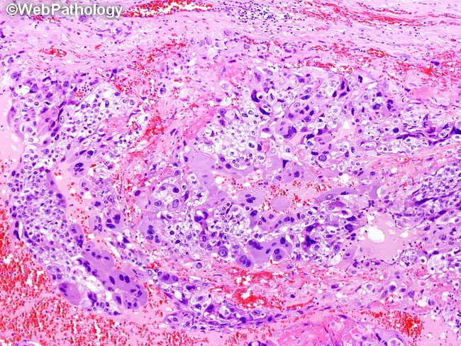
Light microscopy histology of a gestational trophoblastic neoplasm, specifically choriocarcinoma, using hematoxylin and eosin staining. The sampled tissue from the uterus shows a biphasic trophoblastic proliferation with intermixed cytotrophoblasts and syncytiotrophoblasts embedded in a hemorrhagic background. Cytotrophoblasts are mononucleated with clear to pale pink cytoplasm, distinct cell borders, irregular oval to elongate nuclei, prominent nucleoli, and brisk mitotic activity. They form plexiform aggregates and villous-like structures lacking stromal cores. Syncytiotrophoblasts are large, multinucleated or polyploid cells with abundant deeply eosinophilic cytoplasm and nuclei displaying dense, smudgy chromatin; they extend cytoplasmic processes around cytotrophoblast nests and infiltrate clumps of trophoblast. The interplay between these two cell types creates a biphasic architecture characteristic of choriocarcinoma, often accompanied by extensive hemorrhage and necrosis. The tumor lacks organized placental stromal support and shows high cellular pleomorphism and mitotic rate. Clinically, these histologic features support a diagnosis of gestational choriocarcinoma with high malignant potential and propensity for early hematogenous spread. The finding is diagnostic when coupled with elevated serum beta-hCG and pertinent clinical history. Potential uses include confirmation of diagnosis, prognostic assessment, and guidance of chemotherapy regimens for GTN. The description also informs differential considerations from PSTT or epithelioid trophoblastic tumor. Staging.
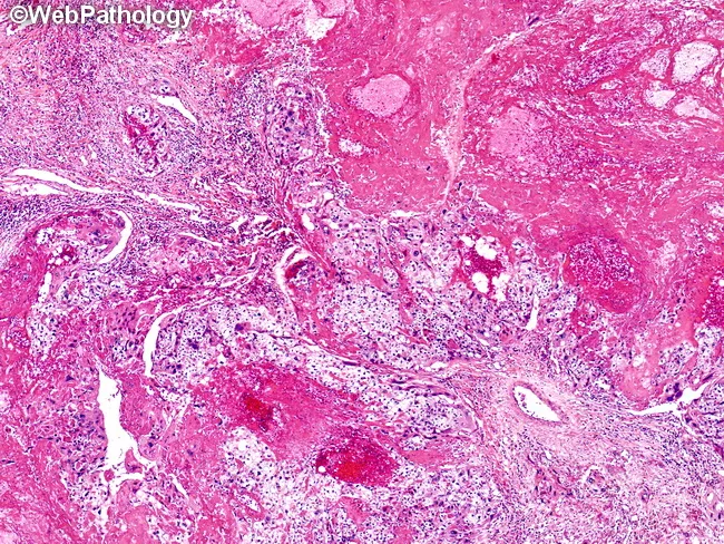
Histopathology image, prepared as a hematoxylin and eosin (H&E) stained tissue section, depicting choriocarcinoma within endometrial/uterine tissue. The lesion displays islands of viable malignant trophoblastic cells embedded in extensive hemorrhage and coagulative necrosis. Trophoblastic proliferation is biphasic, with cytotrophoblasts forming polygonal nests and clusters and syncytiotrophoblasts showing multinucleated, eosinophilic cytoplasm adjacent to fragile hemorrhagic stroma. Nuclear atypia and brisk mitotic activity are evident in viable foci. The surrounding matrix is fibromyxoid with necrotic debris and residual vascular channels, reflecting aggressive, vascular tumor growth. The lack of well-formed villi distinguishes choriocarcinoma from other gestational trophoblastic lesions. Areas of extensive hemorrhage contribute to the gross appearance of reddish, necrotic tissue in vivo. In immunohistochemistry, tumor cells typically express human chorionic gonadotropin (hCG) and cytokeratins, supporting trophoblastic lineage; p63 or inhibin may be variably positive, depending on subtype. Clinically, this histology corresponds to a highly vascular, metastasis-prone neoplasm that requires prompt evaluation of serum hCG levels and imaging for metastatic disease. The diagnostic significance lies in distinguishing choriocarcinoma from non-neoplastic gestational tissue and other germ cell tumors, guiding management including chemotherapy and monitoring for beta-hCG decline. This image is illustrative for educational reference, highlighting histologic hallmarks and diagnostic pitfalls in trophoblastic neoplasia today.
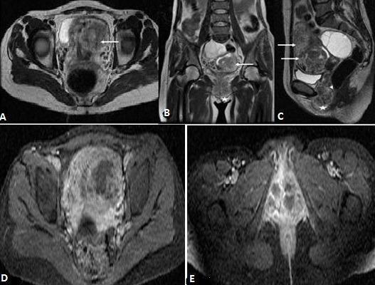
This diagnostic image composite displays pelvic Magnetic Resonance Imaging (MRI) findings in a case of Gestational Trophoblastic Neoplasia (GTN), likely an invasive mole or choriocarcinoma. Panels A, B, and C are T2-weighted sequences in axial, coronal, and sagittal planes, respectively. They reveal an enlarged uterus containing a bulky, heterogeneous tissue mass that extensively invades the myometrium (indicated by arrows). The sagittal view (C) demonstrates downward locoregional extension into the vaginal wall (arrowhead) and the labia majora (star symbol). Panels D and E show post-gadolinium T1-weighted axial images with fat saturation. These images exhibit a markedly heterogeneous enhancement pattern throughout the uterine mass. Prominent, well-demarcated non-enhancing zones are visible within the lesion, characteristic of necrotic regions common in rapidly growing trophoblastic tumors. The imaging highlights the aggressive, hypervascular nature of the tumor and its capacity for deep structural invasion into the pelvic floor, serving as a critical tool for FIGO staging and treatment planning in gynecologic oncology.
Note: Shaw's Textbook of Gynaecology is not available in the medical library. The following note is compiled from Berek & Novak's Gynecology (the premier gynaecology reference) supplemented by Robbins Pathology, covering all the same topics Shaw discusses on GTN.
Gestational Trophoblastic Neoplasia (GTN)
Definition and Overview
- Invasive mole
- Choriocarcinoma
- Placental-site trophoblastic tumor (PSTT)
- Epithelioid trophoblastic tumor (ETT)
- Berek & Novak's Gynecology, p. 2493
Types of GTN
1. Invasive Mole
- A hydatidiform mole that invades the myometrium or vascular spaces
- Locally destructive; may perforate the uterus
- Can metastasize but usually responds well to chemotherapy
2. Choriocarcinoma
- Highly malignant; no chorionic villi - composed of sheets of cytotrophoblasts and syncytiotrophoblasts with hemorrhage and necrosis
- Strong tendency for early hematogenous spread
- Can follow any pregnancy: molar (50%), abortion (25%), normal term (22.5%), ectopic (2.5%)
3. PSTT (Placental-Site Trophoblastic Tumor)
- Arises from intermediate trophoblast at the placental implantation site
- Produces hPL (human placental lactogen) more than hCG
- Relatively chemoresistant - hysterectomy is the primary treatment
4. ETT (Epithelioid Trophoblastic Tumor)
- Rare; arises from chorionic-type intermediate trophoblasts
- Also relatively resistant to chemotherapy
- Hysterectomy is preferred for nonmetastatic disease
Histology of Choriocarcinoma
Clinical Features / Diagnosis of GTN
- Rising hCG on 3 or more values over at least 2 weeks (days 1, 7, 14)
- Plateau of hCG on 4 measurements over 3 weeks (days 1, 7, 14, 21)
- Histologic diagnosis of choriocarcinoma
- hCG elevated 6 months or more after molar evacuation
Sites of Metastasis
| Site | Frequency |
|---|---|
| Lungs | 80% |
| Vagina | 30% |
| Pelvis | 20% |
| Liver | 10% |
| Brain | 10% |
- Berek & Novak's Gynecology, p. 2508-2509
MRI findings in GTN
FIGO Staging (GTN)
| Stage | Description |
|---|---|
| I | Disease confined to the uterus |
| II | GTN extending outside uterus but limited to genital structures (adnexa, vagina, broad ligament) |
| III | GTN extending to lungs with or without genital tract involvement |
| IV | All other metastatic sites (brain, liver, kidneys, GIT) |
- Berek & Novak's Gynecology, p. 2510
WHO Prognostic Scoring System
| Factor | Score 0 | Score 1 | Score 2 | Score 4 |
|---|---|---|---|---|
| Age | ≤39 yr | >39 yr | - | - |
| Antecedent pregnancy | Hydatidiform mole | Abortion | Term | - |
| Interval (end of pregnancy to chemo start) | <4 mo | 4-6 mo | 7-12 mo | >12 mo |
| hCG at diagnosis (IU/L) | <10³ | 10³-10⁴ | 10⁴-10⁵ | >10⁵ |
| ABO blood group | O or A | B or AB | - | - |
| Largest tumor size | <3 cm | 3-5 cm | >5 cm | - |
| Site of metastases | Lung/vagina | Spleen/kidney | GI tract | Brain/liver |
| No. of metastases | 0 | 1-4 | 5-8 | >8 |
| Prior chemotherapy | None | - | Single drug | ≥2 drugs |
- Score ≤6: Low risk - single-agent chemotherapy
- Score >6: High risk - combination chemotherapy required
Management
Diagnostic Evaluation
- Serial serum beta-hCG
- Chest X-ray / CT chest
- MRI pelvis
- CT head, abdomen if metastatic
Protocol for Treatment (Table 41-4)
- If fertility desired: Single-agent chemotherapy (methotrexate or actinomycin-D)
- If no fertility desired: Hysterectomy + adjuvant single-agent chemotherapy
- For PSTT and ETT: Hysterectomy (chemoresistant)
- Resistant disease: Combination chemotherapy, local resection
- Single-agent chemotherapy (~80% remission rate)
- Resistant: Combination chemotherapy
- Intensive combination chemotherapy (EMA-CO: etoposide, methotrexate, actinomycin-D, cyclophosphamide, vincristine) as primary treatment
- Brain metastases: Whole-head radiation (3,000 cGy) + craniotomy for complications
- Liver: Resection or embolization for complications
- Resistant: Second-line combination chemotherapy
Chemotherapy Agents
-
Single agents: Methotrexate (with or without leucovorin rescue), Actinomycin-D
-
Combination (EMA-CO): First-line for high-risk GTN
-
Second-line: BEP (bleomycin, etoposide, cisplatin), VIP, TP/TE regimens
-
Berek & Novak's Gynecology, p. 2512-2514
Follow-Up
- Weekly hCG measurement until normal for 3 consecutive weeks
- Monthly hCG measurement until normal for 12 consecutive months
- Effective contraception throughout the entire follow-up period (to avoid confusion from pregnancy-related hCG rise)
- Berek & Novak's Gynecology, p. 2514
Prognosis
- GTN has an excellent prognosis - even widespread metastatic disease can be cured with chemotherapy
- Low-risk disease: >95% cure rate with single-agent chemotherapy
- High-risk/stage IV disease: ~80-90% cure rate with combination regimens
- After achieving remission, patients can anticipate normal future reproductive outcomes
- Subsequent pregnancies after GTN treatment carry no increased risk of congenital anomalies or obstetric complications
Key Points Summary
| Feature | Detail |
|---|---|
| Marker | Beta-hCG (diagnosis, monitoring, relapse detection) |
| Most common antecedent | Hydatidiform mole |
| Most common metastatic site | Lung (80%) |
| Low-risk treatment | Single-agent MTX or Act-D |
| High-risk treatment | EMA-CO combination chemotherapy |
| Chemoresistant types | PSTT and ETT (prefer surgery) |
| Follow-up duration | 12 months of normal hCG |
| Prognosis | Excellent, even with metastases |
Sources: Berek & Novak's Gynecology (Chapter 41 - Gestational Trophoblastic Disease, pp. 2493-2521); Robbins & Kumar Basic Pathology; Creasy & Resnik's Maternal-Fetal Medicine