
Pt management and assessment for obstructive lung disease
Reading File
Now let me search the medical library for obstructive lung disease - assessment and management. I'll run parallel searches.
Finding Sources
Finding Sources
Searching PubMed
"obstructive lung disease" AND management
Good - found several relevant textbook sections. Let me read the key ones in parallel - Fishman's Pulmonary (comprehensive management) and also search for assessment/spirometry.
Reading File
Reading File
Finding Sources
Finding Sources
Reading File
Finding Sources
Finding Sources
Finding Sources
Finding Sources
Reading File
Reading File
Reading File
Searching Images
COPD GOLD staging spirometry FEV1 classification diagram
Reading File
Searching Images
obstructive lung disease flow volume loop spirometry
I now have comprehensive information to write a thorough response. Let me compile everything.
Loading Image
Loading Image
Excellent - I have comprehensive information and images. Now I'll compile a thorough, well-organized response.
Obstructive Lung Disease: Patient Assessment and Management
Obstructive lung disease is characterized by reduced airflow due to airway narrowing, air trapping, or loss of elastic recoil. The two main conditions are COPD (chronic bronchitis + emphysema) and asthma. COPD is the third most common cause of death in the United States.
1. OVERVIEW: What Is Obstructive Lung Disease?
| Feature | COPD | Asthma |
|---|---|---|
| Age of onset | Older adults (>40) | Any age |
| Reversibility | Not fully reversible | Largely reversible |
| Inflammation type | Neutrophilic | Eosinophilic (T2-high) |
| Progression | Inexorable with smoking | Variable |
| Response to ICS | Poor (unless eosinophilia) | Excellent |
- Katzung's Basic and Clinical Pharmacology, 16th Edition
2. PATIENT ASSESSMENT
2a. History and Symptoms
Key symptoms to elicit:
- Chronic cough with sputum production (defining feature of chronic bronchitis: productive cough ≥3 months/year for ≥2 consecutive years)
- Dyspnea - progressive, worsening on exertion
- Wheeze and chest tightness
- Exercise intolerance (hallmark of advanced disease)
- Exacerbation history: number per year, hospitalizations
Key risk factors:
- Cigarette smoking (primary risk factor - recognized even in smokers with "normal" spirometry)
- Older age
- Environmental/occupational exposures
- Alpha-1 antitrypsin deficiency (genetic)
Assess symptoms using validated tools:
-
mMRC dyspnea scale (0-4)
-
CAT score (COPD Assessment Test, 0-40)
-
Murray & Nadel's Textbook of Respiratory Medicine
2b. Spirometry (Essential for Diagnosis)
Spirometry is the cornerstone of COPD diagnosis. Post-bronchodilator values are used to distinguish COPD from asthma.
Diagnostic threshold:
- FEV1/FVC < 0.70 (GOLD fixed-ratio criteria)
- Alternative: < 5th percentile lower limit of normal (ATS/ERS - avoids overdiagnosis in elderly)
GOLD Severity Classification (by FEV1 % predicted):
| GOLD Grade | Severity | FEV1 % Predicted |
|---|---|---|
| GOLD 1 | Mild | ≥ 80% |
| GOLD 2 | Moderate | 50-79% |
| GOLD 3 | Severe | 30-49% |
| GOLD 4 | Very Severe | < 30% |
Note: GOLD now incorporates both FEV1 AND symptom burden + exacerbation history (GOLD ABE groups) for full staging.
Flow-volume loop in obstructive disease: Shows a characteristic concave (scooped) expiratory curve with reduced flow at all lung volumes. The volume-time curve shows prolonged expiratory time.
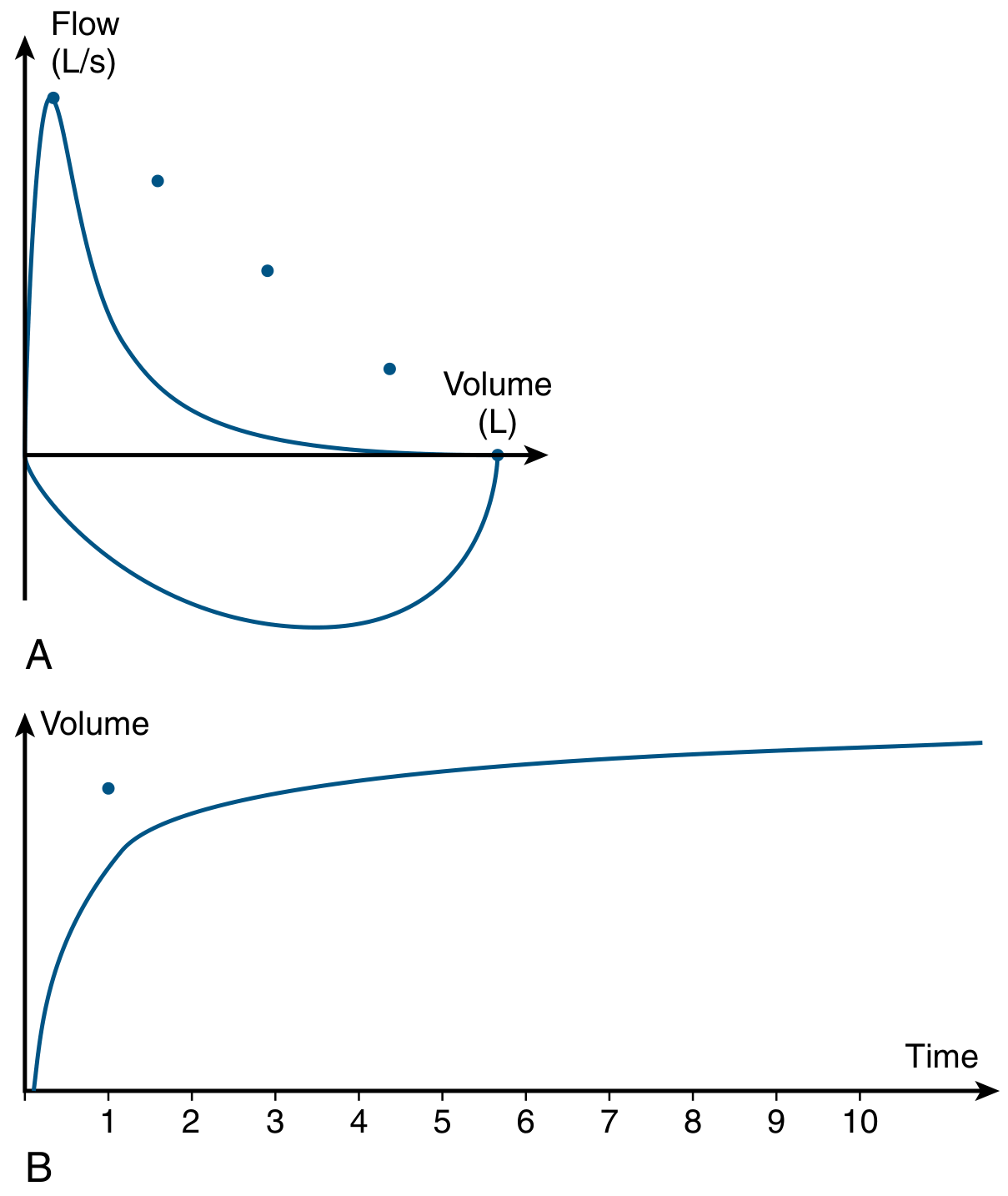
- Murray & Nadel's Textbook of Respiratory Medicine, p. 1471
2c. Lung Volumes
- TLC: Increased (especially in emphysema, due to loss of elastic recoil)
- RV: Increased disproportionately (air trapping)
- RV/TLC ratio: Elevated
- FRC: Increased (static hyperinflation)
- Vital capacity: Decreased due to hyperinflation
- Must measure by body plethysmography (not gas dilution) due to heterogeneous airflow and risk of underestimation
2d. Diffusing Capacity (DLCO)
- Reduced in emphysema (loss of alveolar-capillary surface area)
- Near-normal spirometry + severely reduced DLCO + radiographic emphysema = consider combined pulmonary fibrosis and emphysema syndrome
2e. Exercise Testing
6-Minute Walk Test (6MWT):
- Measures 6-minute walk distance (6MWD)
- Easy to administer, no specialized equipment needed
- Assesses need for supplemental O2 during ambulation
- Component of the BODE index (prognostic tool)
- Better correlates with quality-of-life outcomes than peak VO2
BODE Index (Mortality Predictor):
| BODE Score | 52-Month Mortality |
|---|---|
| 0-2 | 19% |
| 3-4 | 32% |
| 5-6 | 40% |
| 7-10 | 80% |
BODE components: Body mass index, airflow Obstruction (FEV1), Dyspnea (mMRC), Exercise capacity (6MWD)
- Murray & Nadel's Textbook of Respiratory Medicine
2f. Cardiopulmonary Exercise Testing (CPET)
In moderate-to-severe COPD:
- VE/MVV ratio approaches or exceeds 100% (ventilatory limitation)
- Dynamic hyperinflation develops: end-expiratory lung volume (EELV) rises, inspiratory reserve volume (IRV) falls
- PetCO2/PaCO2 may rise with exertion (CO2 accumulation)
- Elevated VE/VCO2 slope and VE intercept (VEint) - reflects increased dead space
- Exercise-related desaturation more common in emphysema than in chronic bronchitis phenotype
2g. Additional Investigations
- Arterial blood gas (ABG): PaCO2 typically does not rise above normal until FEV1 falls to ~20-25% of predicted
- Alpha-1 antitrypsin level: screen in patients <45 years or without clear smoking history
- CBC: polycythemia with chronic hypoxemia; eosinophil count guides ICS use
- CT chest: quantifies emphysema, airway disease; identifies comorbid findings
3. PATHOPHYSIOLOGY REVIEW
Understanding physiology guides management:
-
Chronic bronchitis: mucus overproduction from epithelial metaplasia + submucosal gland expansion + ciliary dysfunction; MUC5AC up 10-fold, MUC5B up 3-fold in severe COPD
-
Emphysema: increased lung compliance, elevated relaxation volume, "static hyperinflation"
-
Dynamic hyperinflation: with exercise or exacerbation, insufficient expiratory time traps air; EELV rises, IRV falls, work of breathing increases
-
Diaphragm dysfunction: flattened in hyperinflation, reduced force generation, paradoxical motion in severe disease
-
Hypercapnia: develops when FEV1 ~20-25%; due to increased dead space, V/Q mismatch, mechanical disadvantage, blunted chemoreceptor response
-
Fishman's Pulmonary Diseases and Disorders
4. MANAGEMENT
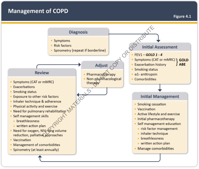
COPD Management Overview
4a. Non-Pharmacological (All Patients)
- Smoking cessation - single most effective intervention; slows progression
- Vaccination: influenza (annual), pneumococcal, COVID-19, pertussis
- Pulmonary rehabilitation: exercise training, self-management education, written action plans
- Supplemental oxygen: indicated if PaO2 ≤55 mmHg or SaO2 ≤88% at rest; prolongs survival
- Active lifestyle and exercise
4b. Pharmacological Management
Step 1 - Acute/Rescue:
- Short-acting beta-2 agonist (SABA): albuterol (salbutamol) - drug of choice for acute bronchospasm; inhaled, lasts several hours
- Short-acting anticholinergic (SAMA): ipratropium bromide
- Combination SABA + SAMA: more effective than either alone for acute relief
Step 2 - Stable Symptoms (persistent dyspnea/activity limitation):
- Long-acting beta-2 agonist (LABA): salmeterol, formoterol, vilanterol - preventive, 12-24 hour duration; NOT for acute exacerbations
- Long-acting anticholinergic/muscarinic antagonist (LAMA): tiotropium, umeclidinium
- LABA + LAMA combination: recommended for patients with persistent symptoms; superior to monotherapy
Step 3 - Role of Inhaled Corticosteroids (ICS):
ICS plays a more limited role in COPD than in asthma. Use is recommended for:
- Severe airflow obstruction (GOLD 3-4)
- History of two or more exacerbations/year OR one hospitalized exacerbation
- Elevated blood eosinophil count (suggests T2-high phenotype - more likely to respond)
- Clear coexisting asthma history
- Caution: ICS use is associated with increased risk of bacterial pneumonia
Agents: fluticasone propionate (commonly combined with LABA as fluticasone/salmeterol or fluticasone/vilanterol)
Triple therapy (LABA + LAMA + ICS): for severe, symptomatic patients with frequent exacerbations and elevated eosinophils
Other agents:
-
Roflumilast (PDE4 inhibitor): improves lung function and reduces exacerbation frequency; approved for severe COPD with chronic bronchitis phenotype
-
Azithromycin (daily): significantly reduces exacerbation frequency in patients with ≥2 exacerbations/year; used as add-on
-
Theophylline: no longer routinely recommended - recent large RCT failed to show benefit on exacerbation frequency
-
Katzung's Basic and Clinical Pharmacology, 16th Edition
4c. COPD Exacerbation Management
Exacerbations are the primary driver of morbidity, mortality, and healthcare costs.
Triggers: Predominantly viral respiratory infections; bacterial infections (especially Haemophilus influenzae) also common.
Treatment:
| Intervention | Role |
|---|---|
| SABA +/- SAMA | First-line bronchodilator therapy |
| Systemic corticosteroids | Reduce inflammation, shorten recovery |
| Antibiotics | Routine use (unlike asthma) - targets H. influenzae; use β-lactams, doxycycline, or azithromycin |
| Controlled oxygen | Target SaO2 88-92% (avoid over-oxygenation) |
| NIV (BiPAP) | For acute hypercapnic respiratory failure; reduces intubation rate |
| Invasive ventilation | For severe respiratory failure |
- Katzung's Basic and Clinical Pharmacology, 16th Edition
4d. Managing Chronic Hypercapnia
- NIPPV (non-invasive positive pressure ventilation) is a cornerstone of management for chronic hypercapnic COPD patients
- Persistent hypercapnia carries poor prognosis
- During REM sleep, diaphragmatic dysfunction is most pronounced - SaO2 drops, PtCCO2 rises
4e. Advanced/Surgical Options
- Lung volume reduction surgery (LVRS): in selected patients with upper-lobe predominant emphysema and low exercise tolerance post-rehabilitation
- Lung transplantation: end-stage disease; 6MWD used during transplant evaluation
- Bronchoscopic lung volume reduction (endobronchial valves)
5. ONGOING REVIEW
Per GOLD guidelines, periodic review should assess:
- Symptoms (CAT, mMRC)
- Exacerbation frequency
- Smoking status
- Inhaler technique and adherence (critical - poor technique negates benefit)
- Physical activity level
- Need for pulmonary rehabilitation
- Need for O2, NIV, or palliative approaches
- Management of comorbidities (cardiovascular disease is common and major contributor to mortality)
- Spirometry at least annually
Summary: Key Points for Assessment
- Spirometry is mandatory for diagnosis - post-bronchodilator FEV1/FVC <0.70 confirms obstruction
- Severity = GOLD 1-4 by FEV1 + GOLD ABE by symptoms and exacerbation history
- 6MWT and BODE index guide prognosis and transplant evaluation
- DLCO distinguishes emphysema from other causes; measure lung volumes by plethysmography
- Blood eosinophil count guides ICS prescribing
- PaCO2 rises late (when FEV1 ~20-25%) - its presence marks severe disease
Sources: Fishman's Pulmonary Diseases and Disorders; Murray & Nadel's Textbook of Respiratory Medicine; Katzung's Basic and Clinical Pharmacology 16th Ed; Costanzo Physiology 7th Ed; Robbins & Kumar Basic Pathology
Physiotherapy management
Physiotherapy is central to obstructive lung disease management - covering pulmonary rehabilitation, airway clearance, breathing techniques, exercise training, and education. Let me pull the best textbook content on this.
Searching sources...
Reading File
Finding Sources
Finding Sources
Searching PubMed
"pulmonary rehabilitation" AND "obstructive lung disease"
Finding Sources
Finding Sources
Finding Sources
Excellent - found the key sections. Let me read the main physiotherapy content from Fishman's and Murray & Nadel's in parallel.
Reading File
Reading File
Finding Sources
Reading File
Reading File
Finding Sources
Reading File
Reading File
Finding Sources
Reading File
Reading File
Searching Images
pulmonary rehabilitation exercise training COPD physiotherapy breathing techniques
I now have comprehensive content to deliver a thorough, well-organized physiotherapy management response.
Physiotherapy Management of Obstructive Lung Disease
Physiotherapy is a cornerstone of obstructive lung disease management. It sits within the broader framework of pulmonary rehabilitation (PR) - a comprehensive, evidence-based program that combines exercise training, breathing retraining, airway clearance, psychosocial support, and patient education.
1. PULMONARY REHABILITATION: Overview
Pulmonary rehabilitation is an established effective management strategy that:
Decreases:
- Dyspnea and breathlessness
- Hospital admissions and emergency visits
- Psychological symptoms (anxiety, depression, fear)
- Medical resource utilization
Increases:
- Exercise tolerance (endurance, maximal activity, strength)
- Health-related quality of life (HRQoL)
- Physical activity levels
- Patient independence and self-efficacy
- Knowledge and self-management skills
- Possible prolonged survival
Key point: Lung function (FEV1) does NOT typically improve with rehabilitation - but patients function far better within their existing limits.
Program structure:
-
Duration: typically 6-12 weeks, 2-3 supervised sessions per week
-
Each session: several hours of supervised exercise + individual/group education and psychosocial support
-
Benefits demonstrated in specialist centers AND community-based settings
-
Fishman's Pulmonary Diseases and Disorders
2. PRE-REHABILITATION ASSESSMENT
Before designing a program, thorough assessment is essential:
Pulmonary Function
- Spirometry and lung volumes - characterize disease and quantify impairment
- FEV1 is most useful for estimating maximal ventilatory capacity during exercise
- Diffusing capacity and maximal respiratory pressures (to assess muscle strength) added as needed
Exercise Testing
- Determines exercise tolerance and physiologic limitations
- Reveals coexisting conditions (e.g., cardiac disease)
- Used to establish a safe training prescription
- Type of test should match planned training modality (e.g., treadmill if walking training is planned)
- Incremental test to symptom-limited maximum → establishes peak capacity
- Steady-state test → preferred for determining training prescription
Blood Gas and Oxygenation Status
- ABG at rest AND during exercise - mandatory, since exercise-induced hypoxemia is common but cannot be reliably predicted from resting values
- In mild COPD: PaO2 may not change or may even improve with exercise
- In moderate-to-severe COPD: PaO2 may increase, decrease, or stay the same - unpredictable
- Pulse oximetry (SpO2) is useful for continuous monitoring but has ±4-5% accuracy limits
- Hypoxemia is not a contraindication to exercise training - portable O2 systems allow safe training
Psychosocial Evaluation
- Screen for depression, anxiety, social isolation, fear of dyspnea, and poor coping
Goal Setting
-
After evaluation, set realistic, individualized goals
-
Include family members and significant others
-
Use standardized outcome measures:
- 6-minute walk distance (6MWD) - exercise tolerance
- mMRC or Borg dyspnea scale - symptoms
- SGRQ or CAT - health-related quality of life
-
Fishman's Pulmonary Diseases and Disorders
3. CORE PHYSIOTHERAPY COMPONENTS
3a. Breathing Retraining Techniques
Breathing retraining aims to relieve and control breathlessness, improve ventilatory pattern, prevent dynamic airway compression, improve thoraco-abdominal synchrony, and enhance gas exchange.
Pursed-Lip Breathing (PLB):
- Technique first observed by Laennec in 1830; many patients adopt it naturally
- Patient tenses lips and narrows mouth opening during expiration
- Slows expiration, maintains positive airway pressure, prevents dynamic airway collapse
- Reduces dynamic hyperinflation
- Reduces respiratory rate, prolongs expiratory time
- Most consistently improves dyspnea symptoms
Diaphragmatic Breathing:
- Patient consciously coordinates abdominal wall expansion with inspiration
- Slows expiration through pursed lips simultaneously
- Primary effect: slows respiratory rate, increases tidal volume
- Improves respiratory synchrony between abdominal and thoracic musculature
- Symptom improvement (dyspnea) is a more consistent finding than measurable physiologic change
Mechanism (why these work in COPD):
In COPD, abnormal airways create a resistive load; hyperinflation creates an elastic load on the inspiratory muscles - increasing work and effort of breathing. Pursed-lip breathing mitigates this by slowing and prolonging exhalation, which reduces dynamic hyperinflation. - Murray & Nadel's Textbook of Respiratory Medicine
3b. Bronchial Hygiene (Airway Clearance)
Patients with obstructive lung disease frequently have abnormal airway clearance mechanisms, retained secretions, and recurrent infections.
Techniques taught in rehabilitation:
| Technique | Description |
|---|---|
| Directed coughing (huff coughing) | Forced expiratory technique - a "huff" (open glottis) to mobilize secretions without precipitating airway collapse |
| Postural drainage | Positioning the patient to use gravity to drain specific lung segments |
| Chest percussion | Manual or mechanical tapping over lung segments to loosen secretions |
| Chest vibration | Applied during expiration over drainage positions to enhance secretion movement |
| Active cycle of breathing technique (ACBT) | Combines breathing control + thoracic expansion exercises + forced expiration |
| Positive expiratory pressure (PEP) devices | Maintain airway patency during expiration to prevent collapse and aid secretion clearance |
These techniques are particularly important for patients with:
- Chronic sputum production
- Excess mucus during exacerbations
- Chronic bronchitis phenotype
Note: Mucolytic agents to reduce sputum viscosity are of questionable benefit.
- Fishman's Pulmonary Diseases and Disorders
3c. Exercise Training
Exercise is the most evidence-based component of pulmonary rehabilitation, with both physiologic and psychological benefits. Patients can increase maximal capacity and endurance even though objective lung function does not change.
Types of exercise used:
| Type | Application |
|---|---|
| Walking programs | Most useful; highly applicable to daily life; can be indoor or outdoor |
| Cycling (stationary or outdoor) | Effective lower limb endurance training |
| Swimming | Effective; breathing pattern benefits |
| Upper extremity training | Critical - see below |
| Resistance/strength training | Significant increases in muscle strength for ADLs |
Lower extremity training is the primary focus (walking, cycling) as it targets the muscles most limiting daily function.
Upper extremity training is specifically important because:
- Many patients report disabling dyspnea at very low levels of upper limb work (lifting, grooming, overhead activities)
- Upper limb exercise creates a higher ventilatory demand per unit of work than lower limb exercise
- Training is largely muscle-specific - upper limb training improves upper limb function in ways lower limb training cannot
Exercise Prescription
-
Start at a level the patient can sustain comfortably for several minutes
-
Progress by increasing duration first, then intensity, guided by symptom tolerance (Borg dyspnea/breathlessness rating) rather than heart rate
-
Target: 15-30 minutes of continuous sustained activity
-
In severe disease with ventilatory limitation: interval training (alternating bouts of high and low intensity) may allow higher total work volumes than continuous training
-
Resistive training added for strength gains relevant to ADLs
-
Encourage patients to incorporate exercise into enjoyable daily activities (gardening, golf, social walking)
-
Fishman's Pulmonary Diseases and Disorders
3d. Ventilatory (Inspiratory) Muscle Training (IMT)
-
Techniques: isocapnic hyperventilation, inspiratory resistive loading, inspiratory threshold loading
-
Can improve respiratory muscle function in isolation
-
However, improvement in general exercise performance from IMT alone has not been demonstrated consistently in COPD
-
Role as a routine component of PR is not clearly established
-
May be tried in patients with demonstrable respiratory muscle weakness alongside nutritional support
-
Evidence for IMT efficacy remains limited
-
Fishman's Pulmonary Diseases and Disorders; Murray & Nadel's Textbook of Respiratory Medicine
3e. Supplemental Oxygen During Rehabilitation
- Long-term continuous oxygen therapy (LTOT): clearly improves survival and reduces morbidity in patients with severe resting hypoxemia (PaO2 ≤55 mmHg or SaO2 ≤88%)
- Benefits for non-hypoxemic patients or those with intermittent hypoxemia (exercise- or sleep-induced) are less clearly defined
- Each patient's oxygen needs should be individually assessed
- Portable liquid oxygen systems are preferred for ambulatory patients - more gas, less weight than compressed tanks
- Oxygen-conserving devices extend the life of portable gas sources
- Transtracheal oxygen delivery: improves compliance in selected patients; requires careful catheter care instruction
- Hypoxemia during exercise is NOT a contraindication - portable O2 allows safe training
3f. Education and Self-Management
Education is integral but insufficient alone - attitude and behavior change requires individualized instruction and reinforcement.
Topics covered:
- Normal lung anatomy and physiology
- Understanding their specific lung disease
- Inhaler technique and medication use (nebulizers, MDIs, DPIs)
- Supplemental oxygen use and equipment management
- Written action plan - recognizing exacerbation signs and when to seek help
- Energy-conservation techniques for ADLs
- Nutrition
- Travel with lung disease
- Stress reduction and relaxation techniques
- Smoking cessation support
- Breathlessness management strategies
Philosophy: patients become partners in their own care, assuming active responsibility rather than passive recipients.
3g. Psychosocial Support
An essential physiotherapy and rehabilitation component. Goals:
- Combat progressive hopelessness and inability to cope with chronic progressive disease
- Depression, anxiety (especially around dyspnea), denial, anger, and isolation are all common
- Sexual dysfunction and fear of physical activity are frequent
- Group sessions provide peer support and reduce isolation
- Desensitization to dyspnea: through exercise training, patients still experience dyspnea but develop less anxiety and fear about it and can persist in activity longer - a key mechanism of rehabilitation benefit
4. REHABILITATION IN SPECIAL SITUATIONS
After Acute Exacerbation
-
Landmark evidence: PR initiated within 10 days of hospital discharge for COPD exacerbation leads to large, clinically meaningful improvements in exercise capacity and HRQoL vs. usual care
-
Reduces hospital readmission rates (supported by Cochrane meta-analysis)
-
May reduce mortality
-
Early post-exacerbation rehabilitation is strongly indicated
-
Fishman's Pulmonary Diseases and Disorders
Pre- and Post-Lung Transplantation
- Pre-transplant: maintain function during waiting period (which may last months to years); monitor disease progression; prevent complications; provide education about transplant procedure and expectations; psychosocial support for patients and families
- Post-transplant: different goals - rebuilding from baseline; managing new medications; functional independence
Pre- and Post-Lung Volume Reduction Surgery (LVRS)
- Rehabilitation improves surgical candidacy and post-operative recovery
- Post-LVRS: reduced hyperinflation means respiratory muscles operate more favorably on their length-tension curve → improved diaphragmatic function → better exercise response to training
5. NON-INVASIVE VENTILATION (NIV) AND PHYSIOTHERAPY
- BiPAP/NIPPV provides inspiratory muscle support, offloads the burden on the respiratory muscles, and reduces work of breathing
- Used acutely in emergency department and ICU for acute hypercapnic exacerbation
- Increasingly used in the outpatient setting for chronic hypercapnic COPD
- Can be combined with exercise training in very severe disease to allow higher training intensities
- Physiotherapists play a key role in NIV setup, mask fitting, and patient education
6. OUTCOMES AND MONITORING
| Outcome Domain | Measure |
|---|---|
| Exercise capacity | 6MWT, CPET (peak VO2, ventilatory threshold) |
| Dyspnea | Borg scale, mMRC, Modified Medical Research Council scale |
| Health-related quality of life | SGRQ, CAT, CRQ |
| Psychological function | HAD scale (anxiety/depression) |
| Functional status | Activities of daily living performance |
| Healthcare utilization | Hospitalization rate, ED visits |
Ongoing review should include spirometry at least annually, reassessment of symptoms, exacerbation history, smoking status, inhaler technique, and whether goals need updating.
Summary Table: Physiotherapy Interventions
| Intervention | Evidence Level | Primary Benefit |
|---|---|---|
| Pulmonary rehabilitation (PR) | Strong (Cochrane meta-analysis) | Exercise capacity, HRQoL, hospitalization ↓ |
| Lower limb endurance training | Strong | Functional exercise capacity |
| Upper limb training | Moderate | Upper limb ADL performance, dyspnea |
| Pursed-lip breathing | Moderate | Dyspnea control, dynamic hyperinflation ↓ |
| Diaphragmatic breathing | Moderate | Respiratory pattern, dyspnea |
| Airway clearance (CPT, PEP, ACBT) | Moderate | Secretion clearance in chronic bronchitis |
| Inspiratory muscle training | Limited (inconsistent) | Muscle strength (not general exercise) |
| Early post-exacerbation PR | Strong | Readmission ↓, survival ↑ |
| Supplemental O2 + exercise | Strong (in hypoxemic pts) | Safe exercise, survival benefit |
Sources: Fishman's Pulmonary Diseases and Disorders; Murray & Nadel's Textbook of Respiratory Medicine; Cochrane Database Systematic Reviews (Puhan et al., 2016 - PR after COPD exacerbations, PMID: 37858727)
This is a shared conversation. Sign in to Orris to start your own chat.