
Hepamerz infusion in non alcoholic hepatic encephalopathy
L-ornithine L-aspartate hepatic encephalopathy non-alcoholic
L-ornithine L-aspartate hepatic encephalopathy
LOLA infusion ammonia cirrhosis randomized trial
PMID: 34822189
PMID: 38403493
PMID: 40370740
PMID: 38789596
hepatic encephalopathy ammonia urea cycle L-ornithine L-aspartate mechanism

This biochemical diagram illustrates the ionization equilibrium of L-ornithine, a basic amino acid involved in the urea cycle and ammonia detoxification. The diagram displays four distinct ionic states (L-ornithine2+, L-ornithine+, L-ornithine±, and L-ornithine-) transitioning via sequential deprotonation. The first transition (K1) shows the deprotonation of the carboxylic acid group (COOH to COO-). The second transition (K2) depicts the deprotonation of the terminal delta-amino group (NH3+ to NH2), leading to the zwitterionic form (L-ornithine±). The final transition (K3) represents the deprotonation of the alpha-amino group on the chiral carbon, resulting in the negatively charged anionic form (L-ornithine-). Each step is labeled with its respective equilibrium constant (K1, K2, K3), illustrating the chemical behavior of the molecule across a pH gradient. This clinical chemistry schematic is essential for understanding the pharmacology of L-ornithine L-aspartate used in treating hepatic encephalopathy.
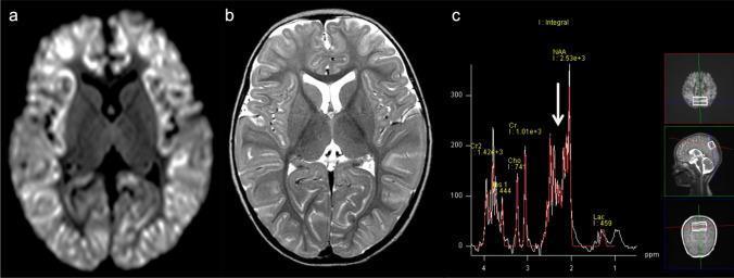
This composite educational image illustrates neuroimaging and spectroscopic findings in a 2-year-old female with Ornithine Transcarbamylase (OTC) deficiency, a urea cycle disorder presenting with acute hyperammonemic encephalopathy. Panel (a) is an axial Diffusion-Weighted Image (DWI) showing a diffuse 'scalloped ribbon' pattern of restricted diffusion throughout the cerebral cortex. Panel (b) is an axial T2-weighted image corroborating these findings with diffuse cortical hyperintensity and edema. The thalami appear relatively spared, a key feature in differentiating urea cycle disorders from hypoxic-ischemic encephalopathy. Panel (c) presents a Proton MR Spectroscopy (1H-MRS) graph (TE = 30 ms) from the bilateral parietal lobes. The spectrum demonstrates a slight elevation in the Glx peak (glutamine-glutamate complex), indicated by a white arrow, alongside standard peaks for NAA, Cr, Cho, and mIns. This diagnostic combination highlights the characteristic imaging signatures of acute metabolic brain injury secondary to neonatal or childhood-onset hyperammonemia.
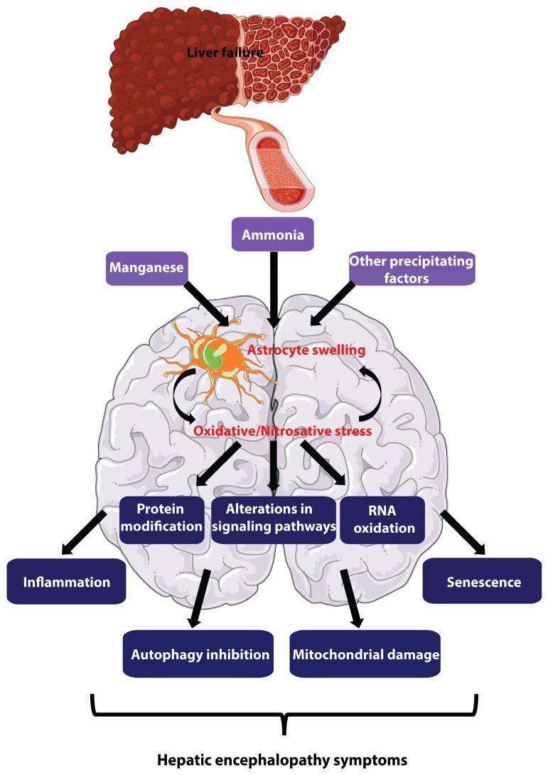
This pathophysiology diagram illustrates the major pathogenesis mechanisms of hepatic encephalopathy (HE). The flow begins with liver failure, shown as a cirrhotic liver, releasing metabolic toxins like ammonia and manganese into the systemic circulation via a blood vessel. These toxins, along with other precipitating factors, target the brain, specifically inducing astrocyte swelling. A central self-amplifying loop is depicted where astrocyte swelling and oxidative/nitrosative stress mutually reinforce each other. This cycle triggers a cascade of detrimental intracellular processes, including protein modification, RNA oxidation, and alterations in signaling pathways. Downstream effects featured include inflammation, autophagy inhibition, mitochondrial damage, and cellular senescence. These multi-factorial changes are linked via brackets to the ultimate manifestation of hepatic encephalopathy symptoms. The illustration uses a combination of anatomical representations (liver and brain) and text-based flow nodes to describe the transition from hepatic dysfunction to neurometabolic impairment, emphasizing the central role of astrocytes in the disease progression.
Hepamerz (L-Ornithine L-Aspartate) Infusion in Non-Alcoholic Hepatic Encephalopathy
What is Hepamerz?
Mechanism of Action
1. Urea Cycle Stimulation (periportal hepatocytes)
- Ornithine is a key intermediate and activator of the urea cycle. It activates carbamoyl phosphate synthetase I (CPS-I), the rate-limiting enzyme, driving conversion of NH3 → urea.
- Aspartate acts as a nitrogen donor in the cycle, combining with citrulline to form argininosuccinate.
2. Glutamine Synthesis (perivenous hepatocytes + skeletal muscle)
- In remaining functional hepatocytes and in skeletal muscle, ornithine and aspartate serve as substrates for glutamine synthetase.
- NH3 + glutamate → glutamine (a non-toxic transport form of ammonia, excreted by kidneys).
- This pathway is particularly important when the urea cycle capacity is reduced due to advanced liver disease.
Classification of Hepatic Encephalopathy
| Grade | West Haven Criteria | Clinical Features |
|---|---|---|
| Minimal (MHE) | Psychometric abnormality only | No clinical signs; detected by testing |
| Grade I | Trivial lack of awareness, shortened attention | Sleep disturbance, mild confusion |
| Grade II | Lethargy, disorientation to time | Asterixis, personality change |
| Grade III | Somnolence, confusion, gross disorientation | Asterixis, bizarre behavior |
| Grade IV | Coma | Unresponsive |
Dosing and Administration
Intravenous (Infusion) - Hepamerz Concentrate
- Standard adult dose: 20-40 g IV per day, diluted in 500 mL of 5% dextrose or normal saline, administered over 4-8 hours (max infusion rate: 5 g/hour to avoid nausea/vomiting)
- For acute/severe OHE (Grade III-IV): 30 g/24 hours as a continuous infusion for 5 days - this regimen was used in the landmark 2022 RCT (PMID: 34822189)
- Duration: Typically 5-7 days for acute episodes; may be extended
- Dilution: Each ampoule (10 g/10 mL or 10 g/20 mL) must be diluted - never administer undiluted
Oral Sachets (maintenance/MHE)
- 3 g sachets, 3 times daily with meals
Clinical Evidence
In Overt/Severe HE (Grade III-IV)
- Design: 140 cirrhotic patients with OHE Grade III-IV randomized to LOLA 30 g/24h continuous IV infusion + lactulose + rifaximin vs. placebo + lactulose + rifaximin
- Results:
- HE grade improvement: 92.5% vs. 66% (p < 0.001) - LOLA group markedly superior
- Time to recovery: 2.70 ± 0.46 vs. 3.00 ± 0.87 days (p = 0.03) - faster recovery
- 28-day mortality: 16.4% vs. 41.8% (p = 0.001) - dramatic mortality benefit
- Significant reductions in ammonia, IL-6, and TNF-alpha in LOLA group
- Conclusion: LOLA + lactulose + rifaximin combination superior to lactulose + rifaximin alone
IV vs. Oral LOLA Comparison
- Mean ammonia reduction Day 1 to Day 5: 55.4 µmol/L (IV) vs. 60.75 µmol/L (oral) - no significant difference (p = 0.511)
- Both routes equally effective in HE grade improvement
- Both significantly reduced ammonia within each group (p < 0.001)
- Implication: IV route preferred in obtunded/comatose patients who cannot take oral medications; once conscious, can transition to oral
In Minimal Hepatic Encephalopathy (MHE)
- LOLA superior to placebo in reversing MHE (RR = 2.26, 95% CI: 1.53-3.35)
- LOLA prevented progression to OHE (RR = 0.22, 95% CI: 0.076-0.637)
- Oral LOLA appeared slightly more effective than IV for MHE reversal in subgroup analysis
- No mortality benefit shown in this MHE population (expected, as MHE is early stage)
LOLA + Lactulose Combination (2025 Meta-analysis)
- LOLA + lactulose combination is 31% more effective than lactulose alone (RR = 1.31, 95% CI: 1.22-1.42)
- Significant reductions in AST, ALT, total bilirubin, and NH3
- Good safety profile
Post-TIPS HE Prevention
Role in Non-Alcoholic Etiology Specifically
-
Pathophysiology is the same - regardless of etiology, the final common pathway of HE involves hyperammonemia, astrocyte swelling, and neuroinflammation. LOLA addresses the ammonia component directly.
-
NAFLD/NASH cirrhosis - these patients often have preserved muscle mass initially, meaning skeletal muscle glutamine synthesis (one of LOLA's mechanisms) may work more effectively.
-
No etiology-specific contraindication - LOLA is used across alcoholic and non-alcoholic etiologies. The Jain 2022 RCT included patients with a wide etiology mix (viral + NAFLD + cryptogenic).
-
Inflammation reduction - IL-6 and TNF-alpha reduction seen with LOLA are particularly relevant in NASH, which has a baseline inflammatory milieu that may exacerbate HE.
Position in Treatment Algorithm
Overt HE (Grade II-IV):
1. Identify & treat precipitants (infection, bleeding, hypokalemia, drugs)
2. Lactulose 30-60 g/day (titrate to 2-3 soft stools/day) - FIRST LINE
3. Add Rifaximin 550 mg BD - especially for recurrence prevention
4. Add LOLA IV infusion (Hepamerz 20-30 g/day) - ADJUNCT, particularly in:
   - Grade III-IV (severe) OHE
   - Post-TIPS HE
   - Inadequate response to lactulose ± rifaximin
   - Unable to take oral medications
5. Branched-chain amino acids - if indicated
6. Treat underlying liver disease / consider transplant evaluation
Safety Profile
- Well tolerated overall
- Nausea/vomiting - most common side effect, especially if infused too rapidly (keep rate ≤ 5 g/hour)
- Contraindicated in severe renal impairment (GFR < 30 mL/min) - risk of glutamine accumulation
- Caution in pregnancy (limited data)
- No significant drug-drug interactions with lactulose or rifaximin
Summary Table
| Parameter | Detail |
|---|---|
| Drug class | Amino acid dipeptide (ammonia detoxifier) |
| Brand name | Hepamerz |
| Mechanism | Urea cycle activation + glutamine synthesis |
| IV dose | 20-30 g/day in 500 mL over 4-8 hours |
| Duration | 5-7 days acute; oral for maintenance |
| Key evidence | 92.5% vs 66% HE improvement (Jain 2022 RCT) |
| Mortality benefit | 16.4% vs 41.8% at 28 days in Grade III-IV |
| MHE | 2.26x more likely to reverse MHE vs placebo |
| Combination benefit | 31% more effective than lactulose alone |
| Use in non-alcoholic HE | Supported - same mechanism, no etiology restriction |
| Main adverse effect | Nausea/vomiting if infused rapidly |
| Contraindication | Severe renal failure |
Can i give it in alf?
L-ornithine L-aspartate acute liver failure
PMID: 37945461
acute liver failure HE management cerebral edema ammonia ICU
PMID: 33029057
Hepamerz (LOLA) in Acute Liver Failure (ALF)
Short Answer: Use with Caution - NOT Standard of Care
Why ALF is Different from Chronic Liver Disease HE
| Feature | Chronic Cirrhotic HE | ALF-HE |
|---|---|---|
| Ammonia source | Gut-derived, portal shunting | Massive hepatocyte necrosis + portal shunting |
| Brain effect | Astrocyte dysfunction, cognitive impairment | Acute cerebral edema + intracranial hypertension |
| Urea cycle capacity | Reduced but present | Near-absent (massive hepatocyte loss) |
| Muscle ammonia handling | Relatively preserved | Overwhelmed |
| Urgency | Subacute | Life-threatening within hours |
| Reversibility | Yes, with treatment | Depends on liver regeneration or transplant |
The Glutamine Paradox - The Core Concern
- Astrocyte glutamine accumulates faster than it can be exported
- Glutamine itself is osmotically active inside astrocytes
- This causes astrocyte swelling - the primary driver of cerebral edema in ALF
- So excess glutamine formation (one of LOLA's mechanisms) can paradoxically worsen cerebral edema
What the Evidence Says
-
Plum & Posner's (Stupor and Coma): LOLA is listed for the chronic liver failure state only. For ALF management, the focus shifts to: mannitol for ICP, N-acetylcysteine (in paracetamol-induced ALF), continuous renal replacement therapy (CRRT), and liver transplant evaluation - LOLA is not mentioned.
-
Sleisenger & Fordtran (GI & Liver Disease): LOLA is noted to activate the urea cycle and enhance ammonia clearance, with a possible beneficial effect on mortality in HE - but this review evidence is for cirrhotic HE, not ALF specifically.
-
INASL Consensus on ALF Management (PMID 33029057): Acknowledges ammonia plays a crucial role and that "the role of newer ammonia-lowering agents is still evolving" in ALF. The emphasis is on early CRRT for ammonia control, lactulose, and transplant - LOLA is not a first-line recommendation.
-
Karvellas et al. 2024 ICU Review (PMID 37945461): LOLA is listed among ammonia-targeted strategies in liver failure (alongside laxatives, rifaximin, CRRT) but the review contextualizes this mainly for ACLF, not pure ALF.
Practical Summary: When LOLA Can/Cannot Be Used in ALF
| Scenario | LOLA Use | Rationale |
|---|---|---|
| True ALF (INR >1.5, encephalopathy, no prior cirrhosis, acute onset <26 weeks) | Not recommended / avoid | Urea cycle non-functional; glutamine accumulation worsens cerebral edema; no evidence of benefit |
| ALF with Grade I-II HE (early, no cerebral edema yet) | Uncertain / no good evidence | Theoretical risk; insufficient RCT data in this group |
| Acute-on-Chronic Liver Failure (ACLF) | Can consider as adjunct | Some residual urea cycle + skeletal muscle function; evidence from chronic HE context applies more |
| ALF requiring ICU / cerebral edema (Grade III-IV HE) | Contraindicated in practice | Mannitol, hypertonic saline, ICP monitoring, CRRT, and transplant are priorities |
What Should You Use Instead in ALF?
- Treat the precipitant - N-acetylcysteine (paracetamol), antivirals (HBV), steroids (autoimmune), etc.
- Lactulose - to reduce gut ammonia production; use cautiously (avoid if ileus or risk of abdominal distension worsening ICP)
- Rifaximin - oral, safe, reduces ammonia-producing gut flora
- CRRT (continuous renal replacement therapy) - the most effective ammonia-lowering strategy in ALF with high ammonia/cerebral edema; removes ammonia directly from blood
- Mannitol / hypertonic saline - for elevated ICP / cerebral edema
- Avoid sedatives, narcotics, nephrotoxins
- Early transplant evaluation - the only definitive therapy for poor-prognosis ALF
- MARS (Molecular Adsorbent Recirculating System) - extracorporeal albumin dialysis; reduces ammonia and improves severe HE in ACLF
Bottom Line
LOLA/Hepamerz is not recommended in true ALF. Its mechanism depends on functional hepatocytes (for the urea cycle) and generates glutamine - which in ALF can worsen the cerebral edema that kills these patients. The priority in ALF-HE is CRRT, ICP management, and liver transplantation. LOLA has a role in chronic cirrhotic HE and ACLF, but not in hyperacute/acute liver failure.
- Sleisenger & Fordtran's GI and Liver Disease, p. 1817 | Plum & Posner's Stupor and Coma, p. 626-628 | INASL ALF Consensus, PMID 33029057
Acute liver failure definition
Acute Liver Failure (ALF) - Definition
Core Definition
- Severe acute liver injury (hepatocyte necrosis/dysfunction)
- Coagulopathy - INR > 1.5
- Hepatic encephalopathy (HE) - any grade
"Acute liver failure is defined as a severe acute liver injury that precipitates the sudden onset of encephalopathy and coagulopathy (INR > 1.5). By definition, acute liver failure results from an insult within the prior 6 months."
- Goldman-Cecil Medicine
"Encephalopathy in the appropriate setting remains the essential requirement for the diagnosis of ALF."
- Sleisenger & Fordtran
Classification by Time Course (O'Grady/King's College Classification)
| Category | Jaundice to Encephalopathy | Cerebral Edema Risk | Prognosis (without LT) |
|---|---|---|---|
| Hyperacute | ≤ 7 days | Very high (~80% in Grade IV HE) | Paradoxically better - higher spontaneous recovery |
| Acute | 8-28 days | Moderate-high | Intermediate |
| Subacute | 4-24 weeks | Low | Worst - less cerebral edema but poor spontaneous recovery |
- Fulminant hepatic failure - encephalopathy within 8 weeks of illness onset
- Subfulminant hepatic failure - liver disease up to 26 weeks before encephalopathy
Key Distinguishing Features (vs. Chronic Liver Disease)
| Feature | ALF | Chronic HE (Cirrhosis) |
|---|---|---|
| Prior liver disease | Absent (by definition) | Present |
| Onset | Rapid (days-weeks) | Gradual |
| HE mechanism | Cerebral edema + intracranial hypertension | Portosystemic shunting, astrocyte dysfunction |
| Portal hypertension | May be absent early | Present |
| Variceal bleeding | Less common | More common |
| Coagulopathy | Severe, rapid | Chronic, compensated |
| Factor V | Rapidly falls (most sensitive marker) | Slowly declining |
| Reversibility | If liver regenerates or transplant done | Chronic but manageable |
"The encephalopathy of acute liver failure is mediated by cerebral edema, not by the portosystemic shunting and portal hypertension of cirrhosis."
- Goldman-Cecil Medicine
Important Nuance: Wilson's Disease and Autoimmune Hepatitis
Causes by Geography
| Cause | UK | USA | India | France |
|---|---|---|---|---|
| Acetaminophen | 54% | 46% | — | 2% |
| Indeterminate | 17% | 14% | — | — |
| Viral hepatitis (A, B, E) | Rare | ~10% | Most common | Variable |
| Idiosyncratic DILI | — | 13% | ATT drugs | — |
| Autoimmune hepatitis | — | 4% | — | — |
Acute-on-Chronic Liver Failure (ACLF) - Distinction
- Liver failure triggered by an acute insult in a patient with pre-existing liver disease / cirrhosis
- Precipitants: portal hypertensive bleeding, sepsis, acute viral hepatitis superimposed on cirrhosis
- Has a different pathophysiology and prognosis from true ALF
Summary
ALF = Coagulopathy (INR > 1.5) + Hepatic Encephalopathy + No prior chronic liver disease + Onset within 26 weeks Encephalopathy is the sine qua non of the diagnosis. The subtype (hyperacute vs acute vs subacute) depends on the jaundice-to-encephalopathy interval and determines prognosis.
- Goldman-Cecil Medicine, p. 174 | Sleisenger & Fordtran, p. 1836 | Current Surgical Therapy 14e, p. 460