
Midgut volvulus
midgut volvulus malrotation pediatric radiology
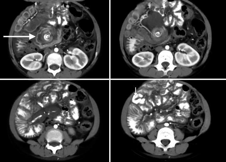
This medical image is a series of four axial slices from a contrast-enhanced abdominal computed tomography (CT) scan illustrating midgut malrotation with volvulus. Key diagnostic features include the 'whirlpool sign' (indicated by a large white arrow), which demonstrates the twisting of the mesenteric vessels and mesentery around the base of the superior mesenteric artery. The duodenojejunal flexure is abnormally positioned on the right side of the spine instead of the left. Additionally, the jejunal loops, identified by their internal iodinated contrast (noted by a smaller arrow), are predominantly located in the right hemiabdomen rather than the left upper quadrant. These findings collectively indicate a failure of normal intestinal rotation and subsequent midgut volvulus, which can lead to intestinal ischemia. The image is a classic example used in pediatric radiology to demonstrate surgical emergencies related to congenital intestinal anomalies.
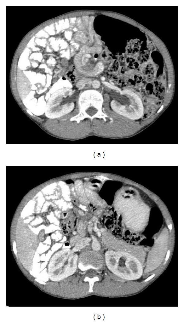
Two axial postcontrast CT images of the abdomen demonstrate findings consistent with midgut malrotation and volvulus. Image (a) reveals the characteristic 'whirl sign,' where the duodenum and a dilated superior mesenteric vein (SMV) wrap in a clockwise direction around the axis of the superior mesenteric artery (SMA). Image (b) shows a reversal of the normal mesenteric vascular anatomy, with the SMV located to the left of the SMA rather than its typical right-sided position. Additional findings include the displacement of jejunal loops to the right side of the abdominal cavity. These radiographic features are hallmark indicators of intestinal malrotation, commonly used in pediatric and emergency radiology to diagnose volvulus and associated mesenteric vascular torsion.
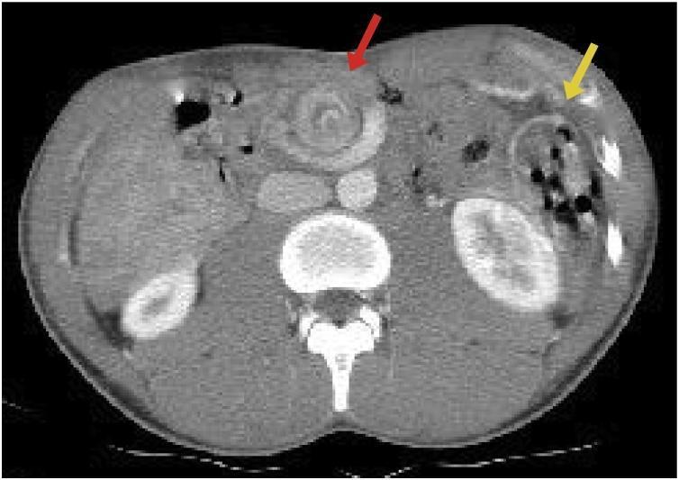
This axial contrast-enhanced abdominal CT scan illustrates pathognomonic findings of intestinal malrotation and midgut volvulus. A prominent 'whirlpool sign' (indicated by a red arrow) is visible in the central abdomen, characterized by the spiral twisting of the superior mesenteric vein and mesentery around the superior mesenteric artery. Additionally, the image demonstrates an aberrant bowel distribution (indicated by a yellow arrow), where small bowel loops are predominantly located in the right hemiabdomen and the colon is displaced to the left, confirming a failure of normal embryological rotation. These radiological findings are critical for the diagnosis of acute midgut volvulus, highlighting the risk of mesenteric vascular compromise and bowel ischemia. The image serves as a high-level educational example for radiology and pediatric surgery trainees regarding congenital gastrointestinal anomalies and acute abdominal emergencies.
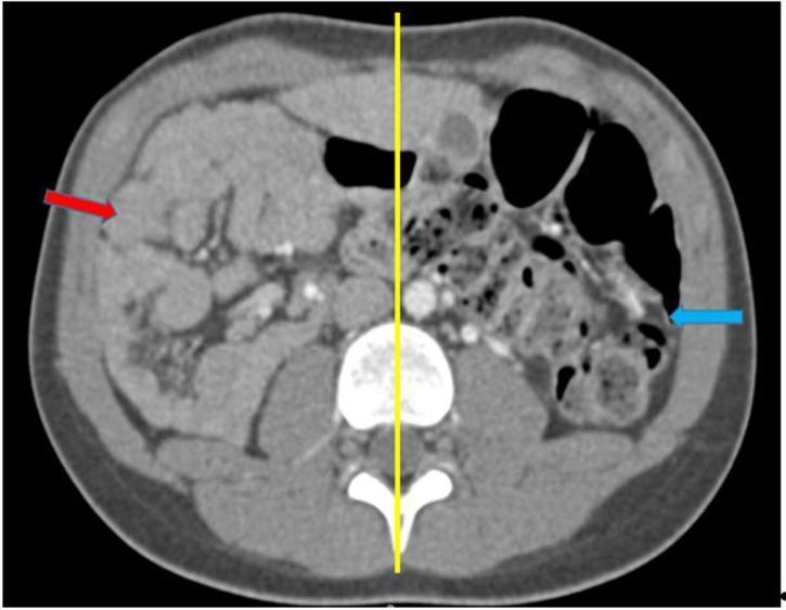
This diagnostic image is an axial contrast-enhanced CT (TDM) scan of the abdomen demonstrating a complete common mesentery, a type of intestinal malrotation. The image shows a distinct anatomical deviation from normal intestinal positioning, highlighted by a yellow vertical bisecting line. The small bowel loops are entirely sequestered on the right side of the abdominal cavity (indicated by the red arrow), while the colon and large bowel segments are located on the left side (indicated by the blue arrow). This non-rotation pattern, where the midgut fails to undergo its normal 270-degree counter-clockwise rotation, results in a narrow mesenteric base. The image is a primary educational tool for radiology and gastroenterology, illustrating the classic presentation of intestinal malrotation in adults or pediatric patients, which carries a clinical risk of midgut volvulus.
midgut volvulus malrotation management
Midgut Volvulus
Definition and Pathophysiology
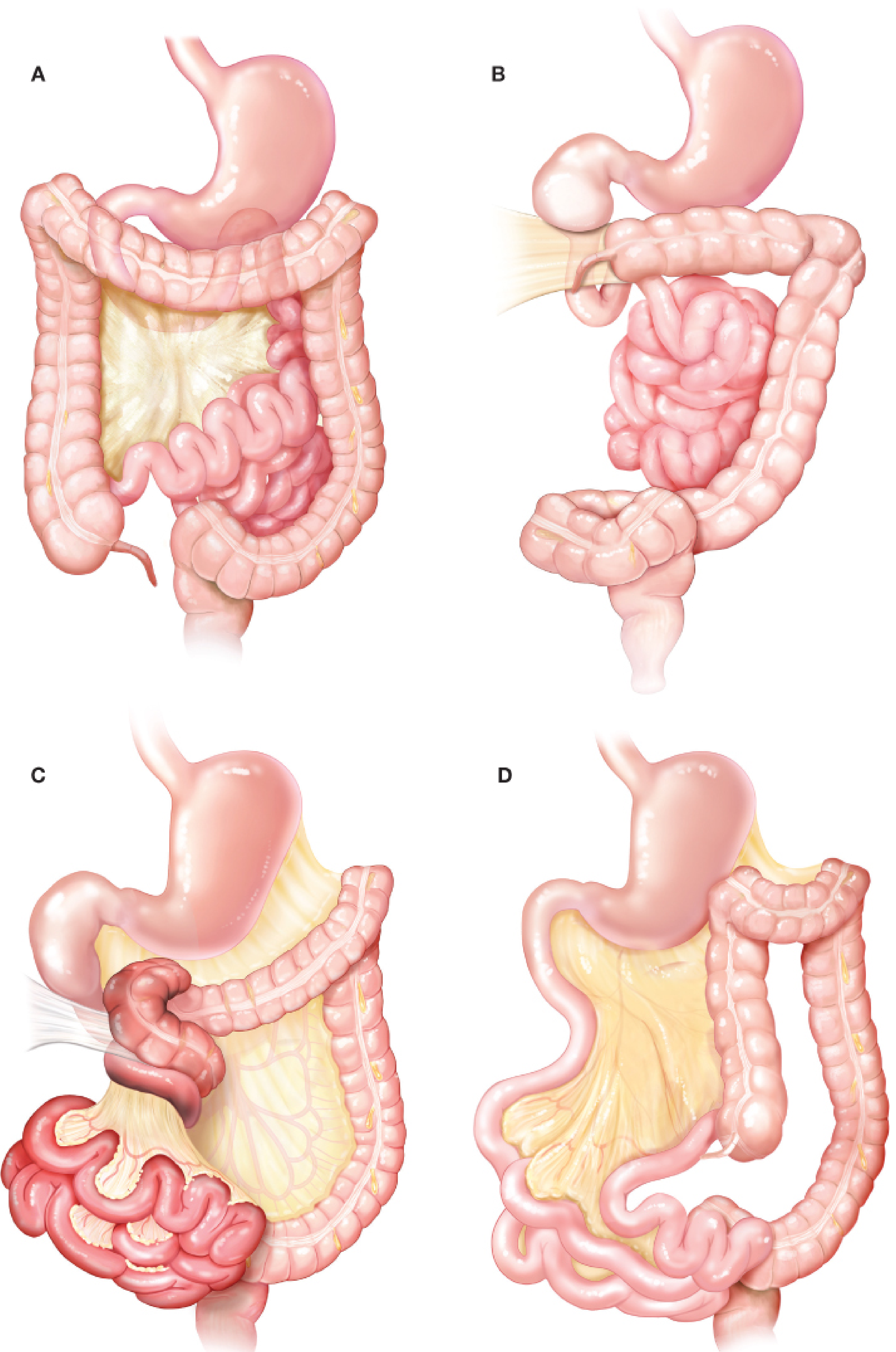
Embryology - Why Malrotation Predisposes to Volvulus
| Week of gestation | Event |
|---|---|
| 6 weeks | Primitive bowel herniates into umbilical cord; rotates 90° CCW |
| 6-10 weeks | Rapid small bowel growth; another 90° CCW rotation |
| 10th week | Bowel re-enters abdominal cavity; final 90° CCW rotation; duodenojejunal junction (DJJ) fixed at ligament of Treitz in the left upper quadrant |
| 10-12 weeks | Cecum descends to right lower quadrant; ascending/descending colon fuse to lateral walls; broad mesentery established |
Incidence
- Rotational anomalies: 2-5 per 1000 live births (autopsy studies)
- Clinically apparent malrotation: ~1 in 6000 live births
- Symptomatic malrotation: estimated as high as 1 in 500 live births
- 90% of cases present before 12 months of age, but up to 1% present in adults - Sabiston Textbook of Surgery, 11e; Fischer's Mastery of Surgery, 8e
Clinical Presentation
Classic Presentation
"Bilious emesis in a newborn = malrotation with midgut volvulus until proven otherwise"
Progression of Symptoms
| Stage | Features |
|---|---|
| Early | Feeding intolerance, irritability, crampy abdominal pain, bilious emesis |
| Intermediate | Guaiac-positive stool (mucosal ischemia), partial obstruction |
| Late | Progressive abdominal distension, hematemesis, hypotension, metabolic acidosis, coagulopathy, shock |
- A distended abdomen is a late sign and, along with metabolic acidosis, may represent the onset of life-threatening intestinal ischemia
- Partial/intermittent volvulus can cause chronic symptoms: mesenteric venous and lymphatic obstruction impairs nutrient absorption, causes protein loss into the gut, mucosal ischemia, and melena
- If untreated: transmural necrosis of the entire midgut (the entire SMA territory)
Diagnosis
Plain Abdominal X-Ray
- Gastric and proximal duodenal distension
- Paucity or absence of distal small bowel gas ("gasless abdomen")
- Nonspecific - early volvulus may have a normal plain film
Upper GI Contrast Series (Gold Standard)
- Study of choice in hemodynamically stable patients
- Classic finding: corkscrew/coiled appearance of the duodenum - the duodenal C-loop does not cross the midline
- "Bird's beak" appearance at the third portion of the duodenum
- Abnormal position of the duodenojejunal junction (to the right of midline)
- Small bowel in the right abdomen, colon/cecum on the left - Mulholland & Greenfield's Surgery, 7e; Sabiston, 11e
In an acutely ill infant with signs of volvulus, immediate operative exploration is warranted without waiting for an upper GI study. - Sabiston, 11e
Ultrasound
- Reversal of the normal SMA/SMV relationship: normally the superior mesenteric vein is to the RIGHT of the SMA
- In malrotation: SMV is ventral to or to the left of the SMA
- "Whirlpool sign" on Doppler: mesenteric vessels and mesentery spiraling around the SMA
CT Abdomen (Contrast-Enhanced)
- Classic "whirlpool sign" - twisting of SMV and mesentery around SMA
- Small bowel loops in the right hemiabdomen, colon displaced to the left
- SMV to the left of SMA
Management
Immediate Steps
- Simultaneous resuscitation + confirmation of diagnosis
- Treat shock: ensure adequate gas exchange, restore intravascular volume
- Urgent laparotomy - do not delay
"Initial assessment, resuscitation, and preoperative preparation in a symptomatic newborn should be conducted simultaneously so that confirmation of malrotation can be followed immediately by laparotomy." - Mulholland & Greenfield's Surgery, 7e
Ladd's Procedure (Operation of Choice)
- Detorsion - deliver the midgut and untwist the volvulus, usually counterclockwise. Use the root of the mesentery and the duodenum as reference points.
- Assess bowel viability - warm sponges on ischemic bowel to improve perfusion. Frankly necrotic bowel is resected. Borderline segments may be left with a second-look laparotomy at 24-36 hours.
- Divide Ladd's bands - lateral peritoneal attachments from the right colon to the lateral abdominal wall (these compress the duodenum and constrict the mesenteric root).
- Mobilize the duodenum (modified Kocher maneuver) - divide all anterior, lateral, and posterior attachments to straighten the duodenum; the DJJ ends up in the right lower quadrant.
- Broaden the mesenteric base - divide the bands at the base of the mesentery across the superior mesenteric vein down to the root, so the mesenteric pedicle is flat and wide.
- Appendectomy - performed because the cecum is repositioned to the left upper quadrant, making future appendicitis difficult to diagnose.
- Final position - small intestine to the right, cecum/colon to the left upper quadrant, with the DJJ and ileocecal junctions as far apart as possible to minimize re-volvulus risk.
- Check for synchronous duodenal obstruction with intraluminal air or saline.
Securing the cecum or duodenum to the abdominal wall by suturing has no proven benefit. - Sabiston, 11e
Complications and Prognosis
| Complication | Detail |
|---|---|
| Recurrent volvulus | Up to 10% after Ladd's procedure |
| Short bowel syndrome | Midgut volvulus causes ~18% of pediatric SBS |
| Prolonged postoperative ileus | Especially after extensive resection |
| Complete midgut infarction | Survivable only with total enterectomy + permanent TPN |
Associated Conditions
- Congenital diaphragmatic hernia
- Gastroschisis
- Omphalocele
- Congenital heart disease with heterotaxy syndrome (atypical malrotation)
- Duodenal atresia/stenosis (can coexist)
Key Points Summary
| Feature | Detail |
|---|---|
| Underlying cause | Intestinal malrotation with narrow SMA mesenteric pedicle |
| Classic sign | Bilious emesis in a neonate |
| Ischemia timeline | Irreversible necrosis after ~6 hours |
| Imaging study of choice | Upper GI contrast series (stable patient) |
| CT sign | Whirlpool sign (SMV wrapping around SMA) |
| US sign | SMV to the left of or anterior to SMA |
| Treatment | Urgent Ladd's procedure |
| Recurrence after surgery | ~10% |
| Peak age | <12 months (90% of cases) |