
SLE & ANA patterns/ interpretation Robbins based md pathology exam answer easy for replicating
Reading File
Reading File
Finding Sources
Finding Sources
Searching PubMed
"systemic lupus erythematosus" AND "antinuclear antibodies"
Searching the Web
Robbins Pathologic Basis of Disease systemic lupus erythematosus ANA patterns interpretation
Finding Sources
Finding Sources
Finding Sources
Finding Sources
Finding Sources
Finding Sources
Reading File
Reading File
SLE and ANA patterns - Robbins-style exam answer
Systemic lupus erythematosus (SLE)
Definition:
SLE is a chronic, multisystem autoimmune disease characterized by production of autoantibodies, especially antinuclear antibodies (ANA). Tissue injury occurs mainly due to:
SLE is a chronic, multisystem autoimmune disease characterized by production of autoantibodies, especially antinuclear antibodies (ANA). Tissue injury occurs mainly due to:
- Immune-complex deposition - type III hypersensitivity
- Autoantibody-mediated injury to cells and tissues - type II hypersensitivity
Commonly affected organs are skin, joints, kidneys, serosal membranes, blood cells, CNS, and heart.
One-line pathology:
Loss of self-tolerance leads to ANA formation, immune-complex deposition, complement activation, and inflammation in multiple organs.
ANA in SLE
What are ANAs?
ANA are antibodies directed against nuclear constituents. By indirect immunofluorescence, the pattern of nuclear staining gives a clue to the likely antigen.
Four broad ANA antigen groups in Robbins:
- Antibodies to DNA
- Antibodies to histones
- Antibodies to non-histone proteins bound to RNA
- Antibodies to nucleolar antigens
Important: ANA pattern is a clue, not a final diagnosis. Mixed patterns can occur because several autoantibodies may coexist.
ANA patterns and interpretation
| ANA immunofluorescence pattern | Main antibody/antigen association | Clinical interpretation |
|---|---|---|
| Homogeneous / diffuse nuclear staining | Anti-dsDNA, anti-nucleosome, anti-histone | Common in SLE. Anti-histone suggests drug-induced lupus. |
| Peripheral / rim staining | Anti-dsDNA | Supports SLE and may be associated with lupus nephritis. |
| Speckled pattern | Anti-Sm, anti-RNP, anti-Ro/SSA, anti-La/SSB | May occur in SLE, Sjögren syndrome, mixed connective-tissue disease. |
| Nucleolar pattern | Antibodies to nucleolar proteins | More suggestive of systemic sclerosis than classic SLE. |
| Centromere pattern | Anti-centromere antibody | Seen mainly in limited cutaneous systemic sclerosis/CREST; not typical of SLE. |
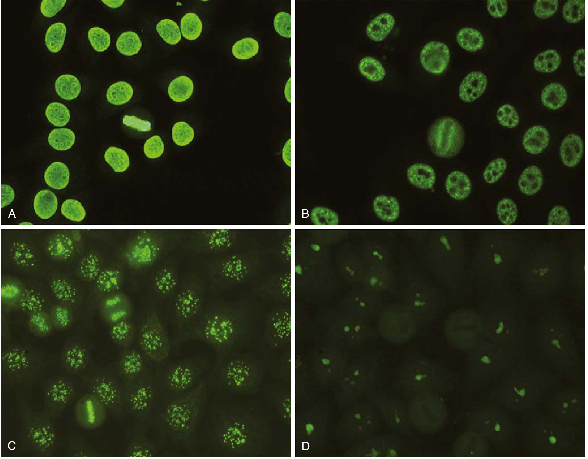
Important antibodies in SLE
| Antibody | Importance |
|---|---|
| ANA | Very sensitive screening test for SLE, but not specific. A positive result alone does not diagnose SLE. |
| Anti-dsDNA | Highly specific for SLE; titre can correlate with disease activity and is associated with lupus nephritis. |
| Anti-Sm (Smith) | Highly specific for SLE. Less sensitive, but its presence strongly supports the diagnosis. |
| Anti-histone | Characteristic of drug-induced lupus. |
| Anti-Ro/SSA | Seen in subacute cutaneous lupus and Sjögren syndrome; maternal antibody can cause neonatal lupus/congenital heart block. |
| Antiphospholipid antibodies | May cause arterial/venous thrombosis, recurrent pregnancy loss, thrombocytopenia, prolonged PTT, and false-positive VDRL test. |
How to interpret ANA in an exam
1. ANA positive
- Supports autoimmune connective-tissue disease only if clinical features are compatible.
- It is sensitive but not specific.
- It may be positive in other autoimmune diseases and sometimes in healthy persons.
2. Homogeneous pattern
- Think: SLE / drug-induced lupus
- Then check:
- Anti-dsDNA
- Anti-Sm
- Anti-histone
3. Peripheral or rim pattern
- Think: anti-dsDNA
- Strongly consider SLE with renal involvement.
4. Speckled pattern
- Think: antibodies to Sm, RNP, Ro, La
- Seen in SLE but also in Sjögren syndrome and MCTD.
- Do specific antibody testing for confirmation.
5. Nucleolar or centromere pattern
- Think mainly systemic sclerosis, not typical SLE.
Ready-to-write 5-mark answer
SLE is a multisystem autoimmune disease characterized by ANA production. Tissue damage is caused by immune-complex deposition and autoantibody-mediated cellular injury. ANA are detected by indirect immunofluorescence. The major ANA patterns are: homogeneous pattern due to antibodies against dsDNA, nucleosomes, and histones, commonly seen in SLE; peripheral/rim pattern due to anti-dsDNA, suggesting SLE and lupus nephritis; speckled pattern due to anti-Sm and RNP antibodies, seen in SLE and other connective-tissue diseases; nucleolar pattern, more suggestive of systemic sclerosis; and centromere pattern, associated with limited systemic sclerosis. ANA is sensitive but nonspecific. Anti-dsDNA and anti-Sm antibodies are highly specific for SLE, with anti-dsDNA also associated with nephritis and disease activity.
Memory trick
“SLE has DNA and Sm”
- D - anti-DsDNA: lupus nephritis, activity
- S - anti-Sm: most specific for SLE
- H - anti-Histone: drug-induced lupus
- R - anti-Ro: subacute cutaneous/neonatal lupus
Robbins emphasizes that homogeneous staining is typical of antibodies against dsDNA, nucleosomes, and histones, while speckled staining is associated with Sm and RNP antibodies. Robbins & Kumar Basic Pathology, section “Spectrum of Autoantibodies in SLE”.
A recent systematic review also confirms that ANA-negative SLE is unusual, so a negative ANA makes conventional SLE less likely but does not make it impossible (2025 systematic review, PMID 40635365).
Also include Pathogenesis morphology and clinical features including diagnosis in short and give combined answer
Reading File
Finding Sources
Finding Sources
Searching PubMed
"systemic lupus erythematosus" AND diagnosis
Searching the Web
2023 EULAR ACR systemic lupus erythematosus classification criteria ANA entry criterion
Finding Sources
Finding Sources
Finding Sources
Systemic Lupus Erythematosus (SLE): Pathogenesis, Morphology, Clinical Features, ANA and Diagnosis
Definition
SLE is a chronic, relapsing, multisystem autoimmune disease characterized by production of autoantibodies, especially antinuclear antibodies (ANA), causing tissue injury mainly by immune-complex deposition.
It commonly affects young women and may involve skin, joints, kidneys, serosa, blood cells, CNS, and heart.
1. Pathogenesis
Basic defect
Loss of self-tolerance to nuclear antigens leads to formation of autoantibodies and immune complexes.
Sequence
-
Genetic susceptibility
- Association with HLA genes and other immune-regulatory genes.
- Deficiency of early complement components, especially C1q, C2, and C4, predisposes to SLE.
- Defective complement function causes poor clearance of apoptotic cells and immune complexes.
-
Environmental triggers
- UV light increases apoptosis and exposes nuclear antigens.
- Infections, certain drugs, and hormonal influences may contribute.
-
Defective clearance of apoptotic nuclear material
- Nuclear antigens such as DNA, histones, and ribonucleoproteins persist in tissues.
-
Activation of autoreactive B and T lymphocytes
- Autoreactive CD4+ T cells help B cells form pathogenic autoantibodies.
- Nuclear DNA/RNA activates Toll-like receptors, enhancing ANA production.
- Type I interferons promote B-cell, T-cell, and dendritic-cell activation.
-
Tissue injury
- Type III hypersensitivity: DNA-anti-DNA immune complexes deposit in glomeruli, vessels, skin, joints, and serosa.
- Complement activation causes inflammation and tissue damage.
- Type II hypersensitivity: antibodies against RBCs, platelets, and leukocytes cause cytopenias.
Flowchart for exam
Genetic susceptibility + UV/infection/drugs
→ defective clearance of apoptotic nuclei
→ loss of self-tolerance
→ autoreactive T cells and B cells
→ ANA and other autoantibodies
→ immune-complex deposition + complement activation
→ multisystem inflammation and damage.
→ defective clearance of apoptotic nuclei
→ loss of self-tolerance
→ autoreactive T cells and B cells
→ ANA and other autoantibodies
→ immune-complex deposition + complement activation
→ multisystem inflammation and damage.
2. Autoantibodies and ANA patterns
| ANA pattern | Antibody association | Interpretation |
|---|---|---|
| Homogeneous/diffuse | Anti-dsDNA, anti-nucleosome, anti-histone | Common in SLE; anti-histone suggests drug-induced lupus |
| Peripheral/rim | Anti-dsDNA | Suggests SLE, especially lupus nephritis |
| Speckled | Anti-Sm, anti-RNP, anti-Ro/SSA, anti-La/SSB | Seen in SLE, Sjögren syndrome, MCTD |
| Nucleolar | Antibodies to nucleolar proteins | More typical of systemic sclerosis |
| Centromere | Anti-centromere antibody | Typical of limited systemic sclerosis/CREST, not SLE |
Important antibodies
| Antibody | Significance |
|---|---|
| ANA | Very sensitive but nonspecific screening test |
| Anti-dsDNA | Highly specific; correlates with disease activity and lupus nephritis |
| Anti-Sm | Highly specific for SLE |
| Anti-histone | Drug-induced lupus |
| Anti-Ro/SSA | Subacute cutaneous lupus, neonatal lupus, congenital heart block |
| Antiphospholipid antibodies | Thrombosis, recurrent abortions, thrombocytopenia, prolonged PTT, false-positive VDRL |
Remember: ANA positive does not itself diagnose SLE. Interpret it with symptoms, specific antibodies, complement level, urine findings, and biopsy where indicated.
3. Morphology
A. Blood vessels
- Immune-complex deposition causes acute necrotizing vasculitis.
- Vessel walls may show fibrinoid necrosis.
- Chronic vascular lesions may show intimal thickening.
B. Kidneys: lupus nephritis
Renal involvement is an important cause of morbidity and mortality.
- Immune complexes deposit in glomeruli.
- Findings include proliferative glomerulonephritis, capillary-wall thickening, and sclerosis.
- Wire-loop lesions: marked capillary-wall thickening due to subendothelial immune-complex deposits.
- Immunofluorescence often shows a full-house pattern: IgG, IgA, IgM, C3, and C1q.
C. Skin
- Erythematous rash with interface dermatitis.
- Degeneration of basal cells and deposition of immunoglobulin/complement at dermoepidermal junction may produce a positive lupus band test.
D. Joints
- Synovitis may occur, but joint disease is classically nonerosive.
E. Heart
- Libman-Sacks endocarditis: sterile verrucous vegetations on either surface of valve leaflets, often mitral valve.
- Pericarditis is common.
F. Hematoxylin bodies and LE cells
- Hematoxylin bodies (LE bodies): homogeneous, basophilic nuclear remnants of damaged cells coated by ANA.
- LE cell: a neutrophil or macrophage that has phagocytosed a denatured nucleus. It is of historical interest and is no longer used routinely for diagnosis.
4. Clinical features
SLE is a relapsing-remitting disease with variable presentation.
Constitutional
- Fever, fatigue, weight loss, malaise.
Skin and mucosa
- Malar or butterfly rash
- Photosensitivity
- Discoid rash
- Oral or nasal ulcers
- Alopecia
- Raynaud phenomenon may occur.
Musculoskeletal
- Arthralgia and arthritis.
- Typically nonerosive arthritis involving small joints.
Renal
- Proteinuria, hematuria, RBC casts.
- Nephritic or nephrotic syndrome.
- Hypertension and renal failure in severe lupus nephritis.
Serosal
- Pleuritis and pleural effusion.
- Pericarditis and pericardial effusion.
Hematologic
- Autoimmune hemolytic anemia.
- Leukopenia/lymphopenia.
- Thrombocytopenia.
Neurologic
- Seizures, psychosis, cognitive dysfunction, neuropathy, stroke.
Antiphospholipid syndrome manifestations
- Recurrent arterial or venous thrombosis.
- Recurrent fetal loss.
- Thrombocytopenia.
- Prolonged PTT in vitro despite thrombotic tendency in vivo.
5. Diagnosis
Diagnosis is clinical plus immunological
Suspect SLE in a young woman with combinations of:
- Malar/photosensitive rash
- Oral ulcers
- Nonerosive arthritis
- Serositis
- Nephritis
- Cytopenias
- Neurological manifestations
Investigations
| Test | Finding/meaning |
|---|---|
| ANA by indirect immunofluorescence | Sensitive screening test |
| Anti-dsDNA | Specific; useful in nephritis and disease activity |
| Anti-Sm | Highly specific for SLE |
| C3 and C4 | Low during active immune-complex disease, especially nephritis |
| CBC | Anemia, leukopenia, lymphopenia, thrombocytopenia |
| Urine examination | Proteinuria, hematuria, RBC casts |
| Direct Coombs test | May be positive in autoimmune hemolysis |
| Antiphospholipid antibody panel | Lupus anticoagulant, anticardiolipin, anti-β2 glycoprotein I |
| Renal biopsy | Required to classify lupus nephritis and guide treatment |
Classification point
For the 2019 EULAR/ACR classification criteria, ANA positivity at a titre of at least 1:80 at least once is an entry criterion. Classification needs clinical and immunological features with an adequate weighted score. These are classification criteria, not a replacement for clinical diagnosis.
Short combined exam answer
Systemic lupus erythematosus is a chronic multisystem autoimmune disease characterized by antinuclear antibodies and immune-complex-mediated injury. Its pathogenesis involves genetic susceptibility, including HLA and complement deficiencies such as C1q, C2, and C4 deficiency, along with environmental triggers such as UV light. Defective clearance of apoptotic nuclear material causes loss of self-tolerance, activation of autoreactive B and T cells, and production of ANA, anti-dsDNA, anti-Sm, and other autoantibodies. Immune-complex deposition causes type III hypersensitivity injury, while antibodies against blood cells cause type II hypersensitivity cytopenias.
Morphological lesions include immune-complex vasculitis with fibrinoid necrosis, lupus nephritis with wire-loop lesions, Libman-Sacks endocarditis, nonerosive synovitis, and hematoxylin bodies. Clinically, SLE presents with fever, malar rash, photosensitivity, oral ulcers, nonerosive arthritis, serositis, nephritis, CNS involvement, and hematological abnormalities. ANA is sensitive but nonspecific. Homogeneous ANA pattern is associated with anti-dsDNA, nucleosomes, and histones; speckled pattern with anti-Sm and RNP; rim pattern with anti-dsDNA; nucleolar and centromere patterns suggest systemic sclerosis. Anti-dsDNA and anti-Sm are highly specific for SLE. Diagnosis is based on compatible clinical findings supported by ANA, anti-dsDNA/anti-Sm antibodies, low complement levels, urine examination, CBC, and renal biopsy when nephritis is suspected.
Robbins & Kumar Basic Pathology, “Systemic Lupus Erythematosus” and “Spectrum of Autoantibodies in SLE”; Robbins, Cotran & Kumar Pathologic Basis of Disease, “Pathogenesis” of SLE.
This is a shared conversation. Sign in to Orris to start your own chat.