
Hi can you explain to me 10 common heart diseases along with their ECG changes and a picture of the ECGs including their management according to latest guidelines please
STEMI ST elevation myocardial infarction ECG
atrial fibrillation ECG irregular rhythm absent P waves
complete heart block third degree AV block ECG
ventricular tachycardia ECG wide complex tachycardia
Wolff-Parkinson-White WPW ECG delta wave pre-excitation
hypertrophic cardiomyopathy ECG left ventricular hypertrophy
pulmonary embolism ECG S1Q3T3 right heart strain sinus tachycardia
pericarditis ECG diffuse ST elevation saddle-shaped PR depression
dilated cardiomyopathy ECG LBBB left bundle branch block low voltage
aortic stenosis ECG left ventricular hypertrophy strain pattern
2024 2025 ACC AHA guidelines STEMI NSTEMI atrial fibrillation heart failure management summary
10 Common Heart Diseases: ECG Changes & Management
1. 🫀 ST-Elevation Myocardial Infarction (STEMI)
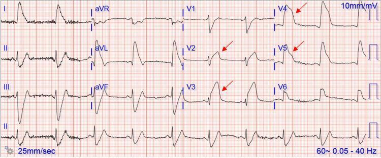
ECG Changes
- Hyperacute T-waves (earliest sign — tall, peaked, asymmetric)
- ST-segment elevation ≥1 mm in ≥2 contiguous limb leads, or ≥2 mm in ≥2 contiguous precordial leads (convex/tombstone morphology)
- Pathological Q waves (transmural necrosis — emerge within hours)
- Reciprocal ST depression in opposing leads
- Lead localization: anterior (V1–V4 = LAD), inferior (II, III, aVF = RCA), lateral (I, aVL, V5–V6 = LCx)
Management (2023 ACC/AHA/ESC Guidelines)
- Primary PCI within 90 min of first medical contact (FMC-to-balloon ≤90 min) — Class I
- Fibrinolysis if PCI not available within 120 min of diagnosis — Class I
- Antiplatelet: Aspirin 325 mg + P2Y12 inhibitor (ticagrelor 180 mg or prasugrel 60 mg preferred over clopidogrel) — Class I
- Anticoagulation: UFH, enoxaparin, or bivalirudin during PCI — Class I
- Beta-blockers: oral within 24 h if no cardiogenic shock — Class I
- ACE inhibitor/ARB: for all STEMI patients, especially with reduced EF — Class I
- High-intensity statin: immediately (atorvastatin 80 mg or rosuvastatin 40 mg) — Class I
- Coronary angiography of non-culprit vessels: staged PCI in haemodynamically stable patients
2. 🫀 Non-ST-Elevation ACS (NSTEMI/Unstable Angina)
ECG Changes
- ST depression ≥0.5 mm (horizontal or downsloping) in ≥2 contiguous leads
- T-wave inversions (symmetric, deep — especially in anterior leads with proximal LAD lesion)
- Transient ST elevation possible (Prinzmetal variant)
- Normal ECG in up to 30–50% of cases — does NOT exclude NSTEMI
- De Winter T-waves (upsloping ST depression + tall symmetric T-waves in V1–V5) — LAD equivalent
Management (2023 ESC NSTE-ACS Guidelines)
- Risk stratification: GRACE score for mortality risk; HEART score for 30-day MACE
- Antiplatelet: Aspirin + ticagrelor (preferred) or clopidogrel — Class I
- Anticoagulation: fondaparinux (preferred), enoxaparin, or UFH — Class I
- Invasive strategy timing:
- Immediate (<2 h): refractory angina, haemodynamic instability, life-threatening arrhythmia
- Early (<24 h): GRACE >140 or dynamic ECG/troponin changes
- Selective (24–72 h): stable intermediate-risk patients
- Beta-blocker, statin, ACE inhibitor/ARB as for STEMI
3. 💓 Atrial Fibrillation (AF)
ECG Changes
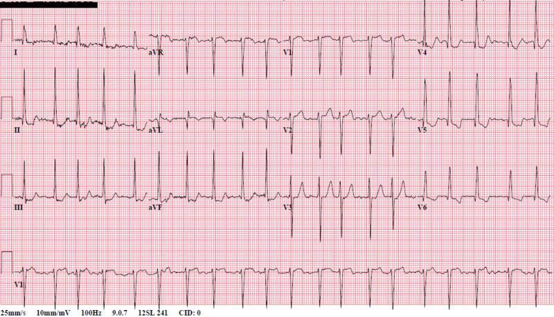
- Irregularly irregular RR intervals (hallmark)
- Absent P waves — replaced by chaotic fibrillatory baseline (best seen in V1)
- Narrow QRS complexes unless aberrant conduction or accessory pathway
- Rapid ventricular response (AF with RVR) if rate >100 bpm
- Variable QRS amplitude
Management (2023 ACC/AHA AF Guidelines)
- Anticoagulation: CHA₂DS₂-VASc score ≥2 (men) or ≥3 (women) → DOACs (apixaban, rivaroxaban, edoxaban, dabigatran) preferred over warfarin — Class I
- Rate control: beta-blockers or non-DHP calcium channel blockers; target HR <110 bpm (lenient) — Class I
- Rhythm control: early rhythm control within 1 year of diagnosis reduces cardiovascular outcomes (EAST-AFNET 4 trial) — Class IIa
- Cardioversion: safe if AF <48 h, or after ≥3 weeks anticoagulation
- Catheter ablation: pulmonary vein isolation — Class I for symptomatic paroxysmal AF after failed antiarrhythmic drug (Class IIa for first-line)
- Treat underlying conditions: hypertension, sleep apnoea, obesity, alcohol
4. 💔 Heart Failure with Reduced EF (HFrEF)
ECG Changes
- Left bundle branch block (LBBB): QRS >120 ms, broad monophasic R in I/aVL/V5–V6, deep S in V1 — strongly associated with DCM
- Left ventricular hypertrophy: high voltage (Sokolow-Lyon: SV1 + RV5 >35 mm)
- Sinus tachycardia: compensatory
- Low voltage: if pericardial effusion or severe LV dilation
- Atrial fibrillation: very common comorbidity
- Ventricular ectopy / NSVT: risk marker
Management (2022 AHA/ACC/HFSA Guidelines — "Fantastic Four")
- ACE inhibitor/ARB/ARNI (sacubitril-valsartan preferred over ACEi) — Class I
- Beta-blocker (carvedilol, metoprolol succinate, bisoprolol) — Class I
- MRA (spironolactone or eplerenone) — Class I
- SGLT2 inhibitor (dapagliflozin or empagliflozin) — Class I (new addition)
- Diuretics for fluid overload: loop diuretics (furosemide) — Class I
- CRT (Cardiac Resynchronisation Therapy): LBBB + QRS ≥150 ms + EF ≤35% — Class I
- ICD: EF ≤35% despite ≥3 months GDMT, NYHA II–III — Class I
- LVAD / Heart transplantation: stage D refractory HF
5. ❤️ Complete (3rd Degree) AV Heart Block
ECG Changes
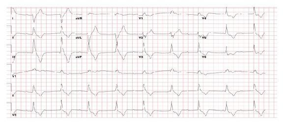
- Complete AV dissociation: P waves and QRS complexes march independently
- Atrial rate > ventricular rate
- Ventricular escape rhythm: junctional (narrow QRS, rate 40–60 bpm) or ventricular (wide QRS, rate 20–40 bpm)
- No fixed PR interval — P waves appear before, within, and after QRS complexes
- Bradycardia (often <40 bpm)
Management (2024 ACC/AHA Bradycardia/CDS Guidelines)
- Urgent temporary pacing: transcutaneous or transvenous if haemodynamically unstable — Class I
- Permanent pacemaker: virtually all patients with acquired complete heart block — Class I
- Dual-chamber pacemaker (DDD): preferred to preserve AV synchrony — Class I
- His bundle pacing / LBBP: preferred over RV apical pacing if available (Class IIa)
- Treat reversible causes: Lyme disease, drug toxicity (digoxin, beta-blockers), inferior MI (temporary — may resolve)
- Atropine (0.5–1 mg IV): temporising measure for vagally-mediated block — Class IIb
- Dopamine/epinephrine infusion: if atropine fails and pacing not immediately available
6. ⚡ Ventricular Tachycardia (VT)
ECG Changes
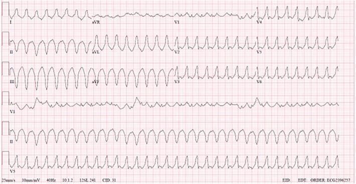
- Wide complex tachycardia: QRS ≥120 ms at rate >100 bpm (usually 150–250 bpm)
- AV dissociation: P waves independent of QRS (pathognomonic) — best seen in long rhythm strip
- Capture beats and fusion beats (highly specific for VT)
- Concordance: positive (all V1–V6 positive) or negative concordance — strongly suggests VT
- Brugada criteria / Vereckei criteria: used to differentiate VT from SVT with aberrancy
- Monomorphic VT: regular, identical QRS morphology
- Polymorphic VT / Torsades de Pointes: twisting QRS axis around baseline, associated with long QT
Management (2022 ESC Ventricular Arrhythmia Guidelines)
- Haemodynamically unstable: immediate DC cardioversion (synchronised) — Class I
- Haemodynamically stable monomorphic VT: IV amiodarone (150 mg bolus then 1 mg/min) or procainamide — Class I
- ICD implantation: secondary prevention after survived cardiac arrest; primary prevention if EF ≤35% — Class I
- Catheter ablation: recurrent VT despite antiarrhythmic drugs, or VT storm — Class I
- Torsades de Pointes: IV magnesium sulphate 2 g; correct QT-prolonging drugs and electrolytes
- Beta-blockers: reduce VT burden in structural heart disease — Class I
- Amiodarone: for VT suppression when ICD therapy is frequent
7. 🔄 Wolff-Parkinson-White (WPW) Syndrome
ECG Changes
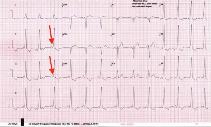
- Short PR interval (<120 ms) — bypass of AV node via accessory pathway (Bundle of Kent)
- Delta wave: slurred upstroke at start of QRS (pre-excitation)
- Wide QRS (>120 ms): fusion of delta wave + normal conduction
- Secondary ST-T changes: discordant repolarisation
- Pseudo-infarct pattern: negative delta waves in inferior leads mimicking Q waves
- AVRT (orthodromic): narrow complex SVT with retrograde P waves after QRS
- AF with WPW: irregular wide complex tachycardia with rapid rate (>250 bpm) → risk of VF
Management (2019 ESC SVT Guidelines, updated recommendations)
- Asymptomatic WPW: risk stratification with electrophysiology (EP) study — Class IIa; ablation if high-risk accessory pathway
- Symptomatic WPW (AVRT): catheter ablation of accessory pathway — Class I (first-line, >95% success)
- Acute AVRT termination: vagal manoeuvres, adenosine (6–12 mg IV) — Class I
- AF + WPW: NEVER use AV nodal blocking agents (adenosine, verapamil, digoxin, beta-blockers) — risk of fatal VF
- AF + WPW acute: DC cardioversion (haemodynamically unstable) or IV procainamide/ibutilide
- Pre-excitation on ECG in athletes: mandatory EP study and ablation if high-risk
8. 🧱 Hypertrophic Cardiomyopathy (HCM)
ECG Changes
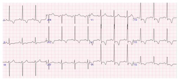
- Left ventricular hypertrophy (LVH): high QRS voltage in precordial leads (Sokolow ≥35 mm)
- Deep, "dagger-like" Q waves: septal hypertrophy in I, aVL, V5–V6
- Apical HCM (Yamaguchi): giant T-wave inversions in V2–V5 (depth ≥10–30 mm)
- ST-segment depression + T-wave inversion (strain pattern) in lateral leads
- P-wave abnormalities: left atrial enlargement
- Absence of septal Q waves in lateral leads (paradoxically absent due to reversed septal depolarisation)
Management (2024 ACC/AHA HCM Guidelines)
- Lifestyle: avoid dehydration, competitive sports (consult cardiologist)
- Obstructive HCM (LVOTO): mavacamten (cardiac myosin inhibitor) — Class I (new guideline addition 2024); first-line before septal reduction
- Beta-blockers or verapamil: for symptom relief, LVOT obstruction — Class I
- Septal reduction: surgical septal myectomy (Class I for refractory NYHA III–IV) or alcohol septal ablation (Class IIa)
- ICD: for primary prevention — risk factors include family history of SCD, LV thickness ≥30 mm, unexplained syncope, NSVT, abnormal BP response to exercise
- HCM Risk-SCD calculator (ESC): used for 5-year SCD risk estimation
- AF management: anticoagulation regardless of CHA₂DS₂-VASc score in HCM + AF
9. 🫧 Acute Pericarditis
ECG Changes (Classic 4-Stage Evolution)
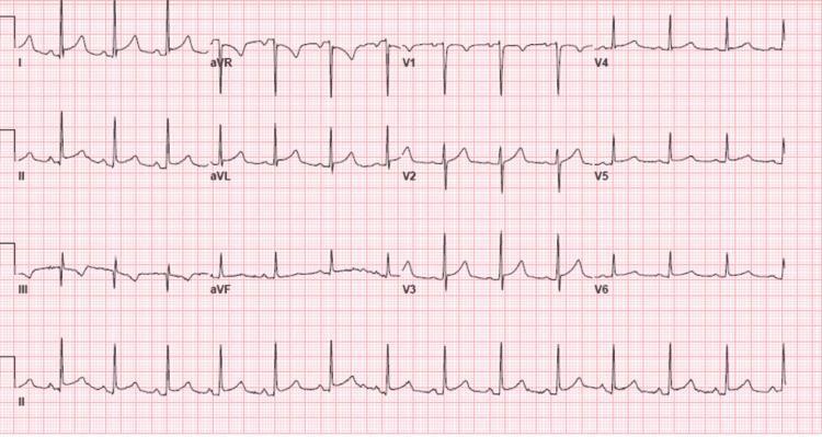
- Stage 1 (days 1–2): Diffuse concave ("saddle-shaped") ST elevation in all leads except aVR and V1; PR depression in II, V4–V6; PR elevation in aVR (pathognomonic)
- Stage 2 (days 3–7): ST normalises, T waves flatten
- Stage 3: T-wave inversion (diffuse)
- Stage 4: ECG normalises
- Spodick's sign: downsloping TP segment
- Key differentiator from STEMI: diffuse (not regional), concave ST, no Q waves, PR depression
Management (2015 ESC Pericardial Disease Guidelines — current standard)
- First-line: NSAIDs (aspirin 750–1000 mg TDS or ibuprofen 600 mg TDS) + colchicine 0.5 mg BD for 3 months — Class I (reduces recurrence by 50%)
- Gastroprotection: proton pump inhibitor with NSAIDs — Class I
- Restrict exercise: until symptoms resolve and CRP normalises (athletes: 3 months minimum)
- Corticosteroids: only if NSAIDs/colchicine contraindicated, or connective tissue disease — low dose (prednisone 0.2–0.5 mg/kg/day), NOT first-line (increases recurrence)
- Recurrent pericarditis: colchicine ± IL-1 antagonist (anakinra, rilonacept) — Class IIa/IIb
- Hospitalisation: for high-risk features (fever >38°C, large effusion, tamponade, immunosuppressed)
- Cardiac tamponade: urgent pericardiocentesis
10. 🩸 Pulmonary Embolism (PE)
ECG Changes
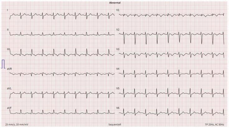
- Sinus tachycardia: most common (>40% of cases)
- S1Q3T3 pattern: S wave in lead I + Q wave in lead III + T-wave inversion in lead III (classic but only in ~20%)
- Right bundle branch block (RBBB): complete or incomplete — right ventricular strain
- T-wave inversions in V1–V4 (right heart strain) — most specific ECG finding for massive PE
- P-pulmonale: tall peaked P waves in II (right atrial enlargement)
- Right axis deviation
- New AF may be precipitated
Management (2023 ESC PE Guidelines)
- Risk stratification: PESI score + RV dysfunction + troponin → massive / submassive / low-risk
- Anticoagulation (start immediately if clinical suspicion high, before confirmed):
- DOACs (rivaroxaban or apixaban) — preferred first-line for most patients — Class I
- LMWH → warfarin (if DOAC contraindicated)
- Duration: 3 months minimum; extended if unprovoked or recurrent
- Massive PE (haemodynamic instability): systemic thrombolysis (alteplase 100 mg over 2 h) — Class I
- Surgical embolectomy or catheter-directed thrombolysis: if thrombolysis fails or contraindicated — Class I/IIa
- Submassive PE (RV dysfunction, no shock): consider thrombolysis if clinical deterioration — Class IIa
- PERC rule: if low pretest probability + all 8 PERC criteria negative → no further workup needed
- CTPA: diagnostic standard; V/Q scan if renal impairment or contrast allergy
- IVC filter: only if anticoagulation absolutely contraindicated
Quick Reference Table
| # | Condition | Key ECG Finding | First-Line Treatment |
|---|---|---|---|
| 1 | STEMI | ST elevation + Q waves | Primary PCI ≤90 min |
| 2 | NSTEMI/UA | ST depression, T-wave inversion | Aspirin + ticagrelor ± early angiography |
| 3 | Atrial Fibrillation | Irregular rhythm, no P waves | DOAC + rate/rhythm control |
| 4 | HFrEF | LBBB, LVH, sinus tachycardia | ARNI + BB + MRA + SGLT2i |
| 5 | Complete Heart Block | AV dissociation, bradycardia | Permanent pacemaker |
| 6 | Ventricular Tachycardia | Wide complex tachycardia | DC cardioversion / amiodarone / ICD |
| 7 | WPW Syndrome | Short PR + delta wave | Catheter ablation |
| 8 | HCM | LVH + giant T-wave inversions | Mavacamten / myectomy / ICD |
| 9 | Acute Pericarditis | Diffuse ST elevation + PR depression | NSAIDs + colchicine |
| 10 | Pulmonary Embolism | S1Q3T3, sinus tachycardia | DOAC ± thrombolysis |
Sources: Braunwald's Heart Disease (12th ed.), Goldman-Cecil Medicine, Rosen's Emergency Medicine; ACC/AHA/ESC Guidelines 2022–2024. ECG images from peer-reviewed clinical literature (PMC).Here's a summary of what was covered across all 10 conditions:
- STEMI — ST elevation/tombstone morphology/Q waves → Primary PCI ≤90 min + dual antiplatelet + statin + ACEI
- NSTEMI/Unstable Angina — ST depression/T-wave inversions/normal ECG → Risk-stratified invasive strategy + ticagrelor + anticoagulation
- Atrial Fibrillation — Irregularly irregular, absent P waves → DOACs + rate/rhythm control; catheter ablation for symptomatic AF
- HFrEF — LBBB, LVH, sinus tachycardia → The "Fantastic Four": ARNI + beta-blocker + MRA + SGLT2 inhibitor
- Complete Heart Block — AV dissociation, escape rhythm → Permanent pacemaker (virtually all acquired cases)
- Ventricular Tachycardia — Wide complex tachycardia, AV dissociation → DC cardioversion if unstable; amiodarone if stable; ICD
- WPW Syndrome — Short PR + delta wave → Catheter ablation (first-line, >95% success); avoid AV nodal blockers in AF+WPW
- Hypertrophic Cardiomyopathy — LVH + giant T-wave inversions → Mavacamten (new 2024 Class I); septal myectomy; ICD for SCD risk
- Acute Pericarditis — Diffuse saddle-shaped ST elevation + PR depression → NSAIDs + colchicine for 3 months
- Pulmonary Embolism — S1Q3T3 + sinus tachycardia + RV strain → DOACs; systemic thrombolysis for massive PE