
Bilateral polycystic ovaries
polycystic ovary ultrasound bilateral multiple follicles
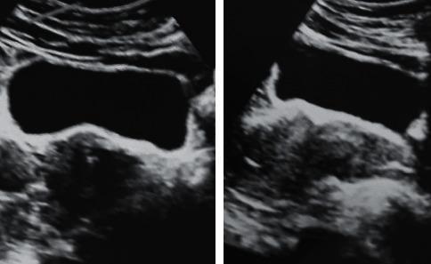
Two diagnostic ultrasound images of the pelvic region demonstrate the classical sonographic findings of Polycystic Ovary Syndrome (PCOS). The images show bilateral ovarian enlargement, with the right ovary measuring 14 ml and the left ovary measuring 17 ml. Both ovaries exhibit a characteristic distribution of multiple small, subcentimeter, hypoechoic follicles located at the periphery, creating a 'string of pearls' appearance. There is a notable increase in the central echogenicity and volume of the ovarian stroma, termed stromal hyperplasia or prominent stroma. These visual features are consistent with the Rotterdam criteria for polycystic ovary morphology in a clinical context of hyperandrogenism. This image serves as an educational reference for identifying gynecological pathology through ultrasonography, specifically highlighting morphological changes in the adnexa associated with endocrine disorders like PCOS and ovarian hyperthecosis.
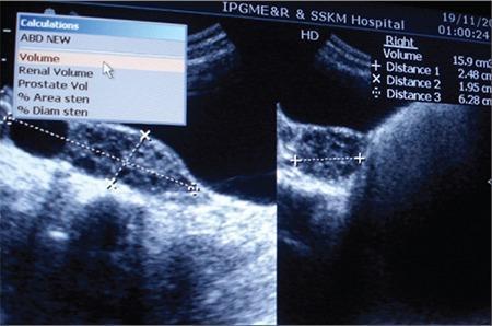
Diagnostic Image: This abdominal ultrasonography scan displays bilateral polycystic ovaries in a 13-year-old patient. The ultrasound reveals an enlarged right ovary with a measured volume of 15.9 cm³, which is significantly increased for the patient's age and pubertal status. Three linear distance measurements are visible on the screen: Distance 1 (2.48 cm), Distance 2 (1.95 cm), and Distance 3 (6.28 cm), used to calculate the ovarian volume and characterize a dominant cyst. Visually, the ovaries exhibit multiple small, peripherally located follicles and increased echogenic stroma, characteristic of polycystic ovarian morphology (PCOM). The clinical context involves a rare case of pediatric hyperinsulinemia and lipodystrophy, where severe insulin resistance leads to ovarian overstimulation and clitoromegaly. This image serves as a teaching tool for recognizing PCOM and understanding the impact of metabolic endocrine disorders on the reproductive system in prepubertal or early adolescent patients.
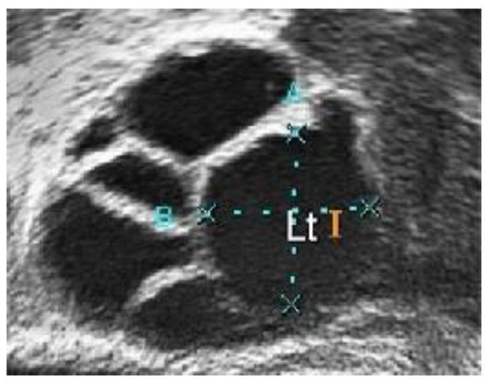
This diagnostic ultrasound image displays a close-up view of an ovary with characteristic features of Polycystic Ovary Syndrome (PCOS). The image shows multiple anechoic (dark), fluid-filled follicles of varying sizes and shapes, primarily circular and oval, distributed within the hyperechoic (bright) ovarian stroma. The largest follicle in the center-right is marked with calipers and annotations ('A', 'B', 'Et I') indicating manual measurement of its dimensions. The follicles exhibit well-defined borders against the surrounding light-pixel tissue, which has a grainy texture due to inherent speckle noise. Clinically, the presence of these numerous developing follicles is a primary diagnostic marker for polycystic morphology. This image is used in medical education to demonstrate the visualization of follicular accumulation and the process of diagnostic measurement during gynecological sonography.
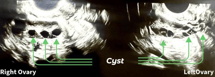
This diagnostic image consists of a grayscale pelvic ultrasound sonography (USG) displaying bilateral ovaries in a female patient. The image is split into two panels, with the right ovary on the left and the left ovary on the right, both exhibiting a speckled stroma. Within the ovarian tissue of both structures, there are multiple anechoic (dark), circular, and ovoid fluid-filled structures identified as cysts. Green arrows highlight these cysts, which vary in size and are scattered throughout the stroma. The appearance is clinically consistent with polycystic ovary syndrome (PCOS) or multifollicular ovaries, where the internal structure shows an increased number of follicles/cysts. The image serves as an educational tool for identifying common sonographic markers of ovarian pathology and follicular monitoring in reproductive medicine.
"polycystic ovary syndrome"[MeSH Terms] AND diagnosis
Bilateral Polycystic Ovaries (PCOS)
Overview
Ultrasound Appearance
- ≥20 follicles in either ovary, each measuring 2-9 mm in diameter, AND/OR
- Increased ovarian volume (>10 mL)
- A single ovary meeting these criteria is sufficient to apply the PCOM label
Diagnostic Criteria (Rotterdam 2003)
| Criterion | Details |
|---|---|
| 1. Menstrual irregularity | Oligomenorrhea, amenorrhea (anovulation/oligo-ovulation) |
| 2. Hyperandrogenism (HA) | Clinical (hirsutism, acne, male-pattern alopecia) and/or biochemical |
| 3. Polycystic ovarian morphology | ≥20 follicles 2-9 mm OR ovarian volume >10 mL on ultrasound |
- HA + ovarian dysfunction + PCO morphology (classic full phenotype)
- HA + ovarian dysfunction (no PCO morphology)
- HA + PCO morphology (ovulatory PCOS)
- Ovarian dysfunction + PCO morphology (no HA)
Pathophysiology
Excess LH → Increased ovarian androgen production
     ↑                          ↓
Hyperinsulinemia          Peripheral conversion to estrogen
     ↑                          ↓
Insulin resistance    Abnormal GnRH pulsatility → elevated LH:FSH ratio
- LH - elevated in ~2/3 of cases; elevated LH:FSH ratio (often >2:1) is a minor criterion
- Androgens - moderately elevated (testosterone, androstenedione, DHEAS)
- Insulin resistance - present in 50-80%; hyperinsulinemia directly stimulates ovarian androgen production and suppresses SHBG, worsening free androgen levels
- FSH - relatively low (prevents follicle maturation and ovulation)
- AMH - elevated; a recent 2024 meta-analysis (PMID: 38944177) confirms AMH as a reliable diagnostic biomarker for PCOS and PCOM
Clinical Features
Reproductive
- Hirsutism - most common manifestation (~70% in the US, 10-20% in Japan due to differences in skin 5α-reductase activity)
- Oligomenorrhea / amenorrhea - from anovulation
- Anovulatory infertility - most common cause of infertility
- Acne, male-pattern alopecia
- Onset typically at or near puberty
Metabolic
- Obesity - >50% of PCOS patients; typically android/central distribution (higher waist-to-hip ratio)
- Insulin resistance / type 2 diabetes - common
- Dyslipidemia - elevated LDL, TG; low HDL
- Hypertension
- Higher risk of metabolic syndrome and cardiovascular disease later in life
Obstetric risks
- Increased risk of early miscarriage
- Gestational diabetes
- Gestational hypertension / preeclampsia
- Preterm birth
Endometrial risk
- Chronic anovulation leads to unopposed estrogen exposure - risk of endometrial hyperplasia and carcinoma if left untreated
Differential Diagnosis (Conditions to Exclude)
- Non-classic congenital adrenal hyperplasia (measure fasting 17-OHP - mandatory in all women with hirsutism)
- Adrenal or ovarian androgen-secreting tumor
- Cushing syndrome
- Hyperprolactinemia
- Hypothyroidism / hyperthyroidism
- Hypo- or hypergonadotropic disorders
Management
Not Seeking Pregnancy
| Goal | Treatment |
|---|---|
| Cycle regulation + HA | Combined oral contraceptive pills (COCPs) - first-line; increase SHBG, reduce androgen levels |
| Persistent hirsutism/acne after 6 months | Add antiandrogens - spironolactone, flutamide |
| Endometrial protection (if not using OCP) | Cyclic progestins (medroxyprogesterone 10 mg OR progesterone 200 mg for 10-14 days every ≥3 months), or levonorgestrel IUD |
| Metabolic risk | Lifestyle modification (diet + exercise); metformin for overweight/obese women for cardiometabolic prevention |
Seeking Pregnancy (Ovulation Induction)
- Letrozole (aromatase inhibitor) - first-line for ovulation induction
- Clomiphene citrate ± metformin - second-line
- Injectable gonadotropins - used judiciously (risk of ovarian hyperstimulation syndrome/OHSS is higher in PCOS)
- Metformin - adjunct with diet/exercise in obese women; NOT recommended alone for infertility
Monitoring at Diagnosis and Regular Intervals
- BMI + waist circumference
- Blood pressure
- Fasting glucose / HbA1c
- Fasting lipid profile (in overweight/obese)
- Screen for depression and anxiety
Genetics
Recent Evidence (2024-2026)
- A 2024 meta-analysis (PMID: 38944177) in Fertility & Sterility confirms AMH as a reliable independent predictor of antral follicle count and a diagnostic biomarker for PCOS/PCOM, supporting its potential use as an alternative to ultrasound.
- A 2024 meta-analysis (PMID: 39178623) in J Diabetes Complications shows GLP-1 receptor agonists (e.g., semaglutide) are effective for weight loss and hormonal regulation in obese women with PCOS - an emerging treatment option not yet in older guidelines.
- A 2024 systematic review (PMID: 39599701) in Nutrients found probiotics, prebiotics, and synbiotics show benefit in managing insulin resistance and hormonal imbalance in PCOS.
In simple terms and severity
🌿 What Is It — In Plain Terms
🟢 Mild / Cosmetic (Annoying, not dangerous)
| Issue | What it means simply |
|---|---|
| Acne | Too much male hormone (testosterone) makes skin oily |
| Unwanted hair (hirsutism) | Hair on face, chest, belly — again from high male hormones |
| Thinning scalp hair | Male-pattern hair loss in women |
| Irregular periods | Egg doesn't release → period doesn't come on time |
🟡 Moderate / Reproductive (Affects having children)
| Issue | What it means simply |
|---|---|
| Difficulty getting pregnant | No egg released = no pregnancy naturally |
| Irregular or absent ovulation | The "egg launch" fails most months |
| Higher miscarriage risk | Even if pregnancy occurs, early loss is more likely |
🟠 Significant / Metabolic (Affects your whole body)
| Issue | What it means simply |
|---|---|
| Insulin resistance | Your body makes insulin but cells ignore it → sugar stays high |
| Weight gain | Especially around the belly — hard to lose |
| Pre-diabetes / Type 2 diabetes | Uncontrolled insulin resistance progresses here |
| High cholesterol / blood pressure | Increases heart disease risk over time |
🔴 Serious / Long-term (If untreated for years)
| Issue | What it means simply |
|---|---|
| Endometrial cancer risk | No period for months = uterine lining keeps thickening → can turn cancerous |
| Heart disease | From years of high insulin, high cholesterol, and high blood pressure |
| Type 2 diabetes | If insulin resistance is never managed |
| Depression & anxiety | Both a symptom AND a consequence of PCOS — very common and often overlooked |
The Core Problem (One Simple Sentence)
PCOS is a hormonal imbalance where too much male hormone + too much insulin prevents normal ovulation, and if left unmanaged, quietly damages the heart, metabolism, and uterus over decades.
The Good News
- PCOS is manageable, not curable but very controllable
- Lifestyle changes (losing even 5-10% of body weight) can restore periods and ovulation naturally
- Medications exist for every symptom - periods, hair, fertility, blood sugar
- Most women live full, normal lives with proper monitoring
Which medications treat PCOS symptoms?
"polycystic ovary syndrome"[MeSH Terms] AND "drug therapy"[MeSH Subheading]
Medications for PCOS — By Symptom
1. 🩸 Irregular Periods & Excess Male Hormones (Not trying to get pregnant)
Combined Oral Contraceptive Pill (COCP) — First-line
- What it does: Regulates your cycle, reduces acne and unwanted hair
- How: The estrogen component raises a protein called SHBG that "mops up" excess testosterone. The progestin protects the uterine lining from thickening
- Timeline: Periods regulate within 1-3 months; hair/acne improvement takes 6+ months
- Choose the lowest effective estrogen dose
- Harrison's 22E
Progestins (if can't take estrogen)
- Drugs: Medroxyprogesterone acetate (10 mg) OR progesterone (200 mg)
- How: Taken for 10-14 days every 3 months to force a withdrawal bleed
- Why it matters: Prevents the uterine lining from building up and becoming cancerous
- A levonorgestrel IUD (Mirena) also works for this purpose
2. 🧔♀️ Unwanted Hair (Hirsutism) & Acne — Antiandrogens
| Drug | How it works | Notes |
|---|---|---|
| Spironolactone | Blocks testosterone from acting on hair follicles and skin | Most commonly used; also lowers blood pressure. Must use contraception - causes birth defects |
| Flutamide | Directly blocks androgen receptors | Effective but risk of liver toxicity; less commonly used |
| Eflornithine cream (topical) | Blocks the enzyme that makes facial hair grow | Applied directly to face; results in 6-8 weeks; hair returns when stopped |
3. 🍬 Insulin Resistance & Metabolic Risk
Metformin — Cornerstone metabolic drug
- What it does: Makes your cells respond better to insulin, lowering insulin and androgen levels
- Who needs it: Overweight/obese women with PCOS; women with pre-diabetes or diabetes
- Bonus effects: Can help restore ovulation, support weight loss, reduce cardiovascular risk
- What it does NOT do: It alone is not enough for endometrial protection, hirsutism, or fertility
- A 2024 meta-analysis (PMID: 37554096) in JCEM confirms metformin + COCP together is better than either alone for metabolic and hormonal outcomes
GLP-1 Receptor Agonists (e.g., Semaglutide / Ozempic, Liraglutide) — Emerging
- What they do: Promote significant weight loss, reduce insulin resistance, improve hormonal balance
- Who: Obese women with PCOS not responding to metformin alone
- A 2024 meta-analysis (PMID: 39178623) confirms they reduce weight AND improve testosterone/LH levels
- Not yet in standard PCOS guidelines but increasingly used
Inositol (Myo-inositol / D-chiro-inositol) — Supplement with evidence
- Acts like a natural insulin sensitizer
- A 2024 systematic review (PMID: 38163998) supporting the updated international PCOS guidelines confirms it improves insulin resistance, ovulation rates, and hormonal profiles
- Considered safe; available OTC
4. 🤰 Fertility / Ovulation Induction (Trying to get pregnant)
Step 1 — Letrozole (Femara) — First-line
- What it is: An aromatase inhibitor (originally a breast cancer drug)
- How it works: Temporarily blocks estrogen production → brain thinks estrogen is low → releases more FSH → egg matures and releases
- Dose: 2.5-5 mg daily for 5 days (day 3-7 of cycle)
- Results: Up to 80% ovulation rate; higher pregnancy and live birth rates than clomiphene; lower risk of twins - Berek & Novak's Gynecology
Step 2 — Clomiphene Citrate (Clomid) — Second-line
- What it is: A selective estrogen receptor modulator (SERM)
- How it works: Fools the brain into producing more FSH by blocking estrogen receptors
- Dose: 50 mg daily for 5 days
- Side effects: Hot flashes, visual disturbances (stop if this happens), risk of twins (~7%)
- Combined with metformin, it works better in insulin-resistant women
Step 3 — Injectable Gonadotropins (FSH/LH injections)
- What they are: Directly inject the hormones that stimulate egg growth
- Used when: Letrozole and clomiphene have failed
- Risk: Ovarian hyperstimulation syndrome (OHSS) - ovaries over-respond and swell. PCOS ovaries are especially sensitive - careful monitoring with ultrasound is mandatory
- Dose: 37-150 IU/day subcutaneous injection, triggered with hCG injection when follicle reaches 16-20 mm
Step 4 — IVF (In Vitro Fertilization)
- If all above fail, eggs are retrieved and fertilized in the lab
- PCOS patients need very careful stimulation protocols to avoid OHSS
5. 🔪 Surgical Option (If medications fail)
Laparoscopic Ovarian Drilling (LOD)
- Not a medication, but worth knowing
- Tiny burns (10-15 punctures per ovary) made with electrocautery needle under laparoscopy
- Destroys some androgen-producing tissue → restores normal hormone balance
- 73% achieve spontaneous ovulation; 72% conceive within 2 years
- Risk: adhesions, reduction in ovarian reserve (AMH drops)
- Used only in clomiphene/letrozole-resistant cases
Simple Summary Table
| Goal | Drug |
|---|---|
| Regulate periods | COCP (pill) |
| Protect uterus (no pill) | Cyclic progestins / Mirena IUD |
| Reduce hair & acne | COCP → add Spironolactone if needed |
| Insulin resistance / weight | Metformin → GLP-1 agonist (obese) |
| Get pregnant (step 1) | Letrozole |
| Get pregnant (step 2) | Clomiphene ± Metformin |
| Get pregnant (step 3) | Injectable gonadotropins (FSH/LH) |
| Supplement support | Myo-inositol |