
Explain SLE from ROBBINS AND COTRANS PATHOGENESIS OF DISEASES in flow chart and tables and points with relevant diagrams and pneumonics
systemic lupus erythematosus pathogenesis mechanism autoimmune
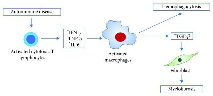
A pathophysiology diagram illustrating the proposed mechanism for coexisting hemophagocytosis and myelofibrosis, specifically in the context of Systemic Lupus Erythematosus (SLE). The process begins with an autoimmune disease trigger that leads to the activation of cytotoxic T lymphocytes. These activated cells release pro-inflammatory cytokines, specifically IFN-gamma, TNF-alpha, and IL-6 (all indicated with upward-facing arrows denoting increased levels). These cytokines then stimulate macrophages, which transition into an activated state. The activated macrophages drive two simultaneous pathways: 1) the direct clinical phenomenon of hemophagocytosis, and 2) the increased production of TGF-beta. The elevated TGF-beta acts upon fibroblasts (depicted as green spindle-shaped cells), stimulating their proliferation and activity, which ultimately results in myelofibrosis. The diagram uses flow arrows to establish a clear sequential relationship between immune activation, cytokine signaling, and tissue-level pathology, highlighting the dual outcomes of hypercytokinemia in hematopoietic and stromal environments.
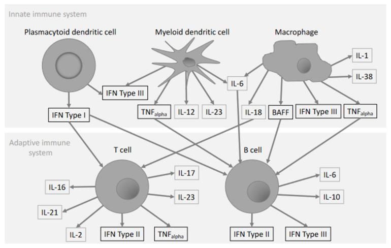
A pathophysiology diagram illustrating the cytokine network in systemic lupus erythematosus (SLE) pathogenesis, divided into innate and adaptive immune system compartments. The upper section (innate) features plasmacytoid dendritic cells (pDC), myeloid dendritic cells (mDC), and macrophages. The pDC is associated with IFN Type I and III production. The stellate mDC produces TNF-alpha, IL-12, IL-23, and IL-6. The irregular-shaped macrophage is shown secreting IL-1, IL-38, IL-18, BAFF, IFN Type III, TNF-alpha, and IL-6. The lower section (adaptive) depicts T cells and B cells. Interconnecting arrows show signaling crosstalk: IFN Type I and BAFF act on both T and B cells; IL-6 and IL-18 target T cells. T cell-derived cytokines include IL-16, IL-21, IL-2, IL-17, IL-23, TNF-alpha, and IFN Type II. B cell-derived cytokines include IL-6, IL-10, IFN Type II, and IFN Type III. This educational schematic highlights the interplay between diverse immune cell subsets and proinflammatory mediators involved in chronic autoimmune inflammation.
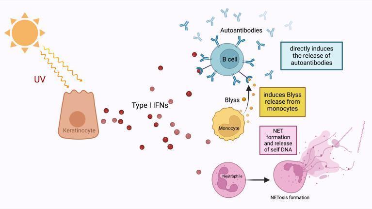
This pathophysiology diagram illustrates the role of Type I Interferons (IFNs) in the pathogenesis of Systemic Lupus Erythematosus (SLE) triggered by ultraviolet (UV) radiation. The process begins with UV light stimulating a keratinocyte in the skin, which subsequently releases Type I IFNs (red circles). These interferons act as central mediators through three primary pathways: (1) Directly stimulating a B cell to release autoantibodies (Y-shaped blue structures); (2) Stimulating a monocyte to release B-lymphocyte stimulator (Blyss), which further promotes B cell autoantibody production; and (3) Stimulating a neutrophil to undergo NETosis. The NETosis pathway shows the transition of a normal neutrophil into a state of 'NET formation and release of self DNA,' where an extracellular web-like fiber network of nucleic acids is expelled. This network serves as a source of autoantigens, reinforcing the autoantibody response. The diagram highlights the transition from an innate immune response (keratinocytes, monocytes, and neutrophils) to an adaptive autoimmune response (B cells and autoantibodies) in a clinical context relevant to photosensitivity and SLE.
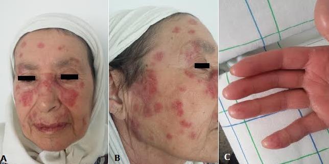
A composite of clinical photographs demonstrating dermatological manifestations suggestive of systemic autoimmune disease, likely Systemic Lupus Erythematosus (SLE). Panel A shows a frontal view of the face with well-demarcated, erythematous, squamous plaques in a malar 'butterfly' distribution across the bridge of the nose and cheeks, sparing the nasolabial folds. Additional discrete discoid-like lesions are visible on the forehead. Panel B presents a lateral view showing multifocal, erythematous, scaly plaques scattered across the cheek and temple areas. Panel C displays the palmar surface of the hand, illustrating digital pulpitis characterized by erythematous, swollen, and shiny fingertips with evidence of localized inflammation and edema. The clinical findings represent classic mucocutaneous involvement, including malar rash and digital vasculopathy, serving as important diagnostic markers in rheumatological and dermatological evaluations for connective tissue disorders.
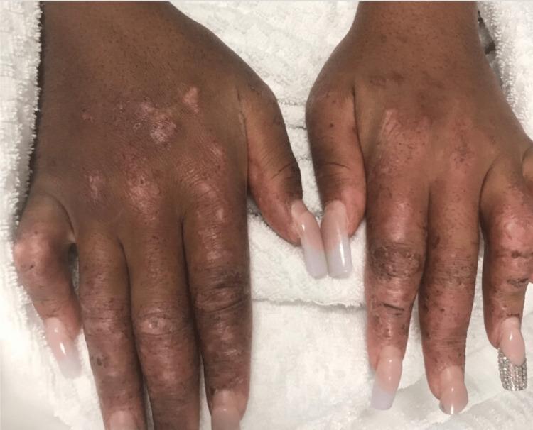
This clinical photograph displays the dorsal surface of the hands and fingers in a patient with a known history of systemic autoimmune disease, including systemic lupus erythematosus (SLE) and scleroderma. The image demonstrates significant cutaneous manifestations characterized by diffuse dyspigmentation. Key features include multiple hypopigmented (leukoderma-like) macules and patches interspersed with areas of hyperpigmentation and scattered dark macules, particularly prominent over the metacarpophalangeal and interphalangeal joints. The skin texture appears thickened and indurated, consistent with sclerodactyly, with visible xerosis and fine scaling. There is evidence of mild soft tissue swelling of the digits, resulting in a slightly tapered or 'sausage-digit' appearance. These findings are clinically significant as they illustrate the overlap features of connective tissue diseases, showing both the pigmentary changes often seen in chronic cutaneous lupus and the structural skin tightening characteristic of systemic sclerosis. The nails feature artificial extensions, which limit the evaluation of the underlying nail beds for splinter hemorrhages or telangiectasia.
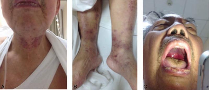
This composite of clinical photographs illustrates multisystemic manifestations of Systemic Lupus Erythematosus (SLE) in a single patient. Panel A displays the head and neck, showing erythematous maculopapular eruptions distributed across the forehead and concentrated densely on the anterior neck and submental region. Panel B focuses on the bilateral lower extremities, revealing scattered, non-blanching purpuric lesions of varying diameters on the ankles and distal shins, characteristic of vasculitic involvement. Panel C provides an intraoral view showing significant mucosal involvement, including a large, deep ulceration on the hard palate and visible hemorrhage from the oral mucosa and tongue surface. Collectively, these images demonstrate the mucocutaneous and vascular signs (purpura, oral ulcers, and malar-like eruptions) that serve as diagnostic criteria for high-activity systemic autoimmune disease, particularly class IV/V lupus nephritis as described in the accompanying clinical context.
SLE lupus nephritis renal histology wire loop lesion
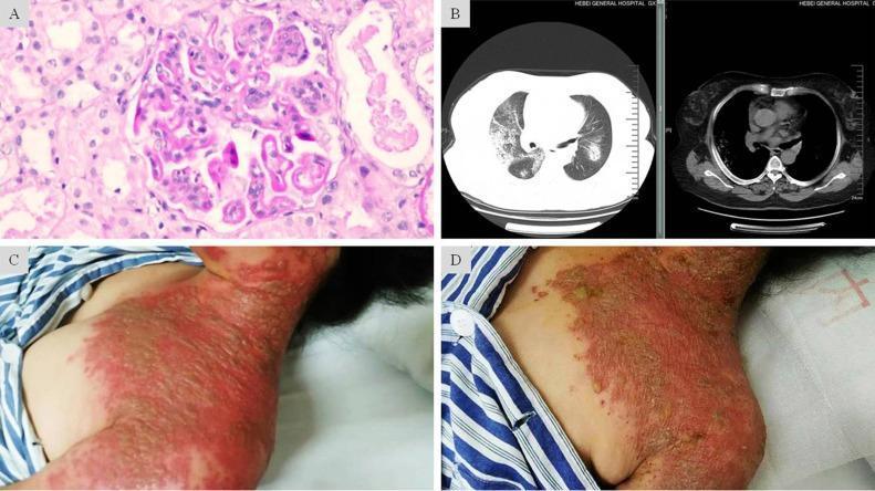
A composite medical image featuring four panels (A-D) illustrating clinical, radiological, and pathological findings of a complex case involving Lupus Nephritis and severe Herpes Zoster. Panel A is a light microscopy image (H&E stain) of a renal biopsy showing glomerular endothelial and mesangial hypercellularity with characteristic 'wire-loop' lesions, consistent with Class IV-G Lupus Nephritis. Panel B presents axial chest CT scans (lung and mediastinal windows) displaying bilateral, scattered patchy and nodular opacities with ill-defined borders, suggestive of diffuse alveolar hemorrhage. Panels C and D are clinical photographs of the neck and chest region showing the progression of skin lesions. Panel C depicts the acute phase of Herpes Zoster, characterized by confluent, grouped vesicles on an intense erythematous base extending across multiple dermatomes. Panel D shows the same region post-antiviral treatment, demonstrating resolving lesions with dried-up exudate and crusting on a fading erythematous base. This figure serves as an educational resource for multi-organ manifestations in immunocompromised patients, specifically highlighting systemic lupus erythematosus complications and disseminated viral infection.
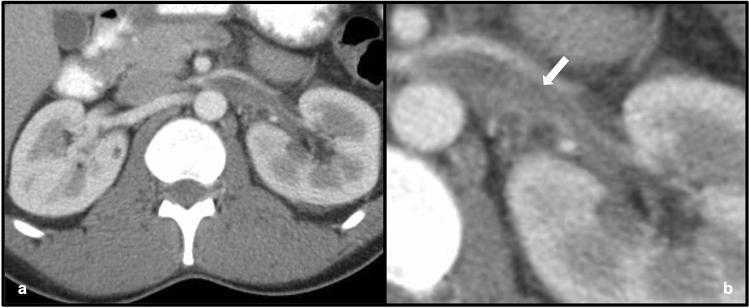
This diagnostic image consists of two axial contrast-enhanced CT views of the abdomen: a wide field-of-view (a) and a magnified narrow field-of-view (b). The images demonstrate the renal anatomy of a 35-year-old male with lupus nephritis. The primary pathological finding is a prominent filling defect within an expanded left renal vein, indicated by the white arrow in image (b). This low-attenuation area within the vessel lumen is characteristic of acute renal vein thrombosis (RVT). For anatomical comparison, the right renal vein appears normal in size and demonstrates uniform contrast enhancement. Both kidneys are visible; while the renal parenchyma is relatively homogenous bilaterally, the left kidney shows subtle enlargement compared to the right. This visual evidence illustrates a significant vascular complication associated with nephrotic-range proteinuria and systemic inflammatory conditions like systemic lupus erythematosus (SLE). The imaging modality effectively distinguishes between the patent right renal vasculature and the obstructive thrombus on the left.
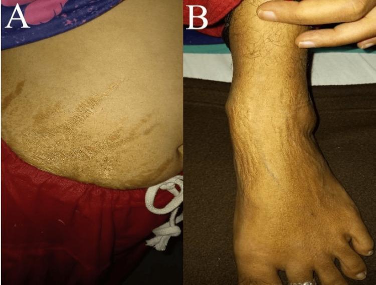
This composite clinical photograph displays dermatological manifestations in a 30-year-old patient with Systemic Lupus Erythematosus (SLE) and Lupus Nephritis (LN). Panel A illustrates the lower abdominal region, showing a series of hyperpigmented, linear striae-like lesions. These markings are irregularly distributed and appear as darker, brownish striations against the surrounding skin, characteristic of atrophic changes or medication-induced striae (often associated with chronic corticosteroid use). Panel B focuses on the anterior aspect of the left lower limb and ankle joint. It reveals significant localized edema (swelling) around the ankle and dorsal foot, accompanied by diffuse cutaneous hyperpigmentation and potentially livedo-like vascular changes. The clinical significance of these images lies in demonstrating the multi-organ involvement of SLE, highlighting both primary skin pathology and secondary changes related to chronic disease management or renal dysfunction (LN). This educational material is intended for clinical training in rheumatology, dermatology, and nephrology.
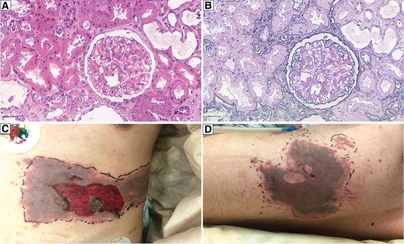
A multi-panel figure containing light microscopy of renal tissue and clinical photographs of dermatological lesions. Panels A and B display renal biopsy specimens at 200x magnification. Image A (Hematoxylin and Eosin stain) shows a glomerulus with mild-to-moderate hypercellularity and mesangial matrix expansion, consistent with lupus nephritis. Image B (Periodic Acid-Schiff stain) highlights the basement membranes and mesangium, demonstrating immune complex deposits in the subendothelial and mesangial regions. Surrounding renal tubules show varying degrees of epithelial changes. Panels C and D are clinical photographs of a patient with systemic lupus erythematosus showing a severe, clustered vesicular rash on the left hypochondrium (C) and left back (D). The lesions exhibit significant confluence, dark necrotic or hemorrhagic centers, and evidence of ruptured vesicles. The affected areas are demarcated by black ink for clinical monitoring, suggesting a dermatomal distribution characteristic of herpes zoster (shingles) occurring in an immunocompromised state. These images collectively illustrate the renal manifestations of SLE and a subsequent secondary viral opportunistic infection.
lupus butterfly malar rash face discoid lupus
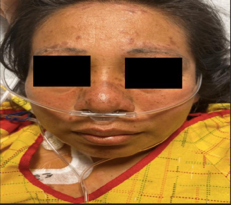
A clinical photograph of a female patient's face demonstrating classic dermatological manifestations of systemic lupus erythematosus (SLE). A confluent, erythematous malar rash is visible in a characteristic 'butterfly distribution,' spanning the bridge of the nose and both cheeks. Notably, the nasolabial folds are spared from the erythema. In addition to the malar rash, there are multiple discrete, raised, hyperpigmented, and round discoid lesions located along the hairline and eyebrows, suggesting discoid lupus involvement. Mild peri-orbital edema is present. The patient is shown with a nasal cannula in place, indicating respiratory support, consistent with potential systemic complications such as pleural effusion. This image serves as a teaching tool for identifying pathognomonic skin signs of autoimmune connective tissue diseases, specifically highlighting the distinction between the malar rash and lesions that involve the forehead and scalp.
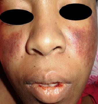
A clinical photograph of a patient's face demonstrating dermatological and mucosal manifestations of Systemic Lupus Erythematosus (SLE). A classic malar rash (butterfly rash) is present, characterized by symmetric, erythematous-to-violaceous patchy lesions over the malar eminences and the bridge of the nose, notably sparing the nasolabial folds. Additionally, the perioral region exhibits significant erythema and mucosal involvement. The lips show evidence of hemorrhagic lesions, with visible blood crusting and a small fissure on the lower lip, suggestive of vasculitis or active systemic inflammation. These visual findings are key diagnostic indicators for SLE, particularly when associated with hematological abnormalities such as thrombocytopenia. The photograph provides a clear example of cutaneous lupus manifestations for clinical diagnosis and medical education.
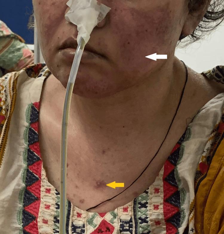
Clinical photograph of a female patient presenting with characteristic dermatological manifestations of Systemic Lupus Erythematosus (SLE). A white arrow points to a malar rash, also known as a butterfly rash, which is characterized by fixed erythema and slight edema across the bridge of the nose and the malar eminences of the cheeks, typically sparing the nasolabial folds. A yellow arrow indicates a discoid rash on the upper chest, appearing as a well-demarcated, erythematous, raised papule/plaque with a slightly hyperpigmented or scarred center. The patient also has a nasogastric tube in place, secured with tape, and appears to have thin hair, potentially representing non-scarring alopecia. The clinical significance of these visual findings is their role as diagnostic criteria in rheumatology for SLE. The presentation is suitable for intermediate to advanced medical education regarding autoimmune connective tissue diseases and cutaneous lupus manifestations.
- Full pathogenesis from Robbins & Cotran (Fig 6.25 - the pathogenesis flowchart image)
- Complete autoantibody spectrum table
- Diagnostic criteria (Table 6.9)
- Clinical manifestations (Table 6.11)
- Morphology for each organ
- Skin histology (Fig 6.26 - H&E + immunofluorescence)
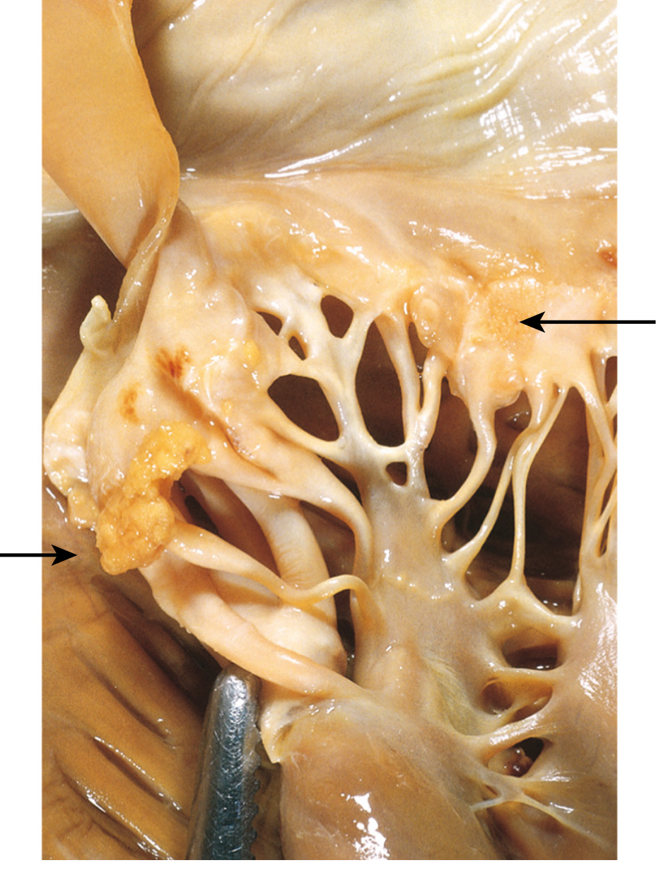
- Libman-Sacks endocarditis (Fig 6.27)
- Clinical images of malar rash, lupus nephritis, cytokine networks
Systemic Lupus Erythematosus (SLE)
Robbins & Cotran Pathologic Basis of Disease
QUICK OVERVIEW MNEMONIC: "SOAP BRAIN MD"
Serositis · Oral ulcers · Arthritis · Photosensitivity · Blood disorders · Renal disorder · Antinuclear antibody · Immunologic (anti-dsDNA, anti-Sm) · Neurologic disorder · Malar rash · Discoid rash
1. DEFINITION & EPIDEMIOLOGY
SLE is a chronic, remitting-relapsing autoimmune disease involving multiple organs, characterized by a vast array of autoantibodies - particularly ANAs - in which injury is caused mainly by deposition of immune complexes and binding of antibodies to various cells and tissues.
- Robbins, Cotran & Kumar Pathologic Basis of Disease, p. 215
| Feature | Detail |
|---|---|
| Prevalence | Up to 1 in 2500 in certain populations |
| Sex ratio | 9:1 F:M (reproductive age 17-55 yr) |
| Sex ratio (childhood / >65 yr) | 2:1 F:M |
| Peak age | 20s-30s (but any age possible) |
| Ethnicity | More prevalent/severe in African, Hispanic, Asian descent vs. European |
2. PATHOGENESIS FLOWCHART (from Robbins Fig. 6.25)
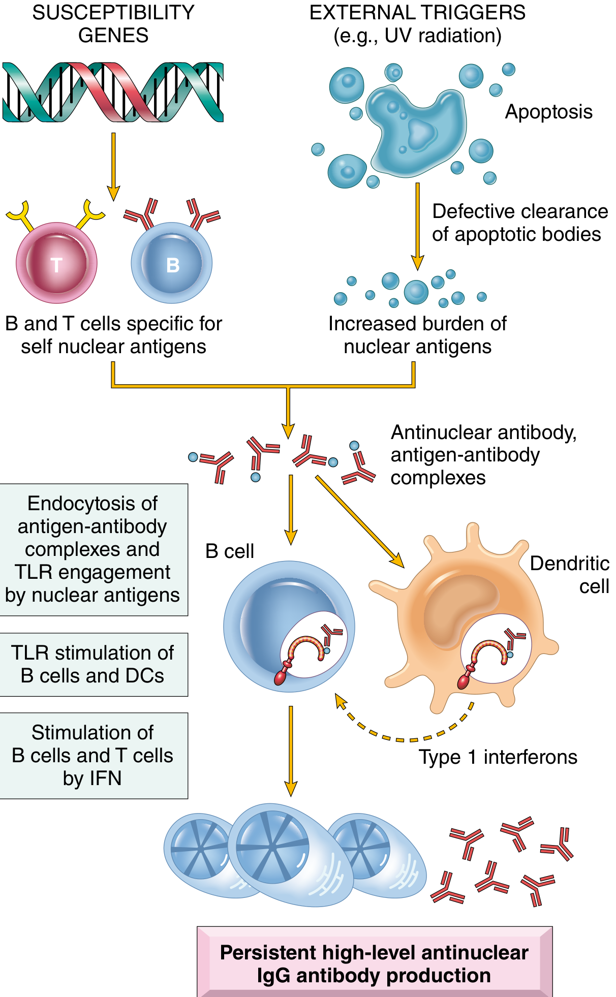
SUSCEPTIBILITY GENES EXTERNAL TRIGGERS (UV, drugs, viruses)
│ │
▼ ▼
B & T cells specific for Apoptosis of cells
self nuclear antigens │
(defective tolerance) ▼
│ Defective clearance of apoptotic bodies
│ │
│ Increased burden of NUCLEAR ANTIGENS
│ │
└──────────────────┬────────────────────────┘
▼
ANTINUCLEAR ANTIBODY + ANTIGEN-ANTIBODY COMPLEXES
│
┌───────────┴───────────┐
▼ ▼
B CELLS DENDRITIC CELLS
(endocytosis of (TLR engagement by
Ag-Ab complexes) nuclear antigens)
│ │
▼ ▼
TLR stimulation Type I INTERFERONS
→ more autoAb → activate B & T cells
└───────────┬───────────┘
▼
PERSISTENT HIGH-LEVEL ANTINUCLEAR IgG PRODUCTION
│
▼
IMMUNE COMPLEX DEPOSITION → TISSUE INJURY
3. GENETIC FACTORS
| Category | Details |
|---|---|
| Family risk | 20% of unaffected first-degree relatives have autoantibodies |
| Twin concordance | Monozygotic >20%, Dizygotic 1-3% |
| HLA associations | HLA-DQ locus → anti-dsDNA, anti-Sm, antiphospholipid antibodies |
| Complement deficiency | C2, C4, C1q deficiency → failure to clear immune complexes & apoptotic cells |
| GWAS loci | Proteins in lymphocyte signaling and IFN responses |
Twins (concordance) · Wild genes (GWAS) · Immune signaling loci · Complement deficiency · Estrogen/sex · HLA-DQ · Clinical relatives affected
4. IMMUNOLOGIC FACTORS (Flowchart)
DEFECTIVE B-CELL TOLERANCE
│
▼
Failure to eliminate self-reactive B cells in bone marrow
OR defective peripheral tolerance
│
▼
CD4+ Helper T cells (specific for nucleosomal antigens)
escape tolerance → help B cells make HIGH-AFFINITY AUTOANTIBODIES
│
▼
TLR ENGAGEMENT by nuclear DNA & RNA in immune complexes
→ B cells get additional activating signals
→ More antinuclear autoantibodies
│
▼
TYPE I IFNs (produced by TLR-stimulated DCs)
→ Activate DCs and B cells
→ Promote helper T cell responses
→ More autoantibodies + more inflammation
5. ENVIRONMENTAL FACTORS
| Trigger | Mechanism |
|---|---|
| UV light | Induces apoptosis; alters DNA for enhanced TLR recognition; stimulates keratinocyte IL-1 |
| Sex hormones (estrogen) | Partly accounts for female predominance |
| Drugs (hydralazine, procainamide, D-penicillamine) | Drug-induced lupus |
| Viruses | May trigger TLR stimulation by nucleic acids |
6. SPECTRUM OF AUTOANTIBODIES (Table from Robbins)
| Antibody | Antigen | Prevalence in SLE | Clinical Significance |
|---|---|---|---|
| Anti-dsDNA | Double-stranded DNA | 70% | Highly specific for SLE; correlates with disease activity/nephritis |
| Anti-Smith (Sm) | Protein complexed to small nuclear RNA | 25-30% | Most specific for SLE (high specificity) |
| Anti-histone | Histone proteins | 70% | Drug-induced lupus (most common here) |
| Anti-Ro (SS-A) | Small cytoplasmic RNA proteins | 30-40% | Neonatal lupus, sicca syndrome, subacute cutaneous lupus |
| Anti-La (SS-B) | RNA polymerase III transcript | 10-15% | Associated with anti-Ro |
| Antiphospholipid (aPL) | Phospholipid-protein complexes | 40-50% | Thrombosis, recurrent miscarriages, false-positive VDRL |
| Anti-RBC | Red blood cell antigens | Variable | Hemolytic anemia |
| Anti-platelet | Platelet antigens | Variable | Thrombocytopenia |
| ANA (generic) | Multiple nuclear antigens | ~100% | Sensitive but NOT specific |
Sm (most specific) · Histone (drug-induced) · ANA (all cases) · Ro/La (neonatal) · Phospholipid (thrombosis) · Double-stranded DNA (activity marker) · SLE = autoimmune
7. MECHANISMS OF TISSUE INJURY
| Mechanism | Hypersensitivity Type | Result |
|---|---|---|
| Immune complex deposition (DNA-anti-DNA) | Type III | Glomerulonephritis, vasculitis, skin lesions, arthritis |
| Autoantibodies against cell surface antigens (anti-RBC, anti-platelet) | Type II | Hemolytic anemia, thrombocytopenia, leukopenia |
| Antiphospholipid antibodies | Type II/III | Thrombosis, recurrent miscarriages, strokes |
| Anti-neuronal antibodies crossing BBB | Type II | Neuropsychiatric SLE |
8. DIAGNOSTIC CRITERIA (1997 Revised ACR Criteria - Table 6.9)
Diagnosis requires 4 of 11 criteria (serial or simultaneous)
| # | Criterion | Definition |
|---|---|---|
| 1 | Malar rash | Fixed erythema over malar eminences ("butterfly"), sparing nasolabial folds |
| 2 | Discoid rash | Erythematous raised patches, keratotic scaling, follicular plugging |
| 3 | Photosensitivity | Skin rash from unusual reaction to sunlight |
| 4 | Oral ulcers | Oral/nasopharyngeal ulceration, usually painless |
| 5 | Arthritis | Nonerosive synovitis, ≥2 peripheral joints |
| 6 | Serositis | Pleuritis or pericarditis |
| 7 | Renal disorder | Proteinuria >0.5 g/24h OR red cell casts |
| 8 | Neurologic disorder | Seizures, psychosis, myelitis, or neuropathy |
| 9 | Hematologic disorder | Hemolytic anemia, leukopenia (<4000), lymphopenia (<1500), thrombocytopenia (<100,000) |
| 10 | Immunologic | Anti-dsDNA, anti-Sm, OR antiphospholipid antibodies |
| 11 | ANA | Abnormal ANA titer by immunofluorescence |
9. MORPHOLOGY - ORGAN-BY-ORGAN
Skin (85% of patients)
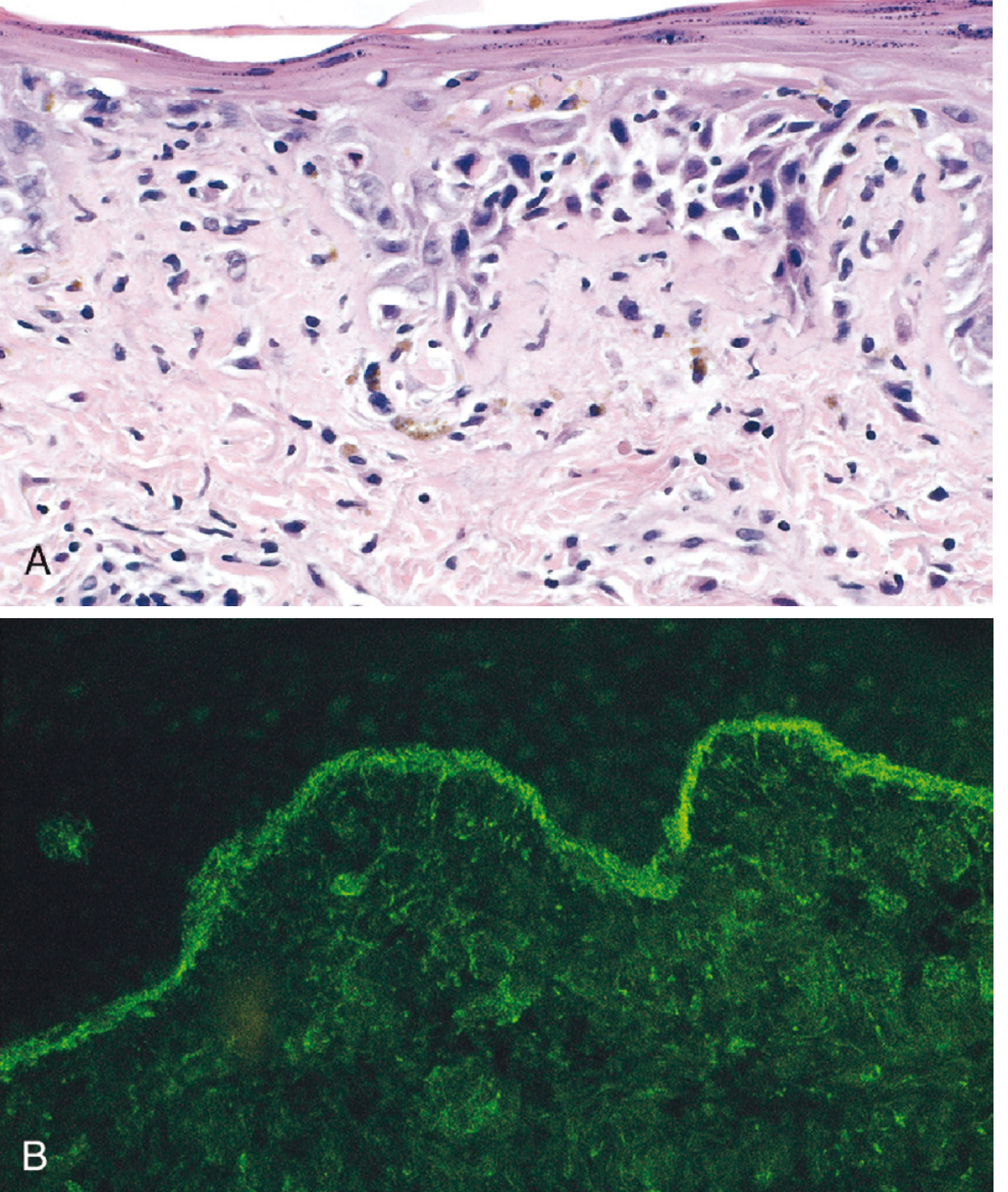
| Gross | Butterfly rash (50%), urticaria, bullae, maculopapular lesions, ulcerations |
|---|---|
| Histology | Vacuolar degeneration of basal epidermis; dermal edema; perivascular inflammation; fibrinoid necrosis of vessels |
| Immunofluorescence | IgG + complement deposits along dermoepidermal junction (even in uninvolved skin = "lupus band") |
Kidney (50-70% of patients) - Lupus Nephritis
| WHO/ISN Class | Name | Features |
|---|---|---|
| I | Minimal mesangial | Normal light microscopy; mesangial deposits on IF |
| II | Mesangial proliferative | Mesangial hypercellularity |
| III | Focal | <50% of glomeruli affected |
| IV | Diffuse (most common/severe) | >50% of glomeruli; "wire-loop" lesions; subendothelial deposits |
| V | Membranous | Subepithelial deposits; nephrotic syndrome |
| VI | Advanced sclerosing | >90% sclerosed; end-stage |
- Wire-loop lesion: Massive subendothelial immune complex deposition → glomerular capillary wall thickening resembling a wire loop
- Complement levels (C3, C4) fall during active nephritis (consumption)
Heart
| Lesion | Feature |
|---|---|
| Libman-Sacks endocarditis | Small, sterile verrucous vegetations on both surfaces of valve leaflets (esp. mitral); NOT infective |
| Pericarditis | Most common cardiac manifestation; fibrinous exudate → pericardial effusion |
| Myocarditis | Less common; may cause arrhythmias |
| Accelerated atherosclerosis | Leading cause of long-term mortality |
Joints (80-90%)
- Nonerosive synovitis - key distinction from Rheumatoid Arthritis (RA)
- Little deformity; no pannus formation
- ≥2 peripheral joints involved; tenderness, swelling, effusion
Blood Vessels
- Acute: fibrinoid necrotizing vasculitis of capillaries, small arteries, arterioles (any tissue)
- Chronic: fibrous thickening with luminal narrowing
Serosal Membranes (Serositis)
- Pleuritis (45%) + Pericarditis (25%)
- Acute: fibrinous exudate on mesothelial surfaces
- Chronic: thickened, opaque, "shaggy" fibrous tissue; possible obliteration of cavity
CNS (25-35%)
- No clear morphologic lesions correlating with neuropsychiatric symptoms
- Noninflammatory occlusion of small vessels by intimal proliferation (from endothelial damage by autoantibodies)
- Anti-neuronal antibodies crossing BBB → cognitive dysfunction, psychosis, seizures
Hematologic (100%)
| Cytopenia | Mechanism | Prevalence |
|---|---|---|
| Anemia (hemolytic or ACD) | Anti-RBC antibodies → opsonization + phagocytosis (Type II); also chronic inflammation + iron deficiency | ~50% |
| Thrombocytopenia | Anti-platelet antibodies → destruction | ~10% |
| Leukopenia/Lymphopenia | Anti-WBC antibodies | Common |
10. CLINICAL FEATURES SUMMARY TABLE (Table 6.11 - Robbins)
| Clinical Manifestation | Prevalence (%) |
|---|---|
| Hematologic | 100 |
| Fatigue | 80-100 |
| Arthritis/arthralgia/myalgia | 80-90 |
| Skin | 85 |
| Fever | 55-85 |
| Weight loss | 60 |
| Renal | 50-70 |
| Pleuritis | 45 |
| Raynaud phenomenon | 15-40 |
| Neuropsychiatric | 25-35 |
| Pericarditis | 25 |
| Gastrointestinal | 20 |
| Peripheral neuropathy | 15 |
| Ocular | 5-15 |
11. CLINICAL FEATURES - KEY POINTS
- Butterfly rash over face
- Fever
- Joint pain (no deformity) in multiple peripheral joints
- Pleuritic chest pain
- Photosensitivity
- Febrile illness of unknown origin
- Fatigue + abnormal urinalysis
- Joint disease mimicking RA or rheumatic fever
12. ANTIPHOSPHOLIPID ANTIBODY SYNDROME (Secondary APS in SLE)
Antiphospholipid Antibodies (40-50% of SLE patients)
│
┌─────────┴─────────┐
▼ ▼
VENOUS THROMBOSES ARTERIAL THROMBOSES
(DVT, PE) (stroke, TIA, AMI)
│
▼
RECURRENT SPONTANEOUS MISCARRIAGES
(fetal loss due to placental thrombosis)
│
▼
FALSE-POSITIVE VDRL/RPR (syphilis test)
"The aPL triad: Clots, Crashes (miscarriages), and False-positive VDRL"
13. COMPREHENSIVE PATHOGENESIS FLOWCHART (All in one)
GENETIC PREDISPOSITION + ENVIRONMENTAL TRIGGERS
│
▼
FAILURE OF SELF-TOLERANCE
(B cells + T cells survive)
│
┌──────────┴──────────┐
▼ ▼
B CELL ACTIVATION CD4+ T CELL ACTIVATION
(bone marrow + (nucleosomal Ags escape
peripheral defects) tolerance)
│ │
└──────────┬──────────┘
▼
ANTINUCLEAR AUTOANTIBODY PRODUCTION
(anti-dsDNA, anti-Sm, anti-histone, aPL)
│
┌──────────┴──────────────────┐
▼ ▼
IMMUNE COMPLEX DIRECT ANTIBODY
FORMATION (Type III) BINDING (Type II)
│ │
▼ ▼
Glomerulonephritis Hemolytic anemia
Vasculitis Thrombocytopenia
Skin lesions Leukopenia
Serositis Antiphospholipid syndrome
Arthritis
14. KEY PATHOLOGIC MNEMONICS
| Mnemonic | Expands to |
|---|---|
| "SOAP BRAIN MD" | ACR diagnostic criteria (need 4/11) |
| "SHARP DS" | Autoantibodies spectrum |
| "Wire Loop = Class IV" | Most severe lupus nephritis |
| "Libman-Sacks = Both sides" | Vegetations on BOTH surfaces (vs. infective = tips only) |
| "Butterfly rash spares nasolabial folds" | Key distinction from rosacea |
| "Lupus band = IF at DEJ" | IgG + complement at dermoepidermal junction |
| "3 No's of SLE arthritis" | No erosion, No deformity, No pannus |
| "9:1 female at childbearing age" | Sex predominance |
| "Anti-Sm = Most specific; ANA = Most sensitive" | Serology memory aid |
| "Anti-dsDNA = Disease Activity marker" | Correlates with nephritis flares |
15. PROGNOSIS & CAUSES OF DEATH
| Phase | Cause |
|---|---|
| Early (active disease) | Renal failure (Class IV nephritis), infections (immunosuppression) |
| Late (chronic) | Accelerated atherosclerosis - leading long-term cause of death |
| Antiphospholipid related | Stroke, pulmonary embolism, MI |
16. UV LIGHT → SLE FLARE (Mechanism)