
21 year male , complains of bleeding per rectum ,while passing motion associated with pain and constipation
21 year male , complains of bleeding per rectum ,while passing motion associated with pain and constipation
anal fissure clinical appearance
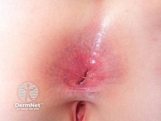
Imaging modality and technique: Clinical photography of external perianal skin; frontal view with standard color imaging under ambient lighting; no magnification, dermoscopy, or contrast agents. Comprehensive description: The image shows localized erythema and irritation around the anal verge with a superficial midline fissure. The perianal skin is reddened with mild edema and subtle desquamation. A small linear slit extends from the anal opening toward the perianal skin, consistent with an anal fissure. Surrounding tissue exhibits shallow excoriations and diffuse inflammatory changes; there is moisture-associated dermatitis and possible contact dermatitis features. No visible purulence or deep ulceration is evident; no evident hemorrhoidal prolapse or conspicuous skin folds are noted. The pattern favors an inflammatory dermatosis of the peri-anal region rather than a deep infectious ulcer. The appearance may reflect irritation from stool moisture, soaps, pads, or friction; secondary infection cannot be excluded if symptoms persist. Clinical significance and use cases: This presentation supports diagnoses such as perianal dermatitis and anal fissure, irritant/contact dermatitis, or candidal dermatitis in the appropriate clinical context. It informs topical therapy (emollients, barrier creams, vasodilators for fissures), hygiene counseling, and follow-up to monitor healing or progression. Differential considerations include hemorrhoids with fissure, lichen sclerosus, or Crohn disease–associated fissure in chronic cases.

A series of four clinical photographs (a-d) showing the chronological progression and healing of a chronic anal fissure (AF) in the perianal region over a six-month period following treatment with incobotulinumtoxinA (incoBoNT/A). Frame (a) represents the baseline, showing a deep, erythematous longitudinal tear in the anoderm with associated inflammation of the surrounding perianal skin. Frame (b) shows the one-month follow-up, demonstrating a persistent fissure but with reduced surrounding erythema. Frame (c) illustrates the three-month follow-up, where the fissure size is significantly reduced and the tissue appears less irritated. Frame (d) represents the six-month follow-up, showing near-complete resolution of the fissure, restoration of skin integrity, and a healthy, non-inflamed perianal appearance. This sequence demonstrates the clinical efficacy of botulinum toxin in promoting the healing of chronic fissures by reducing internal anal sphincter hypertonicity and improving local perfusion.
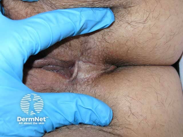
Clinical photography of the perianal region in a male patient. Modality: standard color image acquired with ambient to diffuse white light, no contrast, portable high‑resolution camera. The image shows a close‑up view of the natal/intergluteal cleft with a small, midline, slit‑like opening in the perianal skin. Surrounding skin is hair-bearing, mildly hyperemic, with a subtle scar line and slight pliable edema along the fold. The finger in the frame provides scale and gently retracts surrounding tissue to reveal the external glandular orifice. The tract appears to be a superficial external opening, potentially part of a pilonidal sinus or perianal fistulous communication. No frank purulence or surrounding cellulitis is evident in this snapshot; there is no visible discharge. The appearance is compatible with recognisable dermatologic or colorectal conditions that create sinus tracts or fissures in the anoderm or natal cleft. Clinically, this finding warrants assessment for infection, depth, and possible tracts to guide management. Differential considerations include pilonidal disease, external anal fissure, anal fistula, epidermoid cyst, and hidradenitis suppurativa. Diagnostic significance lies in determining need for drainage, surgical excision, or further imaging such as MRI for tract mapping. This image serves educational, diagnostic, and reference purposes for dermatology and colorectal surgery.
21-Year-Old Male: Bleeding Per Rectum + Pain + Constipation
Most Likely Diagnosis: Anal Fissure
What is an Anal Fissure?
Clinical Features (Matching This Patient)
| Feature | This Patient | Typical Anal Fissure |
|---|---|---|
| Age | 21 years | Young adults |
| Sex | Male | Both sexes |
| Bleeding | Per rectum, during motion | Bright red, streaks on stool/paper |
| Pain | Present during defecation | Severe, "passing razor blades/broken glass" |
| Constipation | Present | Common - hard stools cause/worsen fissure |
Pathophysiology
Acute vs. Chronic Fissure
| Feature | Acute | Chronic |
|---|---|---|
| Duration | < 6-8 weeks | > 6-8 weeks |
| Appearance | Shallow tear in anoderm | Exposed internal sphincter fibers at base |
| Additional findings | None | Hypertrophied anal papilla (proximal) + Sentinel pile/skin tag (distal) |
| Pain severity | Severe, acute | Less severe but unrelenting |
Differential Diagnosis
- Hemorrhoids (internal) - Painless bright red bleeding typically; rarely painful unless thrombosed
- Perianal abscess/fistula - Throbbing pain, may have discharge
- Crohn's disease - If fissure is off midline (atypical location), consider Crohn's, HIV, TB, syphilis, herpes, anal cancer - Sabiston, p. 1835
- Rectal polyp/carcinoma - Less likely at 21, but must exclude if bleeding is persistent
- Inflammatory Bowel Disease - If associated with diarrhea, systemic symptoms, weight loss
Examination
- Gentle separation of buttocks may reveal the fissure or a sentinel skin tag
- Spreading buttocks alone may cause intolerable pain - examination may need to stop
- Gentle pressure at posterior/anterior anal canal reproduces pain
- Digital rectal exam and anoscopy are often deferred due to pain
- If diagnosis is unclear or malignancy suspected: examination under anesthesia (EUA)
Management
Conservative (First-line for Acute Fissure)
- Increase fluid and dietary fiber - to maintain soft, formed stools
- Sitz baths - significant pain relief in >90% of patients with acute fissures
- Stool softeners / laxatives to avoid straining
- Majority of acute fissures resolve with medical management alone - Sabiston, p. 1841
Medical (For Chronic Fissure)
| Drug | Mechanism | Notes |
|---|---|---|
| Topical GTN (nitroglycerin) | NO donor → sphincter relaxation, vasodilation | ~50% healing; headache is major side effect (up to 20% stop therapy) |
| Topical Calcium Channel Blockers (diltiazem, nifedipine) | Sphincter relaxation | Similar efficacy to GTN, fewer headaches - preferred |
| Botulinum toxin injection | Blocks acetylcholine at neuromuscular junctions | ~50% cure rate; temporary incontinence in up to 10% |
- Bailey and Love's Short Practice of Surgery, p. 614; Sabiston, p. 1844-1845
Surgical (When Medical Fails)
- Lateral Internal Sphincterotomy (LIS) - Gold standard surgical treatment; divides part of internal anal sphincter
- Anal sphincter dilatation - rarely indicated due to risk of incontinence at multiple sphincter sites
- Bailey and Love's, p. 642
Summary Box (Bailey & Love's)
- Acute or chronic ulcer in the midline of the anal canal
- Ectopic site suggests a more sinister cause
- Symptoms: Pain on defecation, bright-red bleeding, mucus discharge, constipation
Key Points for This Patient
- Most likely diagnosis: Acute anal fissure triggered by constipation
- Start conservative treatment immediately: fiber, fluids, sitz baths, stool softeners
- If no improvement in 6-8 weeks, move to topical CCB (diltiazem) or botox
- Red flags requiring further evaluation: off-midline fissure, weight loss, family history of IBD/colorectal cancer, no improvement with treatment - consider colonoscopy to rule out IBD or neoplasia
- Reassure the patient - this is a benign, common, and treatable condition
Slightly darks colour blood , mixed with stool, present 2 times since morning
ulcerative colitis colonoscopy findings
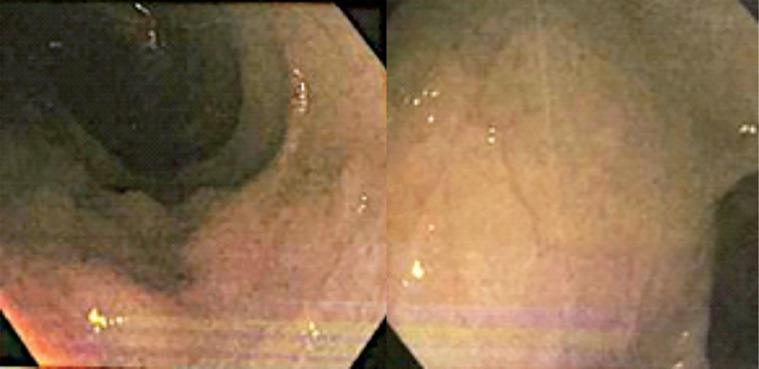
This diagnostic image consists of two endoscope frames from a colonoscopy, depicting the rectum and sigmoid colon of a patient with Ulcerative Colitis. The visual highlights active chronic colitis characterized by diffuse mucosal inflammation. Key features include multiple irregular ulcers appearing as dark, depressed lesions against a background of erythematous (reddened) and edematous mucosa. The ulcerations vary in morphology, ranging from small pinpoint erosions to larger, more confluent serpiginous patterns. There is a noticeable loss of the normal fine vascular pattern, indicative of significant mucosal engorgement and friability. These findings are clinically relevant for assessing disease activity and severity in Inflammatory Bowel Disease (IBD), specifically correlating with high Pediatric Ulcerative Colitis Activity Index (PUCAI) scores in pediatric gastroenterology.
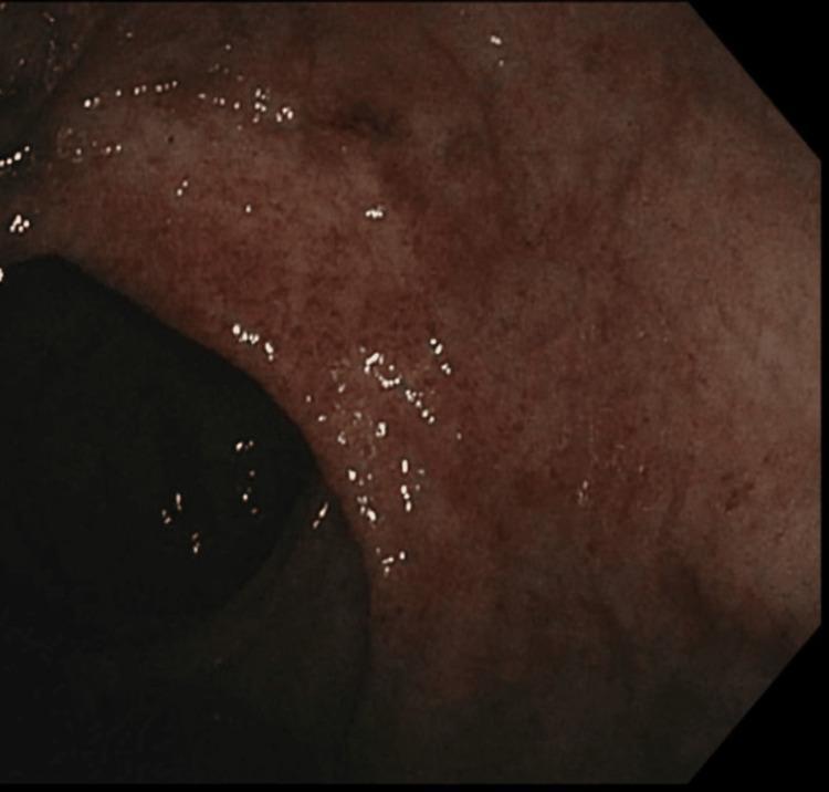
This diagnostic image is a clinical photograph obtained during a colonoscopy, showing the mucosal lining of the rectum. The visual field demonstrates key features of active ulcerative colitis, characterized by moderate endoscopic activity (Mayo Score 2). The rectal mucosa exhibits significant diffuse erythema and a granular, irregular texture, indicating active inflammation. Key pathological findings include a loss of the normal vascular pattern and the presence of numerous small, punctate lesions and friability across the mucosal surface, consistent with superficial ulcerations and erosions. The educational focus of this image is the recognition of endoscopic signs of inflammatory bowel disease (IBD), specifically the transition from healthy tissue to inflamed, erythematous, and damaged mucosa in a patient with left-sided ulcerative colitis. This clinical imaging is essential for medical students and gastroenterology trainees to understand the visual markers used in the classification and monitoring of disease severity in ulcerative colitis.
Updated Clinical Assessment - REVISED DIAGNOSIS
Why This Changes Everything
| Feature | Anal Fissure | This Patient Now |
|---|---|---|
| Blood color | Bright red | Slightly dark |
| Blood location | On surface / toilet paper | Mixed with stool |
| Frequency today | - | 2 episodes this morning |
| Pain | During defecation | Present |
| Constipation | Yes | Yes |
Revised Differential Diagnosis
1. Inflammatory Bowel Disease (IBD) - TOP PRIORITY
- Peak onset: 15-35 years - fits perfectly
- Classic symptoms: bloody diarrhea/constipation, tenesmus, crampy abdominal pain
- Inflammation starts in rectum and extends proximally as one continuous lesion
- Blood is typically mixed with stool or mucus - ROSEN's Emergency Medicine, p. 2747
- Also peaks before age 30
- More likely if: perianal disease (fissures, skin tags, abscesses), skip lesions, small bowel involvement
- Patients may have nocturnal diarrhea, fatigue, fever, weight loss - ROSEN's, p. 2754
2. Infective Colitis - Important to exclude
- Bacterial: Salmonella, Shigella, Campylobacter, E. coli O157:H7, C. difficile
- Amebic colitis
- All can present with dark blood mixed in stool, pain, altered bowel habits
3. Colorectal Polyp / Juvenile Polyp
- Young age group; juvenile polyps can bleed and cause dark blood mixed with stool
- Usually painless, but can cause constipation if large
4. Solitary Rectal Ulcer Syndrome
- Young patients, straining, constipation
- Blood and mucus in stool, rectal pain
5. Intussusception (less likely at 21 years)
- More common in children; dark "currant jelly" stool
Colonoscopy Findings in Active UC
IBD Severity Classification
Ulcerative Colitis
| Severity | Stools/Day | Blood | Systemic Signs |
|---|---|---|---|
| Mild | < 4 | Some | None (no fever, no tachycardia, no anemia) |
| Moderate | > 4 | Present | Minimal |
| Severe | > 6 bloody | Present | Fever, tachycardia, anemia, raised ESR |
Investigations to Order NOW
Bedside / Urgent
- Vitals - pulse, BP, temperature (rule out systemic toxicity)
- Stool examination - microscopy, culture, ova & parasites, C. diff toxin
Blood Tests
- CBC - hemoglobin (anemia?), WBC (infection/inflammation)
- CRP, ESR - inflammatory markers
- LFTs, RFTs - baseline before treatment
- Serum electrolytes
Stool Tests
- Fecal calprotectin - elevated in IBD (differentiates from IBS)
- Stool culture for pathogens
Imaging
- Abdominal X-ray - rule out toxic megacolon (colonic dilation)
- Colonoscopy with biopsy - gold standard for IBD diagnosis; shows continuous mucosal inflammation starting from rectum in UC
Serology (if IBD suspected)
- ANCA (UC associated) vs ASCA (Crohn's associated)
Management Approach
Immediate (Outpatient if Mild)
- Stool tests to rule out infective cause first
- Hydration and dietary modification
- Avoid NSAIDs and antidiarrheals (can worsen IBD or precipitate toxic megacolon)
If UC Confirmed - Mild to Moderate
- 5-ASA (aminosalicylates) are the mainstay - mesalazine orally (≥3 g/day) ± rectal enema/suppository
- Combined oral + topical 5-ASA is superior to oral alone
- If 5-ASA fails: oral/topical steroids (budesonide, hydrocortisone enema/foam)
- Persistent disease: Azathioprine (2 mg/kg/day) as immunomodulator
- Refractory: Infliximab (anti-TNF biologic)
- ROSEN's Emergency Medicine, p. 2884-2889
Indications for Hospitalization
-
6 bloody stools/day
- Systemic toxicity (fever, tachycardia)
- Severe abdominal pain or distension
- Suspicion of toxic megacolon
Surgery
- Reserved for severe disease unresponsive to medical therapy
- Up to 25% of UC patients eventually need colectomy
- Colectomy is curative for UC - ROSEN's Emergency Medicine, p. 2900
Red Flags to Watch For
- Fever > 38°C
- Heart rate > 90 bpm
- Severe abdominal pain or rigidity (perforation?)
- Abdominal distension (toxic megacolon?)
- Significant pallor/anemia
- Weight loss
Summary
A 21-year-old male with dark blood MIXED with stool, pain, and constipation most likely has Ulcerative Colitis or Infective Colitis. Anal fissure is now less likely as the primary diagnosis. Urgent investigations include stool culture, fecal calprotectin, CBC, CRP, and colonoscopy. Infective causes must be excluded before starting immunosuppressive therapy for IBD.