
explain about cerebral edema with simple understanding
cerebral edema brain swelling CT scan diagram
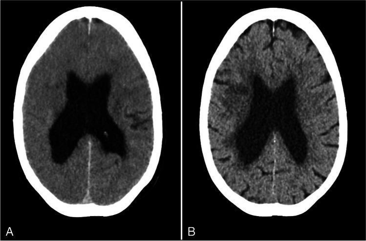
This Comparison Chart displays two axial non-contrast CT scans of the brain, demonstrating the evolution of cerebral edema. Image A (left) was taken immediately after a mechanical thrombectomy, showing significant diffuse brain swelling. Key diagnostic findings include the effacement of the cerebral sulci and cortical crowding, indicating increased intracranial pressure and loss of normal cerebrospinal fluid (CSF) spaces on the brain surface. The lateral ventricles appear prominent. Image B (right) is a follow-up scan from 4 days later, showing the resolution of the swelling. In this follow-up, the cerebral sulci have reappeared and are clearly visible, indicating a reduction in parenchymal pressure. A small hypodense area in the right middle cerebral artery territory is visible, consistent with a localized infarct. This comparison serves as an educational tool for identifying radiological signs of acute brain swelling and monitoring its clinical resolution over time.
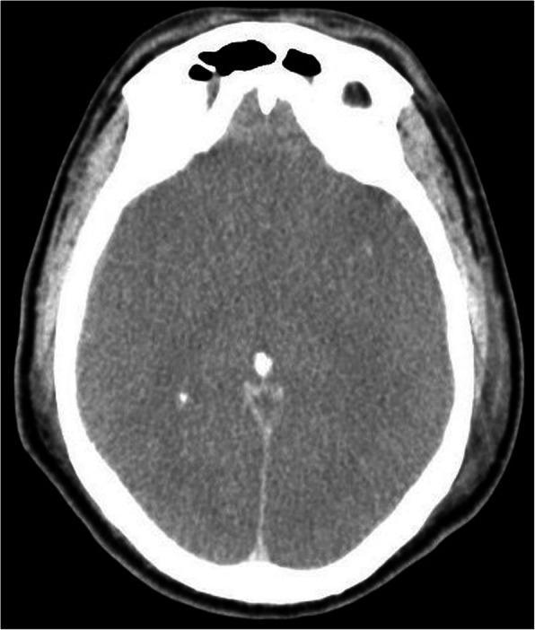
This diagnostic image is an axial non-contrast computed tomography (CT) scan of the brain demonstrating features of diffuse cerebral edema. The scan shows a global loss of the normal grey-white matter differentiation, which is a classic sign of severe brain swelling or global hypoxic-ischemic injury. The cerebral sulci and cisterns are effaced, indicating significant mass effect and increased intracranial pressure as the brain parenchyma expands against the rigid skull. A hyperdense midline structure corresponding to the interhemispheric fissure is visible, along with small hyperdense foci likely representing calcified pineal or choroid plexus structures. The surrounding skull appears intact with normal density. Extracranial soft tissue swelling is also noted. Clinically, these findings are highly significant in the context of severe sepsis or meningitis, where progressive swelling can lead to brain herniation or brain death if not managed aggressively. This image serves as a critical educational example of generalized intracranial hypertension and its radiographic manifestations in emergency medicine and neurology.
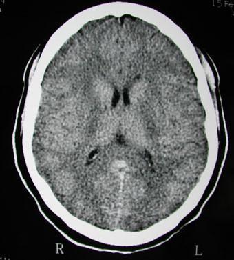
This diagnostic axial non-contrast CT scan of the brain demonstrates classic findings of diffuse cerebral edema. The image reveals a generalized loss of grey-white matter differentiation throughout the cerebral hemispheres, with the brain parenchyma appearing abnormally hypodense. Significant mass effect is evident through the near-complete effacement of the cortical sulci and cisterns, creating a 'smooth' appearance of the brain surface. The ventricular system, including the lateral and third ventricles, appears slit-like and severely compressed due to the increased intracranial pressure and global tissue swelling. These radiological features are characteristic of significant intracranial hypertension and global hypoxic-ischemic injury or inflammatory processes, such as the hypersensitivity syndrome described in the clinical context. The scan is a primary educational example of how acute cerebral swelling alters normal neuroanatomical landmarks on computed tomography.
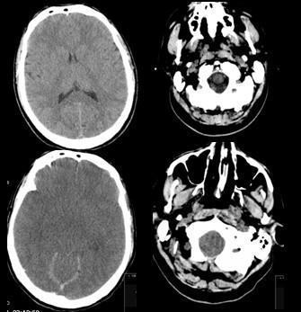
This diagnostic image displays four axial slices of a non-contrast Computed Tomography (CT) scan of the head, focusing on the brain and skull base. The primary finding is diffuse cerebral swelling associated with intracranial hypertension, clinically contexted in this case by cerebral malaria. Key features include generalized effacement of the cerebral sulci and cisterns, indicating significant mass effect from brain edema. The lateral ventricles appear slit-like and compressed. There is a notable loss of normal gray-white matter differentiation throughout the cerebral parenchyma, with a global decrease in tissue density. The lower slices show the posterior fossa and skull base, revealing compression of the subarachnoid spaces and crowding at the foramen magnum. In the most inferior slices, anatomical structures of the facial skeleton, including the nasal cavity, paranasal sinuses, and soft tissues of the neck, are visible with some evidence of overlying soft tissue swelling. This series illustrates the neuroradiological manifestations of acute cerebral edema and the resulting compromise of intracranial CSF spaces.
"cerebral edema" pathophysiology treatment
Cerebral Edema - A Simple, Comprehensive Guide
What Is It?
- Robbins & Kumar Basic Pathology: "Cerebral edema is the accumulation of excess fluid within the brain parenchyma."
The Two Main Types
1. Vasogenic Edema - "The Leaky Wall Type"
- Brain tumors (they inflame and disrupt the BBB around them)
- Brain abscess or infection (meningitis, encephalitis)
- Stroke
- Head injury
- High-altitude cerebral edema (HACE)
2. Cytotoxic Edema - "The Cell-Swelling Type"
- Global brain ischemia (cardiac arrest, drowning)
- Severe hypoxia
- Metabolic toxins
- Stroke (both types often coexist)
In practice, most serious brain injuries involve both types simultaneously. - Robbins, Cotran & Kumar Pathologic Basis of Disease
What Does It Look Like?
- The brain becomes softer than normal
- The gyri (ridges on the brain surface) become flattened and pressed against the skull
- The sulci (grooves between ridges) narrow and disappear
- The ventricles (fluid-filled spaces inside) get compressed
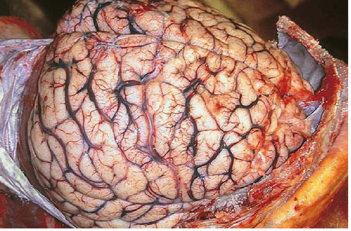
- Loss of normal grey-white matter differentiation
- Effacement (disappearance) of cortical sulci and cisterns
- Slit-like, compressed ventricles
- Increased intracranial pressure signs
Causes at a Glance
| Category | Examples |
|---|---|
| Trauma | Head injury, subdural hematoma |
| Ischemia/Infarction | Stroke, cardiac arrest |
| Infection | Meningitis, encephalitis, abscess |
| Tumors | Primary or metastatic brain tumors |
| Metabolic | Diabetic ketoacidosis, hyponatremia, liver failure |
| Altitude | High-altitude cerebral edema (HACE) |
| Toxins | Carbon monoxide, certain drugs |
Symptoms - What the Patient Feels
- Headache - typically worse in the morning, worse on bending/coughing
- Nausea and vomiting (often projectile)
- Altered consciousness - confusion, drowsiness, progressing to coma
- Blurred vision / papilledema (swelling of the optic disc from raised pressure)
- Focal neurological deficits - weakness, speech problems, depending on the area affected
- Seizures
Management
1. Osmotic Therapy
- Mannitol (0.25-1 g/kg IV) - the standard first-line agent; reduces brain volume, lowers CSF production, improves cerebral blood flow - Bradley & Daroff's Neurology
- Hypertonic saline (3% or 7.5% NaCl) - slightly more effective than mannitol per meta-analysis data; target sodium 145-155 mmol/L - Rosen's Emergency Medicine
2. Reduce Blood Volume
- Head elevation (30 degrees) to improve venous drainage
- Controlled hyperventilation - lowers CO2, causes cerebral vasoconstriction, reduces blood volume. Used as a temporary rescue measure only (too much causes ischemia)
3. CSF Drainage
- Intraventricular catheter (EVD) to drain CSF mechanically - used in head injury or acute hydrocephalus
4. Corticosteroids
- Dexamethasone is highly effective for vasogenic edema around tumors and abscesses (reduces BBB permeability)
- NOT effective for cytotoxic edema from stroke or global ischemia
5. Reduce Metabolic Demand
- Treat fever (fever worsens brain swelling)
- Avoid hypoxia and hypotension
- Treat seizures promptly
6. Surgery
- Decompressive craniectomy - removing part of the skull to give the brain room to swell outward. Lifesaving in malignant middle cerebral artery infarction
- Tumor or hematoma evacuation when these are the cause
The Dangerous Cascade - Why It Matters
Injury → Brain swells → ICP rises → Blood flow to brain decreases
→ More ischemia → More edema → Herniation → Death
Quick Summary
| Feature | Vasogenic | Cytotoxic |
|---|---|---|
| Location of fluid | Extracellular (between cells) | Intracellular (inside cells) |
| BBB | Disrupted | Intact (initially) |
| Main cause | Tumors, infection, trauma | Ischemia, hypoxia, toxins |
| Responds to steroids? | Yes | No |
| CT appearance | White matter edema | Grey and white matter both |
- Robbins & Kumar Basic Pathology - Chapter on CNS pathology
- Robbins, Cotran & Kumar Pathologic Basis of Disease - Chapter 28
- Bradley & Daroff's Neurology in Clinical Practice - Treatment of Brain Edema
- Goldman-Cecil Medicine - Cerebral Edema section
- Rosen's Emergency Medicine - ICP management
- DiMaio's Forensic Pathology, 3rd Edition - Brain swelling after head trauma
cerebral edema explanation
Cerebral Edema - Quick Recap
Two Main Types
| Type | Mechanism | Key Causes |
|---|---|---|
| Vasogenic | Blood-Brain Barrier breaks down → fluid leaks into extracellular space | Tumors, infection, trauma, stroke |
| Cytotoxic | Cell membrane pumps fail → cells swell from inside | Ischemia, hypoxia, cardiac arrest, toxins |
In most serious injuries, both types occur together.
Key Symptoms
- Headache (worse in the morning)
- Nausea and vomiting
- Confusion → drowsiness → coma
- Papilledema (swollen optic disc)
- Focal neurological deficits, seizures
Treatment Summary
- Mannitol or Hypertonic saline - osmotic therapy (pulls water out of brain)
- Head elevation + controlled hyperventilation - reduces cerebral blood volume
- CSF drainage (intraventricular catheter)
- Dexamethasone - for vasogenic edema (tumors, abscesses) only
- Decompressive craniectomy - surgical last resort