
Epidural hemorrhage is it occure between dura matter layers or between the cranium and dura matter ?
Epidural hemorrhage occurs between the cranium (skull) and the dura mater - not between the layers of the dura mater (that would be a subdural hemorrhage location, though subdural is technically between dura and arachnoid).
Searching sources...
Reading File
Finding Sources
Reading File
Here is a clear, textbook-confirmed answer:
Epidural (Extradural) Hemorrhage - Location
Epidural hemorrhage occurs between the cranium (skull) and the outer surface of the dura mater - NOT between the layers of the dura mater.
Anatomical Basis
The dura mater is a two-layered membrane:
- Outer (periosteal) layer - firmly attached to the inner surface of the skull
- Inner (meningeal) layer - merges with the arachnoid below
Normally, there is NO real space between the skull and dura. The outer dural layer is essentially the periosteum of the skull's inner surface. Epidural hemorrhage occurs when blood strips the dura away from the bone, creating an abnormal space.
As described in the textbook:
"At the moment of impact, the skull moves relative to the dura beneath it, and the dura is stripped from the bone. This produces an empty extradural space at the site of trauma."
- The Essentials of Forensic Medicine and Toxicology, 36th ed.
How It Happens
- Trauma causes the skull to shift relative to the dura
- The dura peels away from the bone, forming the epidural space
- A ruptured blood vessel bleeds into this space
- The most common vessel injured is the middle meningeal artery (from a temporal bone fracture)
Quick Comparison of Hemorrhage Locations
| Type | Location |
|---|---|
| Epidural (extradural) | Between skull and outer dura |
| Subdural | Between inner dura and arachnoid |
| Subarachnoid | Between arachnoid and pia mater |
| Intracerebral | Within the brain parenchyma |
Classic Clinical Picture
- Head trauma (often temporal region)
- Brief lucid interval (patient appears fine, then deteriorates)
- Rapid neurological decline as the hematoma expands and compresses the brain
- Ipsilateral pupil dilation (CN III compression from uncal herniation)
The key memory point: "epi" means above/outside the dura, so it sits between the skull and the dura's outer surface.
The space between periosteal and mani geal layer called ?
The space between the periosteal and meningeal layers of the dura mater is called the meningeal (interdural) space - but this needs a precise answer from textbooks.
Searching sources...
Finding Sources
Finding Sources
Reading File
Reading File
Loading Image
Great question - this is a very important anatomical distinction. Here is the precise answer:
Space Between the Periosteal and Meningeal Layers of Dura Mater
In the cranium (skull), these two layers are normally FUSED together - there is no named space between them under normal conditions.
However, where they do separate, they form:
The Dural Venous Sinuses
The only places the periosteal and meningeal layers of the dura split apart in the cranium are to form the dural (venous) sinuses - large endothelium-lined channels that drain venous blood from the brain.
Examples include:
- Superior sagittal sinus
- Inferior sagittal sinus
- Transverse sinus
- Straight sinus
- Cavernous sinus
As confirmed by the textbook:
"These two layers are separated only at the sites of venous (dural) sinuses, which are lined by endothelium. Venous (dural) sinuses serve as the principal channels for blood returning from the brain."
- Histology: A Text and Atlas, with Correlated Cell and Molecular Biology
Important Contrast - Spinal Cord vs. Cranium
| Location | Periosteal vs. Meningeal Layer |
|---|---|
| Cranium | Fused together; separate only at dural venous sinuses |
| Spinal canal | Separated by the true epidural space (contains fat + venous plexuses) |
This is why in the spine there is a real epidural space (used for epidural anesthesia), but in the skull there is no true epidural space - it only forms when the dura is pathologically stripped away (as in epidural hemorrhage).
Here is the anatomical diagram showing exactly this arrangement:
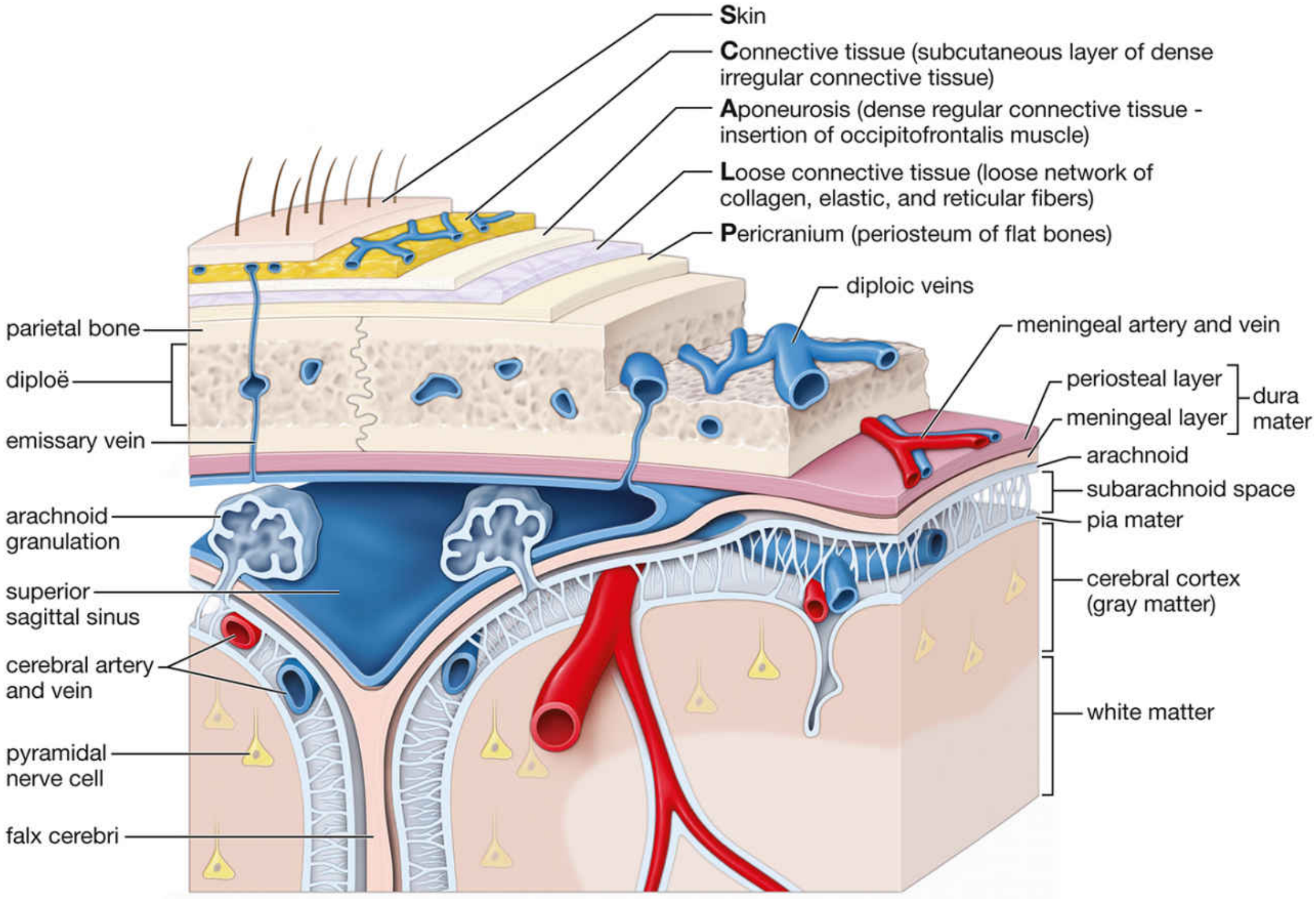
Notice in the diagram how the periosteal layer and meningeal layer are shown as two very thin adjacent lines - tightly fused - and only at the superior sagittal sinus do they open up to form that large blood-filled space.
- Junqueira's Basic Histology, 17th ed., p. 453
- Histology: A Text and Atlas (Wojciech), p. 1003
Blood vessel are present in between intradural space or subdural space
Blood vessels (meningeal arteries and veins) run in the intradural space - between the periosteal and meningeal layers of the dura mater.
Searching sources...
Finding Sources
Finding Sources
Reading File
Here is the precise, textbook-confirmed answer:
Blood Vessels: Intradural Space (Between the Two Dural Layers)
Meningeal blood vessels run in the intradural space - i.e., between the periosteal layer and the meningeal layer of the dura mater.
From Gray's Anatomy for Students:
"The arterial supply to the dura mater travels in the outer periosteal layer of the dura."
And from KJ Lee's Otolaryngology:
"The dura has a periosteal layer and a meningeal layer with an extensive and rich venous plexus running between. Significant bleeding can occur here."
What Runs in Each Space
| Space | Contents |
|---|---|
| Intradural (between periosteal + meningeal layers) | Meningeal arteries (middle, anterior, posterior meningeal) and meningeal veins |
| Subdural (between meningeal dura + arachnoid) | Normally only a thin film of fluid; bridging veins cross this space to reach dural sinuses |
| Subarachnoid (between arachnoid + pia) | CSF, cerebral arteries and veins |
Why This Matters Clinically
Because the middle meningeal artery runs inside the dura (in the intradural/periosteal layer groove), a temporal bone fracture can tear it. The blood then strips the dura off the skull from the outside - producing an epidural hematoma.
The subdural space itself has no arteries - but bridging veins cross it. When these bridging veins tear (usually from deceleration head injury in elderly patients), blood collects in the subdural space - producing a subdural hematoma.
Summary
-
Arteries and veins of the dura = intradural (between the two dural layers, closer to the periosteal/outer side)
-
Bridging veins = cross the subdural space but do not "live" there
-
Subdural space = normally almost no fluid, no resident blood vessels
-
Gray's Anatomy for Students, Cranial dura mater section
-
KJ Lee's Essential Otolaryngology
This is a shared conversation. Sign in to Orris to start your own chat.