
consolidation vs infiltrate va ggo on chest x ray मराठी
consolidation vs ground glass opacity chest x-ray comparison
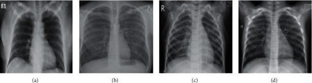
A comparison chart featuring four posterior-anterior (PA) chest X-ray (CXR) samples demonstrating different pulmonary conditions. (a) COVID-19: Shows bilateral, faint ground-glass opacities primarily localized in the lower lung zones. (b) Tuberculosis: Displays significant focal opacity and consolidation in the left upper lung field, suggestive of cavitation or active apical infection. (c) Viral Pneumonia: Exhibits diffuse patchy opacities and areas of consolidation, notably in the right upper and middle lung fields. (d) Normal: Presents as a baseline control with clear lung fields, sharp costophrenic angles, and a normal cardiomediastinal silhouette. The diagnostic images highlight key radiological differences in opacity distribution and morphology between these infectious diseases. This comparison is used to train and evaluate machine learning models for automated respiratory disease classification. The mediastinal structures and heart sizes appear within normal limits across all four examples for comparative purposes.
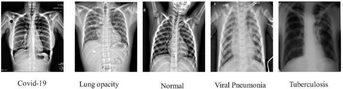
A comparison chart consisting of five posteroanterior (PA) chest X-ray (CXR) images illustrating different pulmonary conditions for educational classification. Each diagnostic image represents a specific clinical entity: (1) COVID-19: Shows subtle, patchy bilateral ground-glass opacities, primarily in the lower lung zones. (2) Lung Opacity: Features a prominent, well-defined consolidation in the right lower lobe, partially obscuring the diaphragmatic contour. (3) Normal: Demonstrates clear lung fields, sharp costophrenic angles, and normal mediastinal and cardiac silhouettes. (4) Viral Pneumonia: Exhibits diffuse, bilateral interstitial infiltrates and peribronchial thickening throughout the lung parenchyma. (5) Tuberculosis: Displays characteristic fibrotic changes and suspected cavitary lesions localized to the upper lobes. The images highlight key radiological differences in pulmonary pathology, including distribution (focal vs. diffuse), density, and anatomical predilection, which are essential for differential diagnosis in respiratory medicine and radiology training.
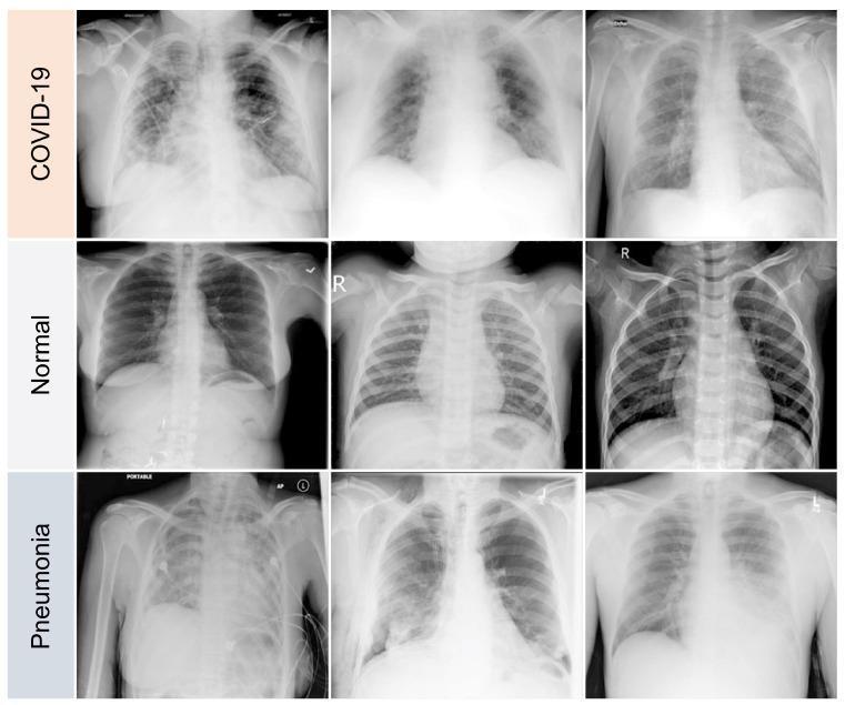
This diagnostic comparison chart presents a grid of nine posterior-anterior (PA) or anterior-posterior (AP) chest X-ray (CXR) images, categorized into three distinct clinical conditions: COVID-19, Normal, and Pneumonia. The top row, labeled 'COVID-19', demonstrates characteristic multifocal and bilateral ground-glass opacities (GGOs) with a predominantly peripheral and basal distribution. These hazy opacities obscure pulmonary vascular markings without completely concealing the underlying lung parenchyma. The middle row, labeled 'Normal', shows clear lung fields with sharp costophrenic angles, well-defined hemidiaphragms, and normal bronchovascular markings, serving as a baseline for anatomical comparison. The bottom row, labeled 'Pneumonia', illustrates varying degrees of pulmonary consolidation and dense opacification. Unlike the diffuse pattern often seen in the COVID-19 samples, these pneumonia images show more localized, dense lobar consolidations and air bronchograms. Some clinical images in this category also show medical interference like ECG leads and portable imaging markers. This comparison highlights key radiological features used in differential diagnosis, focusing on opacity distribution, density, and the presence of interstitial versus alveolar patterns.
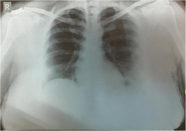
This diagnostic image is a posteroanterior (PA) chest X-ray demonstrating significant pulmonary pathology. The primary findings include extensive, bilateral ground-glass opacities (GGOs) distributed throughout both lung fields. These opacities appear as hazy areas of increased attenuation that do not completely obscure the underlying bronchial or vascular structures, though they exhibit a heterogeneous distribution with a apparent predilection for the middle and lower lung zones. In the left lung, there is a distinct area of consolidation, characterized by a more dense and confluent opacity compared to the surrounding hazy ground-glass areas. This pattern of multifocal ground-glass opacities and consolidation is highly characteristic of viral pneumonia, such as that seen in COVID-19. The mediastinal contour appears within normal limits, though the lower lung details are partially obscured by the increased density of the inflammatory process and potentially overlying soft tissue. The image serves as an educational example of typical radiographic manifestations of acute respiratory infections and the differentiation between hazy GGOs and dense pulmonary consolidation.
ground glass opacity GGO chest CT scan radiology
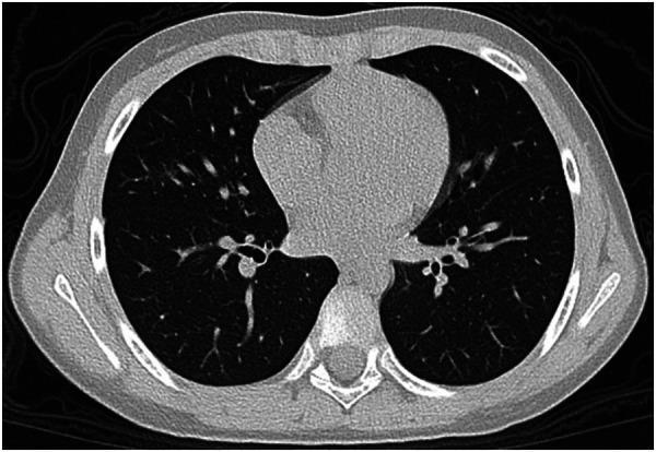
This unenhanced axial chest CT scan demonstrates a focused area of ground-glass opacity (GGO) located within the right middle lobe of the lung in a pediatric patient. The pathology is characterized by a hazy increase in lung density that does not obscure the underlying bronchovascular structures, indicative of partial filling of air spaces or interstitial thickening. The mediastinum, including the heart and great vessels, appears normal in size and position for the patient's age. No evidence of dense consolidation, pleural effusion, or lymphadenopathy is visible at this level. The finding is typical of a mild viral pneumonia, such as that seen in SARS-CoV-2 (Omicron variant) infections, and is consistent with a low CT severity score. This image serves as a clinical example of early or mild pulmonary involvement in viral respiratory diseases, highlighting the importance of identifying subtle GGOs in diagnostic radiology.
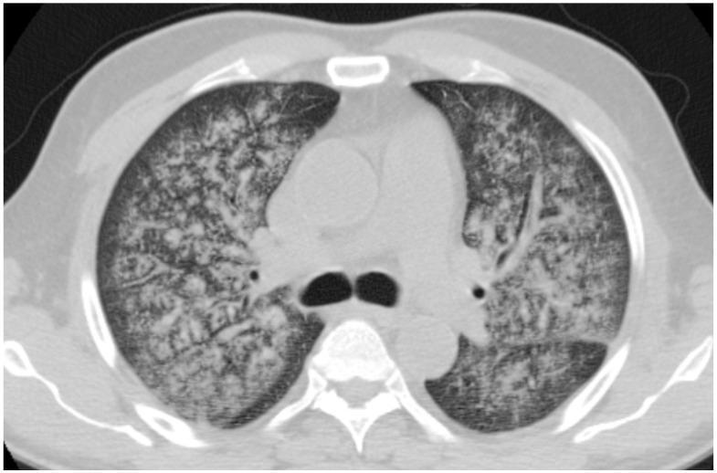
This diagnostic axial CT scan of the chest reveals diffuse bilateral lung parenchymal abnormalities in a patient with Granulomatosis with Polyangiitis (GPA). The primary imaging finding is a widespread ground-glass opacity (GGO) pattern, characterized by a hazy increase in lung attenuation that does not obscure underlying vascular structures. The distribution is predominantly central and perihilar, with notable relative sparing of the subpleural regions. Superimposed on the ground-glass opacities, there is evidence of subtle reticular thickening, suggesting interlobular septal involvement, though distinct consolidations or large pulmonary nodules are absent in this slice. Despite the extensive parenchymal involvement, lung volumes appear preserved. These findings are clinically representative of diffuse alveolar hemorrhage (DAH) secondary to pulmonary vasculitis (hemorrhagic alveolitis). The image serves as a high-yield educational example of the thoracic manifestations of ANCA-associated vasculitides for radiology and rheumatology residents.
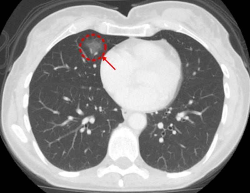
This diagnostic image is an axial slice from a computed tomography (CT) scan of the chest, presented in a lung window setting. The primary finding is a focal area of ground-glass opacity (GGO) in the right lung, specifically located within the right middle lobe near the anterior cardiophrenic angle. The lesion is highlighted by a red dashed circle and indicated by a red arrow. Visually, the GGO presents as a region of hazy, increased attenuation that does not obscure the underlying pulmonary vasculature or bronchial walls, a hallmark of alveolar or interstitial lung processes. The mediastinal structures, including the heart and great vessels, appear within normal limits on this slice. The surrounding lung parenchyma is clear, with no signs of pleural effusion or significant lymphadenopathy visualized at this level. This imaging finding is clinically significant for evaluating pulmonary manifestations of systemic inflammatory diseases, such as Crohn's disease, or assessing early-stage neoplastic processes. The image is appropriate for medical education focusing on thoracic radiology and the interpretation of non-specific alveolar opacities.
Chest X-Ray वर Consolidation vs Infiltrate vs GGO (Ground-Glass Opacity)
🫁 तिन्ही संकल्पना सोप्या भाषेत
1. 📌 Infiltrate (इन्फिल्ट्रेट)
- हा एक umbrella term आहे - म्हणजे "फुफ्फुसात काहीतरी आहे" एवढंच सांगतो
- Radiologist सहसा हा शब्द टाळतात आणि अधिक specific शब्द वापरतात
- थोडक्यात: Infiltrate = एकतर GGO किंवा Consolidation असू शकते
2. 🌫️ Ground-Glass Opacity (GGO) - धुक्यासारखी छाया
फुफ्फुसाच्या भागात हलकी, धुक्यासारखी (hazy) अपारदर्शकता जिथे रक्तवाहिन्या (vessels) आणि ब्रॉन्कस च्या भिंती अजूनही दिसतात.
GGO ची वैशिष्ट्ये:
| वैशिष्ट्य | GGO |
|---|---|
| Density | कमी - हलकी धुक्यासारखी |
| Vessels दिसतात का? | होय ✅ (vessels अजून visible) |
| Air bronchogram | नसतो |
| X-ray वर | अस्पष्ट, पण फार dense नाही |
| CT वर | Hazy attenuation - bronchovascular markings obscure होत नाहीत |
GGO कशामुळे होते?
- Viral pneumonia (COVID-19, Influenza)
- Pulmonary edema (सुरुवातीला)
- Alveolar hemorrhage
- Interstitial lung disease
- Hypersensitivity pneumonitis
3. ⬛ Consolidation (कन्सॉलिडेशन) - घट्ट पांढरी छाया
"Consolidation म्हणजे CXR किंवा CT वर फुफ्फुसाची density वाढणे ज्यात vessels आणि airways च्या कडा अदृश्य होतात. Air spaces मधील हवा replace झालेली असते (inflammatory cells, blood किंवा tumour ने)."
Consolidation ची वैशिष्ट्ये:
| वैशिष्ट्य | Consolidation |
|---|---|
| Density | जास्त - दाट, घट्ट पांढरी |
| Vessels दिसतात का? | नाही ❌ (vessels obscure होतात) |
| Air bronchogram | होय ✅ (महत्त्वाचे sign!) |
| X-ray वर | Dense, homogeneous opacity |
| Volume loss | Acute stage मध्ये नसते |
Consolidation कशामुळे होते?
- Bacterial pneumonia (Lobar pneumonia - S. pneumoniae)
- Pulmonary edema (advanced)
- Pulmonary infarction
- Alveolar hemorrhage
- Bronchoalveolar carcinoma
- ARDS
🔑 Air Bronchogram Sign म्हणजे काय?
हे consolidation चे characteristic sign आहे. याचा अर्थ airway patent आहे पण आजूबाजूचे alveoli भरले आहेत.
📊 तुलनात्मक तक्ता (Summary Table)
| मुद्दा | GGO | Consolidation | Infiltrate |
|---|---|---|---|
| Opacity density | हलकी (हazy) | घट्ट (dense) | दोन्ही असू शकते |
| Vessels दिसतात? | होय | नाही | - |
| Air bronchogram | नाही | होय (बऱ्याचदा) | - |
| Pathology | Partial alveolar filling / interstitial | Complete alveolar filling | Non-specific |
| विशिष्ट कारण | Viral, edema (early) | Bacterial pneumonia, ARDS | - |
| Specificity | Specific sign | Specific sign | Non-specific term |
🖼️ Images
🧠 लक्षात ठेवण्यासाठी Trick
| GGO = "धुकं" - आत दिसते, पण धुक्यातून | Partial filling |
| Consolidation = "पांढरा दगड" - आत काहीच दिसत नाही | Complete filling |
| Infiltrate = "सरकारी भाषा" - काहीतरी आहे एवढंच | Non-specific |