
35 year old female vesicular pimple on buttock ! With pain and fever what to do
herpes zoster shingles vesicular rash buttock
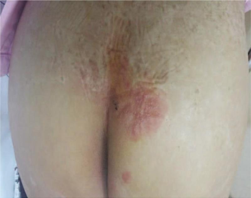
This clinical photograph displays a dermatological presentation of Herpes Zoster (shingles) on the right buttock and sacral region. The image shows a cluster of herpetiform vesicles—small, fluid-filled, and raised lesions—situated on a well-defined erythematous (reddened) base. The distribution of the rash is characteristic of a dermatomal pattern, specifically involving the S2–S4 sacral dermatomes. Some lesions appear to be coalescing, while others show signs of early crusting or excoriation. The surrounding skin exhibits varying degrees of inflammation, with the most intense erythema concentrated beneath the central vesicle cluster. This presentation is a classic visual indicator of acute Varicella-Zoster Virus reactivation in an adult patient, often associated with localized neuropathic pain and, in the case of sacral involvement, potential autonomic dysfunction such as voiding difficulties.
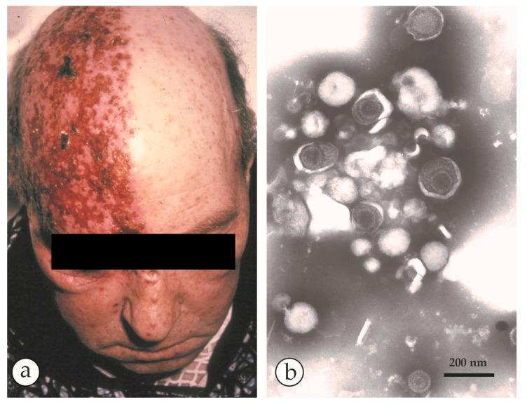
This composite educational image illustrates the clinical and microscopic features of shingles (Herpes Zoster). Panel (a) is a clinical photograph of an elderly patient demonstrating a classic unilateral vesicular rash. The lesions follow a specific dermatomal distribution on the right side of the scalp and forehead, sharply demarcated at the midline. The skin shows confluent erythematous plaques with clustered vesicles and areas of crusting, characteristic of a reactivated Varicella-Zoster Virus (VZV) infection. Panel (b) is a negative-stain electron micrograph (DEM) of fluid collected from a vesicle. It reveals an aggregate of herpesvirus particles. The virions exhibit typical morphology: a 110 nm icosahedral capsid core surrounded by a protein tegument and a loose outer envelope, totaling approximately 150-180 nm in diameter. Some capsids appear dark or 'empty' due to PTA-stain penetration. The presence of these spherical particles alongside cellular detritus confirms the diagnosis. A 200 nm scale bar is provided for size reference. This material is designed for dermatology and infectious disease studies.
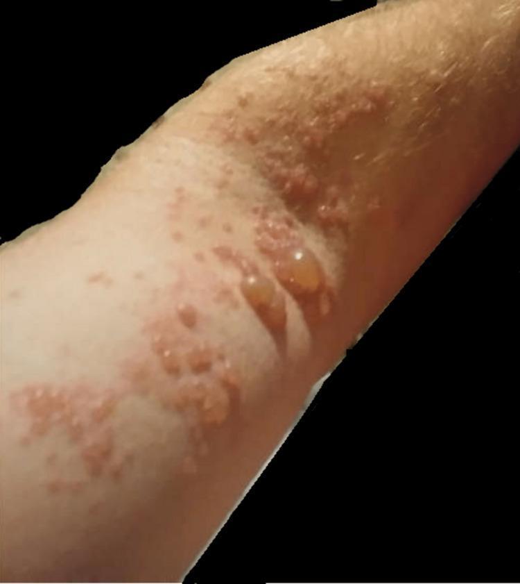
A clinical photograph of the left upper extremity demonstrating a vesicular and bullous eruption characteristic of Herpes Zoster (shingles). The rash exhibits a dermatomal distribution, featuring clusters of lesions on an erythematous base. Morphologically, the lesions range from small, grouped vesicles filled with clear fluid to larger, tense, dome-shaped bullae measuring over one centimeter. Several lesions appear ruptured or umbilicated, resulting in shallow erosions and crusting. The surrounding skin shows signs of inflammation and localized hyperemia. This visual presentation is indicative of Varicella-Zoster Virus (VZV) reactivation, illustrating the classic presentation of painful cutaneous lesions confined to a specific nerve root territory (C5-C6), often associated with neuropathic pain and potential motor weakness in the affected limb.
35-Year-Old Female: Vesicular Lesions on Buttock + Pain + Fever
Most Likely Diagnosis: Herpes Zoster (Shingles)
Why Herpes Zoster?
- Dermatomal pain (often burning/stabbing) precedes or accompanies the rash
- Vesicles are grouped, unilateral, clustered on an erythematous base - not scattered diffusely
- Fever occurs in the prodromal and acute phase from viral replication
- The buttock sits in the S2-S4 sacral dermatomes - a common zoster site
- Age 35 is young but NOT too young - zoster can occur in immunocompetent adults
"The erythematous maculopapular and vesicular lesions of herpes zoster are clustered within a single dermatome, because VZV reaches the skin via the sensory nerve from the single ganglion in which latent VZV reactivates, and not by viremia." - Fitzpatrick's Dermatology
Differential Diagnosis to Consider
| More Likely | Consider |
|---|---|
| Herpes Zoster (VZV reactivation) | Zosteriform Herpes Simplex (HSV-2) |
| Zosteriform HSV-2 (identical appearance, especially on buttock) | Contact dermatitis |
| Insect bites | |
| Impetigo | |
| Burns |
A figure in Fitzpatrick's specifically shows: "This immunocompetent adult with no history of genital herpes presented with clustered vesicular lesions in her left S2/S3 dermatomes. PCR and viral culture both revealed HSV-2." - Fitzpatrick's Dermatology
What To Do: Management
Step 1 - Confirm the Diagnosis
- This is primarily a clinical diagnosis - you do NOT need to wait for lab results to start treatment
- If uncertain: VZV/HSV PCR swab of vesicle fluid is the gold standard (much more sensitive than Tzanck smear)
Step 2 - Start Antiviral Therapy ASAP (within 72 hours of rash onset)
| Drug | Dose | Duration |
|---|---|---|
| Valacyclovir (preferred) | 1 g orally every 8 hours | 7 days |
| Famciclovir (preferred) | 500 mg orally every 8 hours | 7 days |
| Acyclovir (alternative) | 800 mg orally 5 times/day | 7 days |
"Famciclovir or valacyclovir are preferred because of their thrice-daily dosing schedule, their greater oral bioavailability, and the higher and more reliable blood levels of antiviral activity achieved." - Fitzpatrick's Dermatology
Start within 72 hours - the utility of antiviral agents is unproven if treatment is initiated more than 72 hours after rash onset.
Step 3 - Pain Management
- NSAIDs (ibuprofen) or paracetamol for fever and mild-moderate pain
- Gabapentin / Pregabalin if neuropathic pain is prominent
- Opioids for severe acute pain (short course)
- 5% Lidocaine patch for localized pain relief
- Burrow's solution compresses (aluminum acetate) applied locally to hasten drying and soothe lesions
Step 4 - Wound/Lesion Care
- Keep lesions clean and dry
- Avoid scratching or rupturing vesicles
- Cover with loose, non-adhesive dressings
- The patient is contagious to individuals who have never had chickenpox - vesicle fluid contains live VZV
Red Flags - When to Escalate / Admit
- Sacral involvement with urinary retention or bowel dysfunction (S2-S4 zoster can cause neurogenic bladder - check if she can void normally)
- Immunocompromised state (steroids, HIV, malignancy, chemotherapy) - IV acyclovir may be needed
- Disseminated zoster (>20 lesions outside the primary dermatome)
- Ocular involvement (eye pain, redness, visual change)
- Very high fever / signs of encephalitis / severe systemic illness
Important Note on Sacral Zoster Specifically
- Any difficulty urinating (neurogenic bladder is a rare but real complication of sacral zoster)
- Perineal or anal numbness/tingling
Complication to Watch For: Postherpetic Neuralgia (PHN)
- Occurs in ~15% of patients, more common in elderly
- Pain persists weeks to months after rash heals
- Early antiviral treatment is the best way to reduce its risk
- If it develops: gabapentin, pregabalin, topical capsaicin, topical lidocaine, or tricyclic antidepressants
Summary Action Plan
- Swab a vesicle for PCR (VZV/HSV) - confirms diagnosis
- Start Valacyclovir 1g TDS x 7 days immediately (do not wait for results if clinical picture is clear)
- Paracetamol/NSAIDs for fever and pain
- Assess for urinary symptoms (S2-S4 involvement risk)
- Educate patient: contagious to unvaccinated/immunocompromised contacts; avoid close contact until lesions crust over
- Follow up in 5-7 days to assess healing and screen for PHN
Disclaimer: This information is for educational purposes. A qualified healthcare provider should examine the patient and make the final clinical decision. Do not self-treat without medical evaluation.
Not vesicular but a single pustule
furuncle boil skin pustule abscess buttock
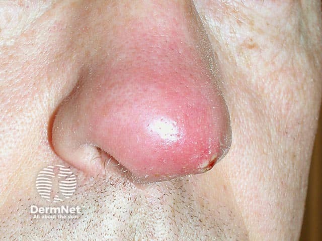
Clinical photography of a single nasal skin lesion showing an acute furuncle (boil) on the nasal vestibule/dorsum. The image depicts a solitary, erythematous, dome-shaped nodule with a shiny surface and surrounding mild edema. A palpable tenderness is suggested by the lesion’s conspicuous inflamed halo. A central pustule or crust may be present at the core, consistent with progression toward abscess formation within a pilosebaceous follicle. The lesion is located on the external nasal skin near the nostril, with frontal/anterior perspective offering clear visualization of the nasal bridge region. The clinical diagnosis is furunculosis of the nose, most often caused by Staphylococcus aureus; in this case PVL (Panton-Valentine leukocidin)–producing strains are implicated, which are associated with more aggressive skin and soft tissue infections and a higher risk of complications. Imaging is not radiologic; this is a dermatologic/clinical photograph used for documentation, teaching, and correlating with microbiology culture results. Management implications include incision and drainage if fluctuant, culture to guide antibiotics, and consideration of toxin-producing strains. The image serves educational purposes for dermatology, infectious disease, microbiology, and medical training in recognizing PVL-associated nasal furunculosis and differentiating it from impetigo, cellulitis, and Sty.
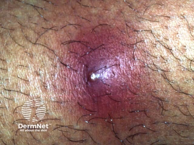
Clinical photograph of a single inflamed dermal lesion consistent with a boil (furuncle) on hairy skin. Modality: clinical photography with macro close-up; high magnification reveals a dome-shaped, erythematous nodule centered on a pus-filled core. The lesion demonstrates a shiny surface, surrounding perilesional edema, and visible follicular structures nearby. The central pale-yellow-to-white pustule indicates active purulent discharge; the erythematous halo suggests acute bacterial infection commonly due to Staphylococcus aureus. There may be crusting at the lesion rim if drainage has begun. The image captures surface features without depth assessment; no clearly delineated surrounding abscess cavity is evident, though edema indicates inflamed dermis and perifollicular inflammation. Clinically this lesion is typically painful, tender, and warm to touch, with potential progression to cellulitis if untreated. Management includes warm compresses, analgesia, and consideration of antibiotic therapy or incision and drainage for larger or fluctuant boils; culture may be performed in recurrent or non-resolving cases. This image is educational for dermatology training, infectious disease differential, and clinical photography repositories, aiding recognition of the pustule-with-edema configuration and localized follicular infection.
35-Year-Old Female: Single Pustule on Buttock + Pain + Fever
Most Likely Diagnosis: Furuncle (Boil)
"A furuncle or boil is a deep-seated inflammatory nodule that develops around a hair follicle, usually from a preceding, more superficial folliculitis and often evolves into an abscess... Furuncles typically arise in hair-bearing sites, particularly regions subject to friction, occlusion, and perspiration, such as the neck, face, axillae, and buttocks." - Fitzpatrick's Dermatology
What is Happening Clinically?
- Folliculitis (superficial infection of a hair follicle) → progresses to
- Furuncle (deep abscess around a follicle with a central pus core) → if untreated, can progress to
- Carbuncle (multiple confluent furuncles, deeper, more dangerous)
Differential Diagnosis
| Condition | Key Distinguishing Feature |
|---|---|
| Furuncle (boil) - Most likely | Single, follicle-centered, tender pustule/nodule |
| Infected epidermoid (sebaceous) cyst | Nodule pre-existing for months/years before infection |
| Hidradenitis suppurativa | Recurrent; affects axilla/groin/buttock folds; multiple lesions |
| Pilonidal abscess | Located at natal cleft/sacrococcygeal area specifically |
| Perianal abscess | Located very close to anus, with perianal pain |
| Infected insect bite | History of bite; no follicular center |
What To Do: Step-by-Step Management
Step 1 - Assess the Lesion
- Is it fluctuant (soft, fluid-filled, "pointing")? → Ready for incision and drainage
- Is it still firm/indurated (early stage)? → Warm compresses first
- Is there surrounding cellulitis (spreading redness, warmth, swelling beyond the lesion)?
- Check temperature - fever >38°C is an indication for antibiotics
Step 2 - Local Treatment (First Line for Small, Uncomplicated Furuncle)
- Promotes spontaneous drainage and maturation of the lesion
- Very effective for early, non-fluctuant furuncles
- Can be enough for small lesions without fever or cellulitis
Step 3 - Incision and Drainage (I&D) if Fluctuant
- Small incision over the center of the lesion under local anesthesia
- Express and irrigate the pus cavity
- Wick placement for continued drainage (if cavity is large)
- Send pus for culture and sensitivity - important to detect MRSA
"Abscesses should be treated with incision and drainage... Most patients (~90%) with fluctuant skin abscesses respond to drainage, and antibiotic therapy does not significantly improve the outcome in uncomplicated cases." - Textbook of Family Medicine
Step 4 - Antibiotics (When Are They Needed?)
- Fever >38°C ✓ (present here)
- Rapid progression with surrounding cellulitis
- Difficult-to-drain location
- Diabetes, immunosuppression, HIV
- Multiple lesions
- Age extremes
| Drug | Dose | Notes |
|---|---|---|
| TMP-SMX (Cotrimoxazole) - First choice | DS tablet (160/800mg) twice daily x 5-7 days | Best for CA-MRSA |
| Doxycycline | 100 mg twice daily x 5-7 days | Good MRSA coverage |
| Clindamycin | 300-450 mg three times daily x 5-7 days | Check local resistance (up to 50% CA-MRSA resistant) |
| Flucloxacillin / Dicloxacillin | 500 mg four times daily x 5-7 days | For MSSA only; if MRSA suspected, avoid |
"TMP-SMX and clindamycin have demonstrated efficacy in cure rates following incision and drainage." - Rosen's Emergency Medicine
Step 5 - Pain and Fever Management
- Paracetamol (1g every 6-8 hours) for fever and pain
- Ibuprofen (400mg every 8 hours with food) as an anti-inflammatory
- Avoid tight clothing over the area
Red Flags - When to Refer/Admit Urgently
- High fever / rigors / feeling very unwell - may indicate bacteraemia/sepsis
- Rapidly spreading redness (cellulitis progressing despite treatment)
- No improvement after 48 hours of antibiotics + warm compresses
- Lesion in a high-risk area (near perineum, close to spine, near genitalia)
- Suspected necrotizing fasciitis - severe disproportionate pain, skin changes, systemic toxicity (this is a surgical emergency)
- Patient is immunocompromised (diabetes, steroids, HIV, chemotherapy)
Also Consider: Hidradenitis Suppurativa
Summary Action Plan
| Priority | Action |
|---|---|
| 1 | Examine - is it fluctuant or firm? |
| 2 | Warm compresses 3-4x/day immediately |
| 3 | If fluctuant: Incision & Drainage + send pus for culture |
| 4 | Start TMP-SMX (Cotrimoxazole) given the fever |
| 5 | Paracetamol/Ibuprofen for fever & pain |
| 6 | Review in 48 hours - if worsening, escalate |
| 7 | Rule out MRSA on culture; adjust antibiotics accordingly |
Disclaimer: This is educational information only. A healthcare provider should examine the patient in person to confirm the diagnosis and manage appropriately. Do not delay seeking care if the patient develops high fever, rapidly spreading redness, or worsening pain.
Dose of acetazolamide and dexamethasone for hight altitude mountains
Acetazolamide & Dexamethasone for High-Altitude Illness
Understanding the Three Syndromes
| Syndrome | Altitude | Key Features |
|---|---|---|
| AMS - Acute Mountain Sickness | >2500 m | Headache, nausea, fatigue, insomnia |
| HACE - High-Altitude Cerebral Edema | >3500 m | Ataxia, confusion, altered consciousness → coma |
| HAPE - High-Altitude Pulmonary Edema | >3500 m | Dyspnea, cough, tachycardia (treated differently - nifedipine) |
Golden rule: Descent is always the definitive treatment. Drugs are adjuncts, not substitutes for descent.
ACETAZOLAMIDE
Doses (Adults)
| Indication | Dose | Route | Duration |
|---|---|---|---|
| Prevention of AMS | 125 mg twice daily (some use 62.5 mg BD) | Oral | Start 1-2 days before ascent; continue 2 days after reaching max altitude |
| Treatment of mild AMS | 125-250 mg twice daily | Oral | Until symptoms resolve |
| Treatment of moderate/severe AMS | 250 mg twice daily | Oral | Until symptoms resolve |
| Adjunct in HACE (alongside dexamethasone) | 250 mg twice daily | Oral | - |
| Night-only (for sleep disturbance at altitude) | 125 mg at bedtime | Oral | As needed |
Doses (Pediatric)
- 2.5 mg/kg every 12 hours (for both prevention and treatment)
Key Points
- Start before ascent for prophylaxis (ideally 1-2 days prior)
- Side effects: tingling in fingers/toes (paresthesias), altered taste of carbonated drinks, increased urination
- Avoid if: sulfa antibiotic anaphylaxis history (contains sulfhydryl moiety - rare cross-reactivity)
- Carbonated drinks (beer, soda) will taste "flat" - this is harmless
"Famciclovir or valacyclovir are preferred... A dose of 125 mg PO twice daily is effective for most individuals, whereas others may require 250 mg PO twice daily." - Tintinalli's Emergency Medicine
DEXAMETHASONE
Doses (Adults)
| Indication | Dose | Route | Notes |
|---|---|---|---|
| Prevention of AMS/HACE | 2 mg every 6 hours OR 4 mg every 12 hours | Oral | Use only when rapid unavoidable ascent; not first-line for prophylaxis |
| Treatment of moderate/severe AMS | 4 mg every 6 hours | PO / IM / IV | Best reserved for moderate-severe cases |
| Treatment of HACE | 8 mg ONCE (loading dose), then 4 mg every 6 hours | PO / IM / IV | Immediate use; combine with descent + O2 |
| Prevention of HAPE (in susceptible individuals) | 8 mg twice daily | Oral | Special high-risk situations only |
Doses (Pediatric)
- 0.15 mg/kg/dose every 6 hours
- Not recommended for prophylaxis in children
Key Points
- Does NOT speed acclimatization - it only masks/treats symptoms
- Stopping abruptly can cause rebound symptoms - taper if used >24-48 hours
- Often combined with acetazolamide: dexamethasone treats the acute symptoms rapidly, acetazolamide drives acclimatization
- For HACE: give dexamethasone immediately while arranging descent - do not wait
"It is often clinically useful to provide simultaneous therapy with acetazolamide (to speed acclimatization) and a brief course of dexamethasone (to treat symptoms)." - Tintinalli's Emergency Medicine
Complete Reference Table (Goldman-Cecil Medicine)
| Drug | Indication | Route | Dose |
|---|---|---|---|
| Acetazolamide | Prevention AMS/HACE | Oral | 62.5-125 mg twice/day (adult); 2.5 mg/kg q12h (child) |
| Acetazolamide | Treatment AMS | Oral | 250 mg twice/day (adult); 2.5 mg/kg q12h (child) |
| Dexamethasone | Prevention AMS/HACE | Oral | 2 mg q6h OR 4 mg q12h (adults only) |
| Dexamethasone | Treatment AMS | PO/IV/IM | 4 mg every 6 hours |
| Dexamethasone | Treatment HACE | PO/IV/IM | 8 mg loading, then 4 mg q6h |
Quick Decision Guide
Ascending to >2500m?
  → PREVENT: Acetazolamide 125mg BD starting 1-2 days before

Headache + nausea at altitude (AMS)?
  → MILD: Acetazolamide 125-250mg BD + halt ascent
  → MODERATE/SEVERE: Acetazolamide 250mg BD + Dexamethasone 4mg q6h

Ataxia or confusion (HACE)?
  → IMMEDIATE DESCENT + O2
  → Dexamethasone 8mg STAT then 4mg q6h IV/IM/PO
  → Acetazolamide 250mg BD as adjunct
  → Hyperbaric bag if descent not immediately possible
Other Drugs Worth Knowing
| Drug | Use | Dose |
|---|---|---|
| Nifedipine SR | HAPE prevention & treatment | 30 mg SR q12h or 20 mg SR q8h |
| Tadalafil | HAPE prevention | 10 mg twice daily |
| Ibuprofen | AMS headache | 600-800 mg q8h |
| Ondansetron | AMS nausea/vomiting | 4-8 mg ODT q4-6h |
| Salmeterol | HAPE prevention | 125 mcg inhaled twice daily |
Disclaimer: These are reference doses for educational purposes. Drug choice and dosing in an actual patient should be guided by a qualified physician, taking into account individual factors, contraindications, and current guidelines.