
It's a pathology of benign prostatic hyperplasia and fibroadenoma and adenocarcinoma, seminoma
Reading File
Now I'll search the medical library for all four conditions in parallel.
Finding Sources
Finding Sources
Finding Sources
Finding Sources
Good. Now I'll read the Robbins Pathology sections for BPH and Fibroadenoma, plus the Campbell-Walsh section for Seminoma and the Robbins/Cotran Pathologic Basis section for Fibroadenoma - all in parallel.
Reading File
Reading File
Reading File
Reading File
Now let me get the prostate adenocarcinoma pathology and check for images from the textbooks.
Finding Sources
Finding Sources
Finding Sources
Loading Image
Now let me read the Bailey & Love section on prostate carcinoma histology, and also get the Robbins BPH image and the seminoma image.
Reading File
Finding Sources
Now let me get the Robbins prostate carcinoma section and BPH images, and also the seminoma image from Campbell-Walsh.
Reading File
I now have comprehensive content from the textbooks. Let me fetch the key histology images to include in the answer.
Loading Image
Loading Image
I now have all the key information and images needed. Let me also read the prostate adenocarcinoma morphology section from Robbins to complete the picture.
Reading File
Loading Image
I now have all the information needed for a comprehensive answer. Let me compile the complete pathology notes for all four conditions.
Pathology of Four Key Conditions
1. Benign Prostatic Hyperplasia (BPH)
Epidemiology
The most common benign prostatic disease in men over 50. Frequency rises progressively with age, reaching 90% by the eighth decade.
Pathogenesis
- Driven by dihydrotestosterone (DHT) - synthesized from testosterone in the prostate by 5α-reductase type 2
- DHT binds nuclear androgen receptors and promotes growth/survival of epithelial and stromal cells
- DHT-induced growth factors increase stromal cell proliferation and decrease epithelial cell apoptosis
- Does not occur in castrated males or those with genetic androgen insensitivity
- With aging, declining testosterone + unchanged/increased estrogens may synergize with DHT to drive growth
Morphology (Gross)
- Prostate weight increases 3- to 5-fold (60-100 g or more)
- Affects the periurethral transition zone - compresses the urethra to a slit-like orifice
- Hyperplastic nodules vary in color/consistency; may be solid or cystic (dilated glands)
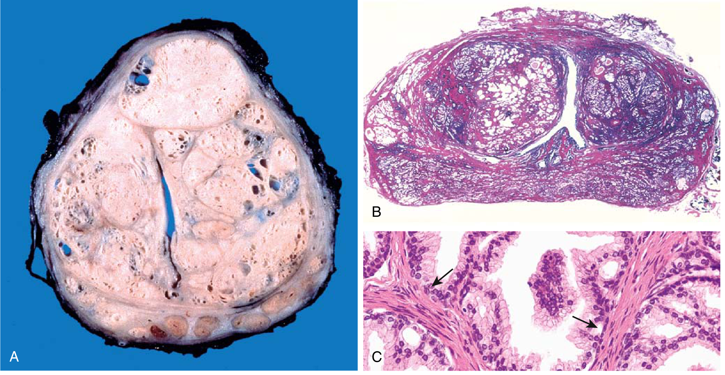
Morphology (Microscopic)
- Nodules composed of variable proportions of glandular + fibromuscular stroma
- Hyperplastic glands lined by two layers:
- Inner: tall columnar epithelial cells
- Outer: flattened basal cells (KEY distinction from malignant glands)
- Glandular lumina often contain corpora amylacea (laminated proteinaceous concretions)
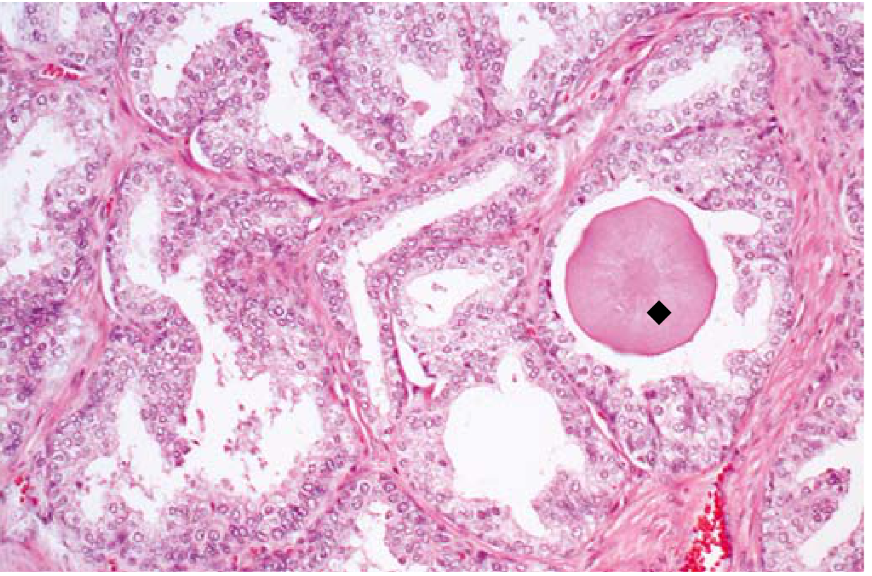
Clinical Features
- Increased urinary frequency, nocturia, hesitancy, poor stream, overflow dribbling, dysuria
- Residual urine predisposes to UTIs
- Complete obstruction can cause hydronephrosis
- Treatment: α1-adrenergic blockers (relax smooth muscle) + 5α-reductase inhibitors (↓ DHT); surgical options (TURP, laser, HIFU) for refractory cases
2. Fibroadenoma
Definition & Epidemiology
The most common benign stromal tumor of the female breast. Most cases occur in women in their 20s and 30s. Often multiple and bilateral.
Pathogenesis (Molecular)
- Two-thirds harbor driver mutations in MED12 (encodes a subunit of the mediator complex, regulating RNA Pol II-dependent gene transcription)
- ~1/3 also have mutations in RARA (retinoic acid receptor alpha - an estrogen target gene)
- MED12 mutations alter expression of sex hormone-regulated genes controlling stromal cell proliferation/survival
- Hormonally responsive: grow during pregnancy, regress after menopause
Morphology (Gross)
- Well-circumscribed, rubbery, gray-white nodules that bulge above surrounding tissue
- Contain slit-like spaces lined by epithelium
- Sizes range from < 1 cm to large masses replacing most of the breast
- In older women: stroma is densely hyalinized, epithelium often atrophic
Morphology (Microscopic)
Two classic patterns:
- Pericanalicular pattern: delicate myxoid stroma surrounds patent ducts
- Intracanalicular pattern: stroma compresses and distorts ductal elements into cleft-like spaces
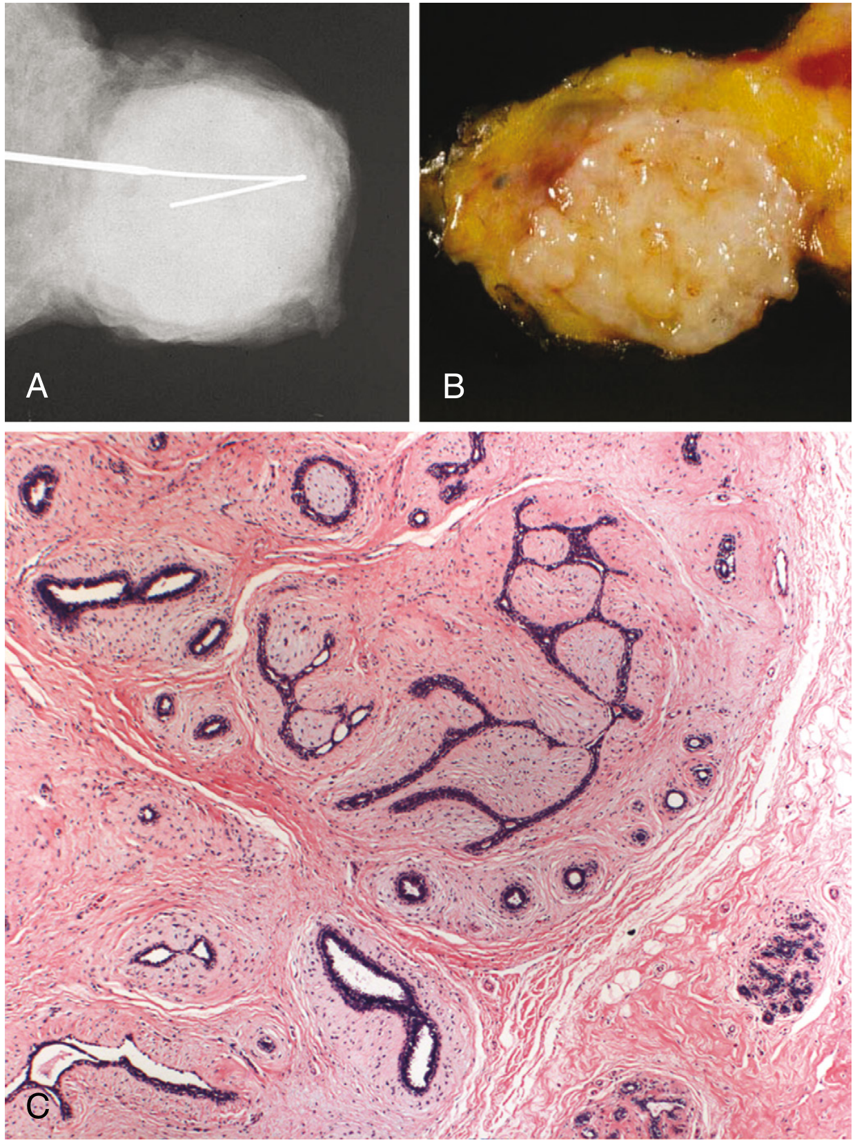
Clinical Notes
- Palpable rubbery, mobile mass ("breast mouse") in young women
- Rapid growth in pregnancy may falsely suggest carcinoma (can cause infarction)
- Cyclosporin A use (post-renal transplant) associated with multiple bilateral fibroadenomas
- Special subtypes: complex fibroadenoma (contains cysts, sclerosing adenosis, papillary apocrine change) - mildly increased cancer risk
3. Adenocarcinoma of the Prostate
Epidemiology
The most common cancer in men (~21% of male cancers in the US). Second leading cause of cancer death in men. Largely a disease of aging (incidence ~20% in 50s, ~70% in 70s-80s).
Pathogenesis
Key molecular events:
- TMPRSS2-ETS fusion: most common alteration; chromosomal rearrangement places an ETS oncogene under control of the androgen-regulated TMPRSS2 promoter → overexpression in androgen-dependent fashion
- PTEN deletion and MYC amplification: accelerate growth, contribute to antiandrogen resistance
- Late-stage: loss of TP53, RB deletions, androgen receptor gene amplifications
- Epigenetic silencing of GSTP1 (glutathione-S-transferase Pi1) - early event, reduces xenobiotic detoxification
- Androgen dependence: cancers often regress with surgical/chemical castration initially, then develop resistance
Morphology (Gross)
- ~70% arise in the peripheral zone (posterior location - palpable on DRE)
- Gritty, firm, gray-white lesion with ill-defined margins infiltrating adjacent gland
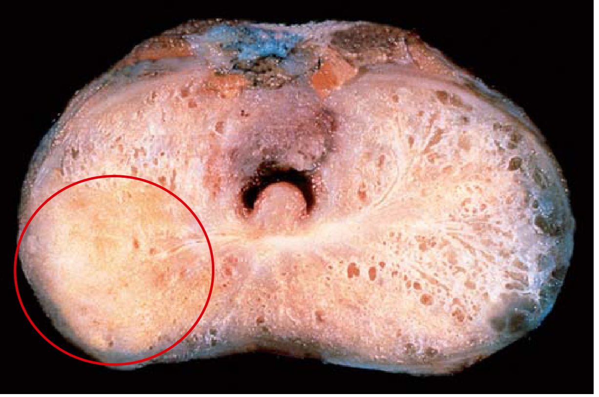
Morphology (Microscopic)
- Single uniform layer of cuboidal/low columnar epithelium - NO basal cell layer (critical distinction from BPH)
- Glands smaller than benign, crowded, lack branching and papillary infoldings
- Nuclei enlarged, prominent nucleoli
- Malignant glands appear confluent; loss of myoepithelial/basement membrane layer
- More poorly differentiated tumors show solid sheets of carcinoma cells
Gleason Grading / ISUP Grade Groups
The histological pattern is graded based on degree of glandular dedifferentiation:
| ISUP Grade Group | Gleason Score | Significance |
|---|---|---|
| 1 | 3+3 = 6 | Low risk |
| 2 | 3+4 = 7 | Intermediate-favorable |
| 3 | 4+3 = 7 | Intermediate-unfavorable |
| 4 | 4+4=8, 3+5=8, 5+3=8 | High risk |
| 5 | ≥9 (4+5, 5+4, 5+5=10) | Very high risk |
Grades 1 and 2 are no longer reported as their outcome is similar to grade 3. Grade 3 cancers almost never metastasize. Higher grade group = poorer prognosis.
Tumor Markers & Treatment
- PSA (prostate-specific antigen): screening (controversial) and monitoring
- Treatment: Androgen deprivation (surgical or chemical castration) ± radiation; resistance develops via AR amplification or AR mutations allowing ligand-independent activation
4. Seminoma
Definition & Epidemiology
The most common germ cell tumor (GCT) of the testis. Most cases diagnosed in the fourth to fifth decade (older average age than non-seminomatous GCTs). Arises from germ cell neoplasia in situ (GCNIS) and is considered the common precursor for other GCT subtypes.
Morphology (Gross)
- Soft, tan to white, diffuse or multinodular mass
- Necrosis may be present but is typically focal (less prominent than other GCTs)
Morphology (Microscopic)
- Sheet-like arrangement of cells with polygonal nuclei and clear cytoplasm
- Cells divided into nests by fibrovascular septae containing lymphocytes (characteristic!)
- Lymphocytic infiltrates and granulomatous reactions are common
- Syncytiotrophoblasts (stain hCG+) present in ~15% of cases but no clear prognostic significance
Immunohistochemistry
| Marker | Seminoma |
|---|---|
| CD30 | Negative |
| CD117 (c-Kit) | Positive |
| PLAP (placental alkaline phosphatase) | Strongly positive |
| OCT3/4 | Positive |
| PAS (glycogen) | Positive (unlike spermatocytic tumor) |
Key Differentials
- Solid-pattern embryonal carcinoma (CD30+, CD117-)
- Yolk sac tumor
- Sertoli cell tumor
Clinical Notes
- Arises from GCNIS (unlike spermatocytic tumor, which does NOT arise from GCNIS)
- Associated with history of cryptorchidism and increased incidence of sarcoidosis
- Ability to transform into NSGCT elements has important therapeutic implications
- Highly radiosensitive; excellent prognosis
Sources:
- Robbins & Kumar Basic Pathology (BPH, Prostate Adenocarcinoma sections)
- Robbins, Cotran & Kumar Pathologic Basis of Disease (Fibroadenoma section)
- Campbell-Walsh-Wein Urology (Seminoma section)
- Bailey and Love's Short Practice of Surgery, 28th ed. (Prostate carcinoma grading)
This is a shared conversation. Sign in to Orris to start your own chat.