
Limbic system
Reading File
The limbic system is a collection of brain structures involved in emotion, memory, motivation, and autonomic regulation. Key components include the hippocampus (memory), amygdala (fear/emotion), hypothalamus (homeostasis), cingulate gyrus, and olfactory areas.
Searching sources...
Finding Sources
Searching the Web
limbic system anatomy components functions
Excellent sources found. Let me read from the most comprehensive textbooks in parallel.
Reading File
Reading File
Reading File
Good content and images found. Let me read more from Kaplan & Sadock and also fetch the key diagrams.
Reading File
Loading Image
Loading Image
Excellent content and images. Let me read the remaining sections from Kaplan & Sadock on the amygdala, hippocampus functions, and Papez circuit, plus fetch the anatomy figure.
Reading File
Loading Image
Excellent material gathered. Now let me read the Papez circuit and functional sections.
Reading File
Reading File
I now have rich content from multiple authoritative sources. Let me compile the comprehensive answer.
The Limbic System
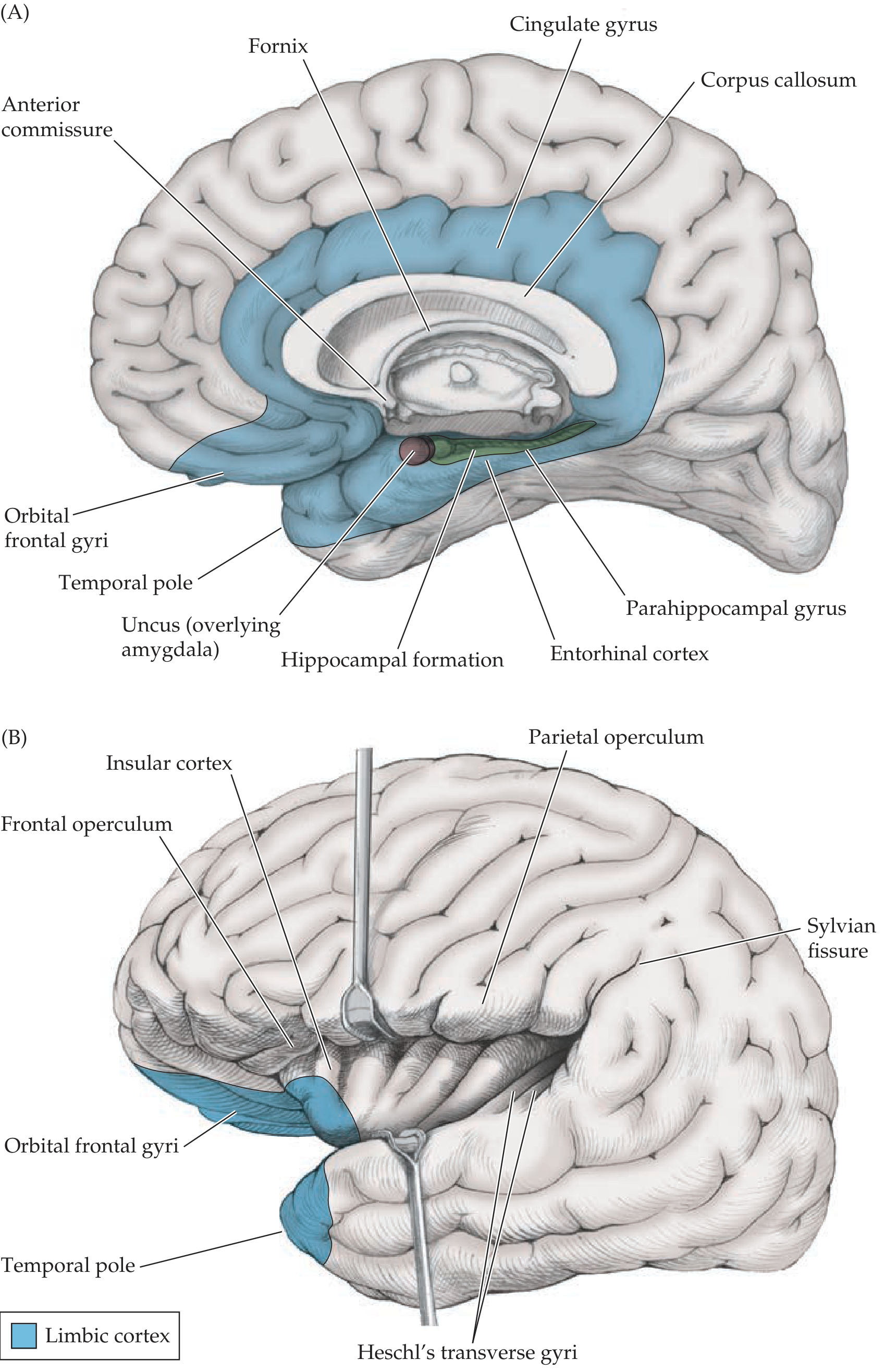
Limbic cortex (blue) shown in medial view (A) and lateral view with Sylvian fissure opened (B). Key structures include the cingulate gyrus, fornix, hippocampal formation, entorhinal cortex, parahippocampal gyrus, and uncus (overlying the amygdala). - Neuroanatomy through Clinical Cases, 3rd Ed.
Etymology & History
The term "limbic" comes from the Latin limbus, meaning border - it describes the curved rim of cortex at the junction of the diencephalon and cerebral hemispheres. Key milestones:
- Paul Broca (late 19th century) - coined "limbic lobe" for the cingulate and parahippocampal gyri
- James Papez (1937) - proposed a circuit linking these cortical areas with the hippocampus, mammillary body, and anterior thalamus as the neural substrate for emotional behavior
- Klüver & Bucy - showed temporal lobe lesions (disrupting the circuit) alter affective responses in primates
- Paul MacLean (1952) - coined "limbic system" to describe Broca's limbic lobe + related subcortical nuclei as the substrate for emotion
(Kaplan & Sadock's Comprehensive Textbook of Psychiatry)
Components
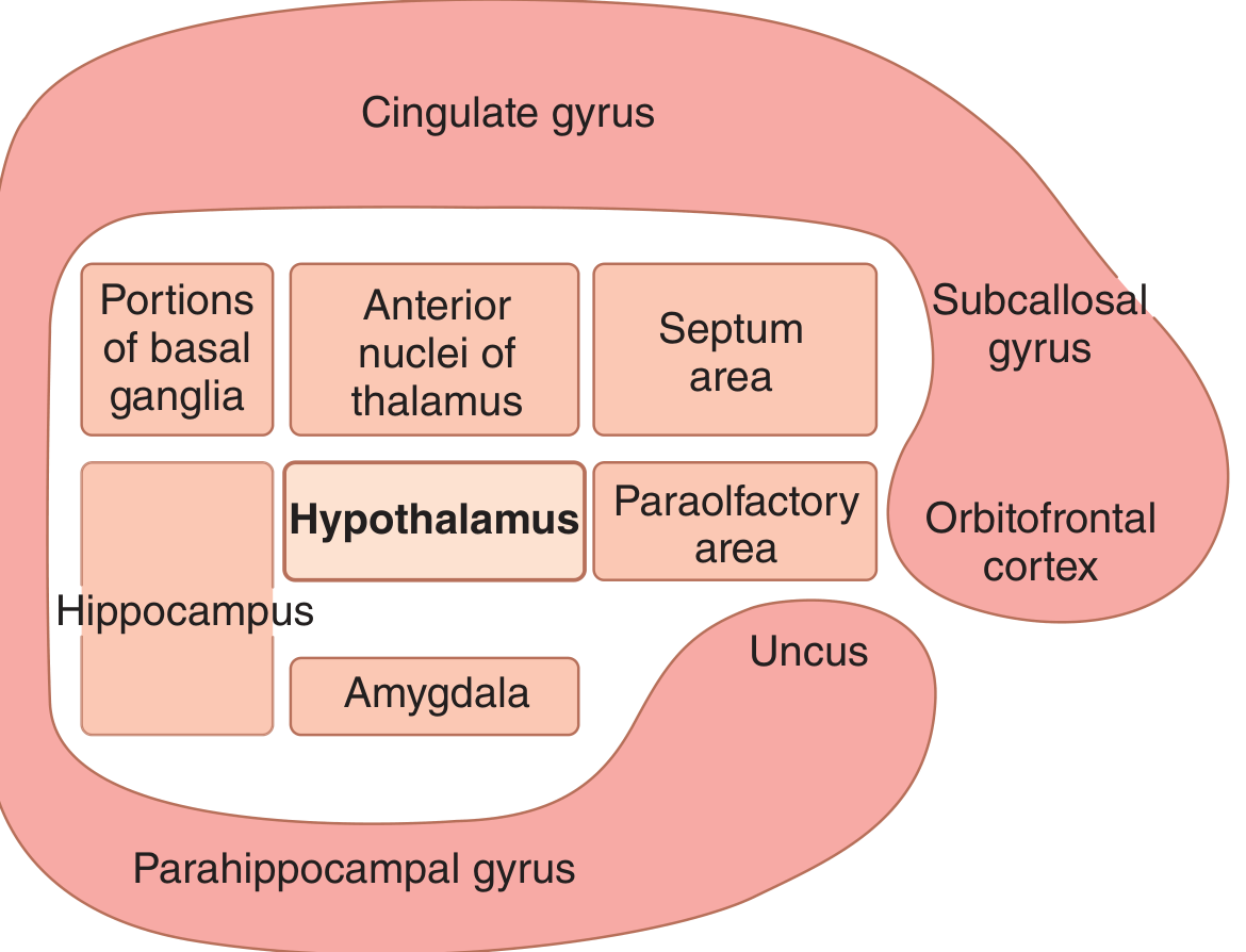
Schematic showing the key position of the hypothalamus at the center of the limbic system, surrounded by the hippocampus, amygdala, septal area, anterior thalamic nuclei, basal ganglia portions, and limbic cortex (cingulate gyrus, parahippocampal gyrus, subcallosal gyrus, orbitofrontal cortex). - Guyton & Hall Medical Physiology
No single universally agreed-upon list exists, but the most commonly cited structures are:
Limbic Cortex
A ring of predominantly paleocortex on the medial and ventral surface of each hemisphere:
- Orbitofrontal cortex - ventral surface of the frontal lobe
- Subcallosal gyrus - extends upward from orbitofrontal area
- Cingulate gyrus - C-shaped cortex dorsal to the corpus callosum; divided into subgenual anterior (BA 25), pregenual anterior, midcingulate, posterior, and retrosplenial zones. The subgenual anterior cingulate (BA 25) is overactive in depression and is a target for deep brain stimulation (DBS).
- Parahippocampal gyrus - in the medial temporal lobe; contains the entorhinal cortex, which funnels processed cortical information into and out of the hippocampal formation
- Uncus - the hook-shaped anterior end of the parahippocampal gyrus (overlies the amygdala)
(Kaplan & Sadock; Guyton & Hall)
Deep Limbic Structures
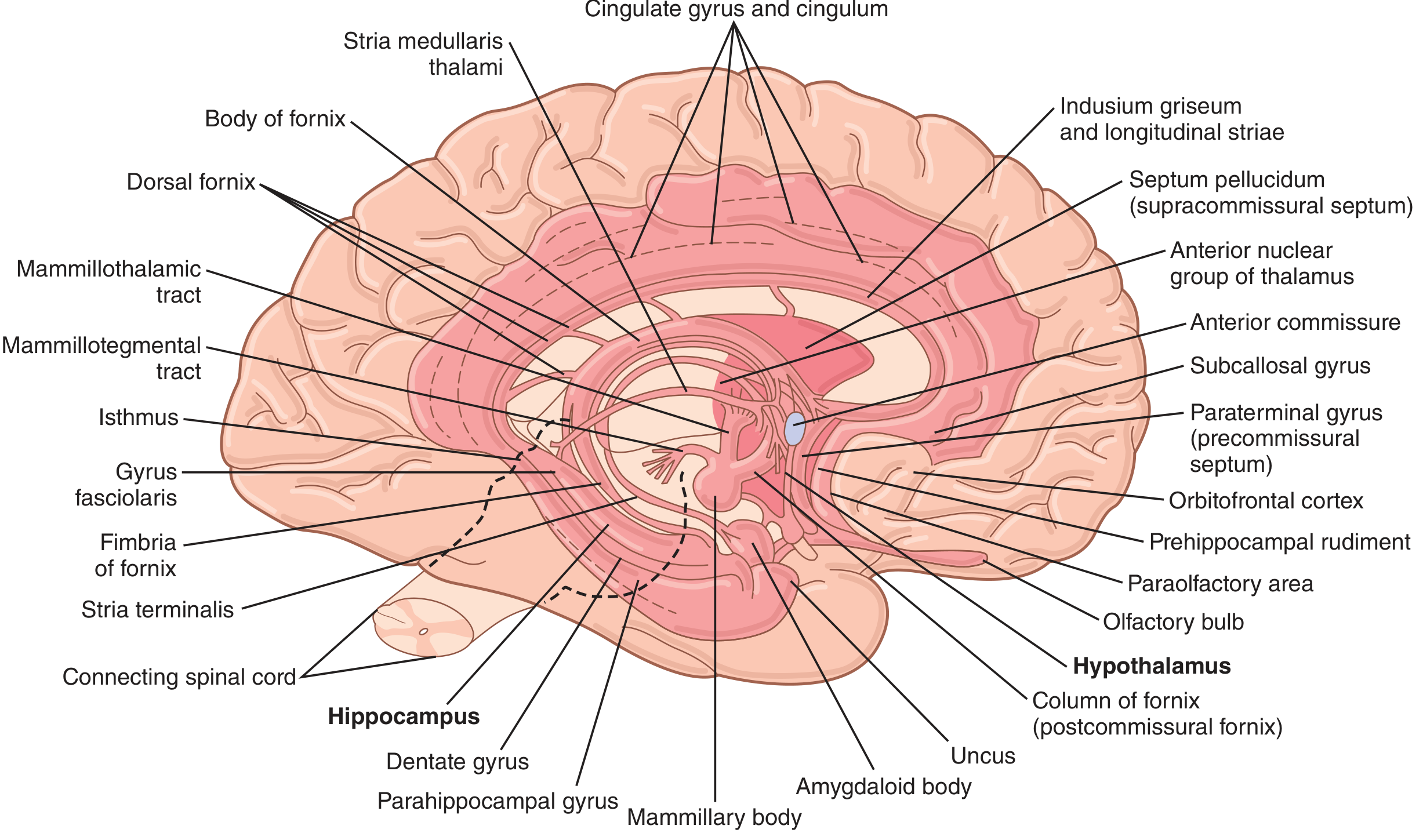
Comprehensive labeled anatomy of the limbic system showing hippocampus, dentate gyrus, parahippocampal gyrus, mammillary body, amygdaloid body, fornix, anterior commissure, cingulate gyrus, hypothalamus, and connecting tracts. - Guyton & Hall Medical Physiology
| Structure | Location | Key Role |
|---|---|---|
| Hippocampal formation | Floor of temporal horn, lateral ventricle | Memory consolidation |
| Amygdala | Medial temporal lobe, anterior to hippocampus | Emotion, fear, threat appraisal |
| Hypothalamus | Diencephalon | Autonomic, endocrine, and vegetative control |
| Septal area | Above anterior commissure | Anxiety regulation (septohippocampal pathway) |
| Anterior thalamic nuclei | Thalamus | Relay in Papez circuit |
| Mammillary bodies | Posterior hypothalamus | Memory; receive hippocampal input via fornix |
| Habenula | Epithalamus | Mood regulation via raphe (serotonin) and VTA (dopamine) |
| Basal ganglia (portions) | - | Behavioral drives |
The Hippocampal Formation
Composed of three adjacent, folded cortical strips arranged in a C-shape:
- Dentate gyrus - three layers (molecular, granule cell, polymorphic); granule cell axons form the mossy fiber projection
- Hippocampus proper - divided into fields CA1, CA2, CA3 (cornu ammonis) and the hilar region (sometimes called CA4, but more properly part of the dentate gyrus)
- Subicular complex - major output zone
The primary information flow is: Entorhinal cortex → Dentate gyrus → CA3 → CA1 → Subiculum → Entorhinal cortex (and back to hypothalamus/thalamus via the fornix)
Lesions in the hippocampal formation cause deficits in consolidating immediate recall into long-term memories - patients can recall remote events but cannot form new memories (anterograde amnesia). (Neuroanatomy through Clinical Cases, 3rd Ed.)
The Amygdala
Located in the medial temporal lobe, anterior to the hippocampus, the amygdala is divided into:
- Basolateral complex - largest group; reciprocally connected with temporal, insular, and prefrontal cortices; functions more like a multimodal cortical region; rich in CB1 cannabinoid receptors
- Centromedial group - connected to brainstem viscerosensory/visceromotor regions and lateral hypothalamus; part of the larger extended amygdala (continuous through the sublenticular substantia innominata with the bed nucleus of the stria terminalis)
- Olfactory/cortical group
The amygdala is central to fear conditioning, threat detection, and emotional memory. (Kaplan & Sadock)
The Papez Circuit
The classic circuit for emotion and memory:
Hippocampus → (via fornix) → Mammillary bodies → (via mammillothalamic tract) → Anterior thalamic nucleus → Cingulate gyrus → Parahippocampal gyrus → Entorhinal cortex → back to Hippocampus
Key White Matter Pathways
| Tract | Connects |
|---|---|
| Fornix | Hippocampal formation ↔ Mammillary bodies, septal nuclei, hypothalamus |
| Stria terminalis | Amygdala ↔ Hypothalamus and septal area |
| Medial forebrain bundle | Septal/orbitofrontal cortex ↔ Hypothalamus ↔ Brainstem reticular formation |
| Cingulum bundle | Cingulate gyrus ↔ Parahippocampal gyrus (continuous white matter) |
| Mammillothalamic tract | Mammillary bodies → Anterior thalamus |
| Uncinate fasciculus | Frontal lobe ↔ Temporal pole (includes amygdala connections) |
The Hypothalamus - Central Hub
The hypothalamus weighs only ~4 grams but represents a critical control center. It sends output in three directions:
- Downward to the brainstem (reticular areas of mesencephalon, pons, medulla → autonomic nervous system)
- Upward to the anterior thalamus and limbic cortex
- Into the infundibulum to control pituitary secretion (both anterior and posterior)
Key hypothalamic nuclei and their functions:
| Nucleus | Function |
|---|---|
| Suprachiasmatic | Circadian rhythm regulation |
| Supraoptic & Paraventricular (magnocellular) | Oxytocin and vasopressin release (→ posterior pituitary) |
| Paraventricular (parvocellular) | CRF release → anterior pituitary control; descending autonomic projections |
| Ventromedial & Arcuate | Anterior pituitary function; reproductive and ingestive behavior |
| Lateral hypothalamic area | Thirst, hunger, emotional drives |
| Posterior nucleus | Increases BP and HR; reciprocal connections with extended amygdala |
| Mammillary bodies | Memory; receive hippocampal input via fornix, project to anterior thalamus |
(Guyton & Hall Textbook of Medical Physiology; Kaplan & Sadock)
Functions of the Limbic System
| Function | Structures Involved |
|---|---|
| Emotional experience & expression | Amygdala, cingulate gyrus, prefrontal cortex |
| Fear conditioning | Amygdala (basolateral complex) |
| Memory consolidation | Hippocampal formation, mammillary bodies, anterior thalamus |
| Motivation & drives | Lateral hypothalamus, septal area |
| Autonomic regulation | Hypothalamus, cingulate gyrus, amygdala (centromedial) |
| Endocrine control | Hypothalamus → pituitary axis |
| Olfactory processing | Entorhinal cortex, uncus, piriform cortex |
| Anxiety regulation | Septohippocampal pathway |
| Mood regulation | Subgenual anterior cingulate (BA 25), lateral habenula |
| Circadian rhythm | Suprachiasmatic nucleus |
Clinical Correlates
- Bilateral hippocampal lesions (e.g., Herpes simplex encephalitis, Korsakoff syndrome): Anterograde amnesia - inability to form new memories; remote memory is relatively preserved.
- Temporal lobe epilepsy (TLE): Arises most commonly from medial temporal limbic structures; seizures may begin with emotional auras (fear), déjà vu, or olfactory hallucinations.
- Klüver-Bucy syndrome (bilateral amygdala damage): Hyperorality, hypersexuality, visual agnosia, emotional placidity, and loss of fear responses.
- Depression: Overactivity of the subgenual anterior cingulate (BA 25); targeted by DBS in treatment-resistant depression.
- PTSD and anxiety disorders: Hyperactive amygdala, impaired prefrontal modulation.
- Wernicke-Korsakoff syndrome: Mammillary body damage (thiamine deficiency) → disrupts Papez circuit → anterograde amnesia.
- Alzheimer's disease: Early neurodegeneration in the entorhinal cortex and hippocampal CA1 field.
Sources:
- Neuroanatomy through Clinical Cases, 3rd Edition (Blumenfeld)
- Kaplan & Sadock's Comprehensive Textbook of Psychiatry
- Guyton and Hall Textbook of Medical Physiology
Basal ganglia role
The basal ganglia are a group of subcortical nuclei primarily involved in motor control, action selection, procedural learning, and habit formation. They work through direct and indirect pathways to facilitate desired movements and suppress unwanted ones, heavily modulated by dopamine.
Searching sources...
Finding Sources
Searching the Web
basal ganglia role function direct indirect pathway motor control
Great sources found. Let me read from the most comprehensive textbooks in parallel.
Reading File
Reading File
Reading File
Excellent content. Now let me get the detailed circuit diagrams and pathway details from Neuroanatomy and Kandel.
Reading File
Reading File
Good. Let me now get the circuit details, pathway images, and clinical disorders section from Neuroanatomy through Clinical Cases.
Reading File
Reading File
Now let me get the circuit pathway details and images from Neuroanatomy through Clinical Cases.
Reading File
Good detail. Let me now fetch key images and read the direct/indirect pathway circuit section.
Loading Image
Reading File
Excellent detail on pathways. Let me fetch the circuit diagram and read the direct/indirect pathway section.
Loading Image
Reading File
Let me fetch the direct/indirect pathway circuit diagram and get the clinical correlates section.
Loading Image
Reading File
I now have comprehensive content and excellent diagrams. Let me compile the full answer.
The Basal Ganglia - Role and Function
Overview
The basal ganglia are a collection of gray matter nuclei located deep within the white matter of the cerebral hemispheres. They do not project directly to the periphery. Instead, they modulate motor activity via reentrant loops through the thalamus back to the cortex, and they also influence cognitive, limbic, and oculomotor functions. Basal ganglia lesions produce either hypokinetic (too little movement, e.g., Parkinson's disease) or hyperkinetic (too much movement, e.g., Huntington's disease) disorders - quite distinct from cerebellar or pyramidal tract lesions.
Anatomy: Components and Terminology
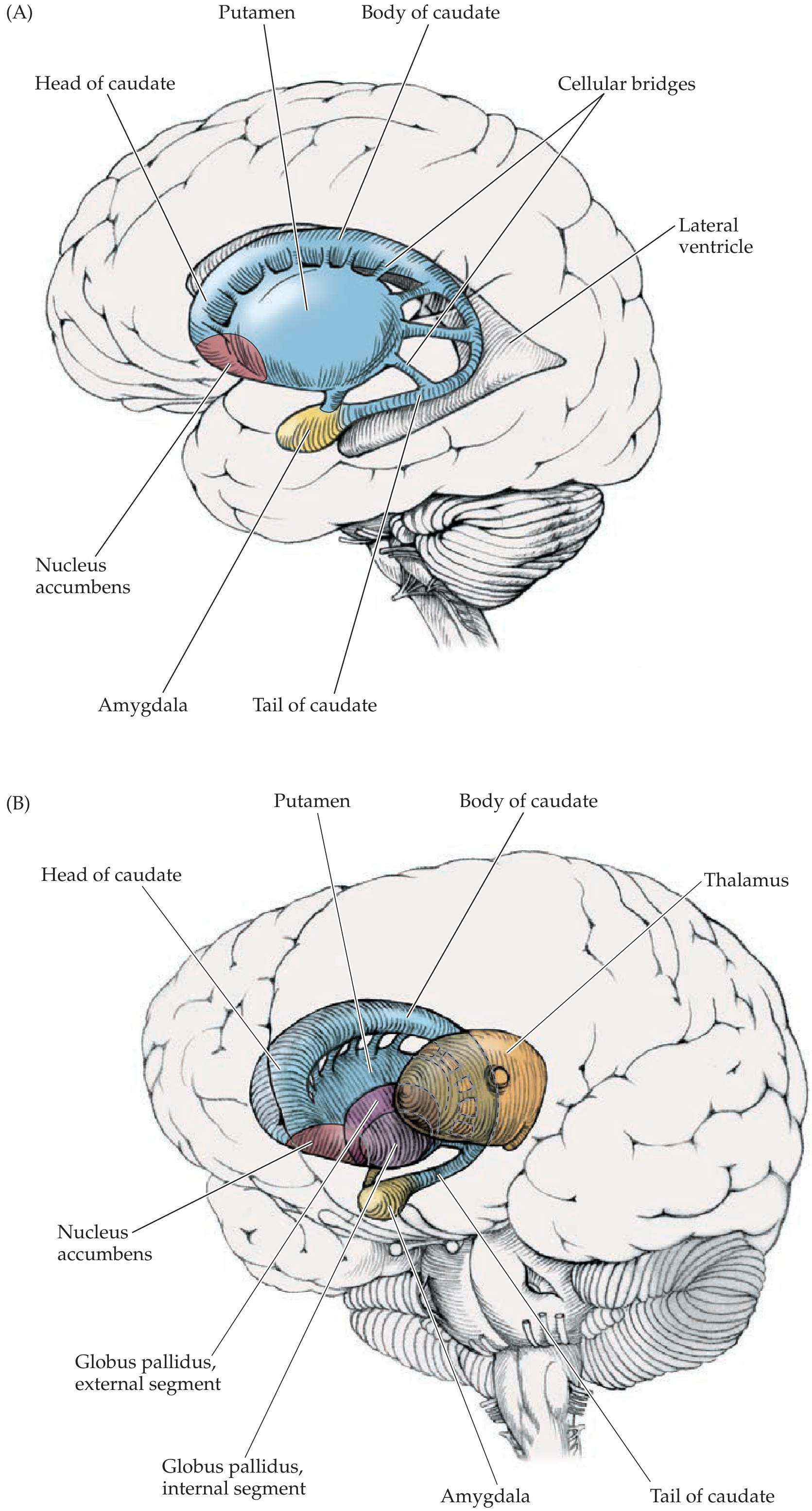
| Structure | Subdivisions | Notes |
|---|---|---|
| Caudate nucleus | Head, body, tail | C-shaped; follows lateral ventricle; joined to putamen by cellular bridges |
| Putamen | - | Largest structure; main motor input nucleus |
| Globus pallidus | External (GPe), Internal (GPi) | Main output nucleus; GPi = inhibitory output to thalamus |
| Substantia nigra | Pars compacta (SNc), Pars reticulata (SNr) | SNc = dopaminergic neurons; SNr = inhibitory output (like GPi) |
| Subthalamic nucleus (STN) | - | Diencephalon origin; excitatory (glutamate) relay in indirect pathway |
| Nucleus accumbens | - | Ventral striatum; reward/limbic circuitry |
Groupings:
- Striatum (neostriatum) = Caudate + Putamen (input nuclei)
- Lentiform/lenticular nucleus = Putamen + Globus pallidus
- Ventral striatum = Nucleus accumbens + ventral caudate/putamen junction
Inputs to the Basal Ganglia
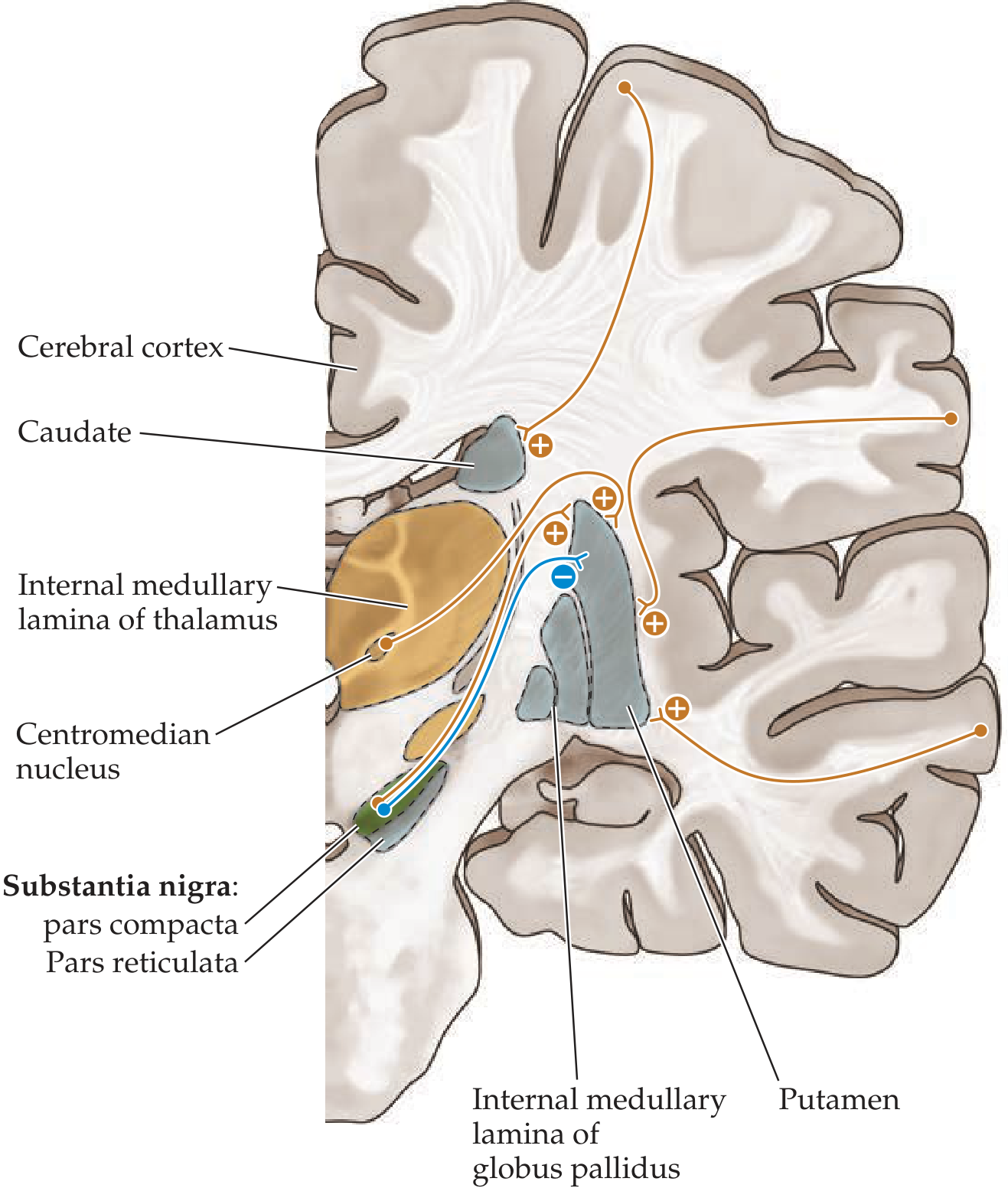
The striatum is the main entry point - it receives:
- Entire cerebral cortex (massive, excitatory, glutamatergic) - the putamen is the key motor input nucleus
- Substantia nigra pars compacta (SNc) - dopaminergic nigrostriatal pathway (modulatory)
- Intralaminar thalamic nuclei (centromedian & parafascicular) - excitatory, glutamatergic
- Modulatory inputs: serotonin (raphe), noradrenaline (locus coeruleus), acetylcholine (intrinsic interneurons), histamine
Outputs from the Basal Ganglia
Outputs arise from two structures:
- GPi (Internal segment of globus pallidus) - for body/limb motor control
- SNr (Substantia nigra pars reticulata) - for head and neck / eye movements
All outputs are inhibitory (GABAergic). They project via the thalamic fasciculus to:
- VL/VA thalamic nuclei → premotor cortex, SMA, primary motor cortex (motor channel)
- Mediodorsal (MD) nucleus → prefrontal/limbic cortex (cognitive/limbic channels)
- Intralaminar nuclei → back to striatum (feedback loop)
The Direct and Indirect Pathways
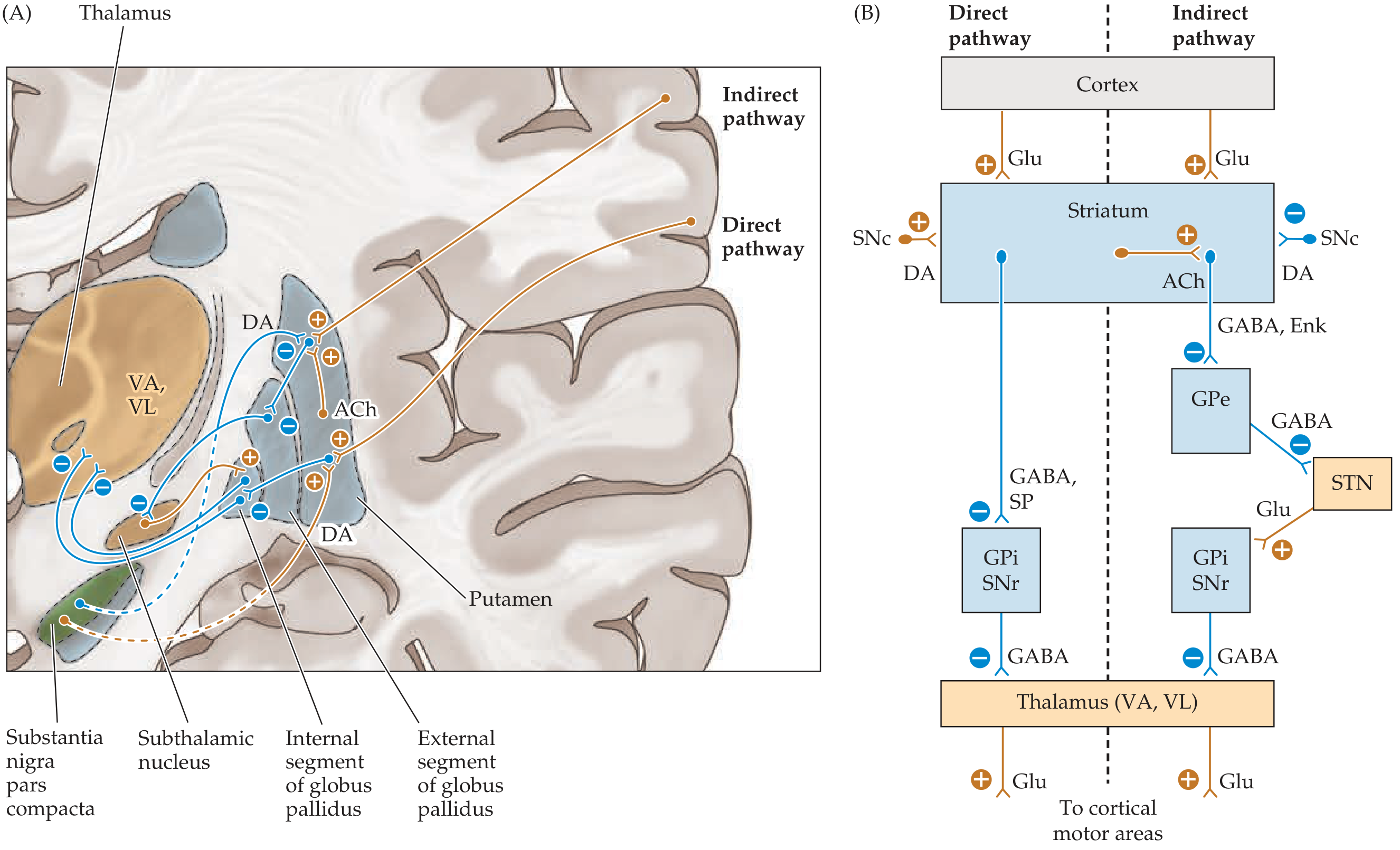
Direct Pathway - "Facilitates Movement"
Cortex → (+)Striatum (Glu) → (-)GPi/SNr (GABA+SP) → (-)Thalamus (GABA) → (+)Cortex (Glu)
- Striatum inhibits GPi/SNr
- GPi/SNr can no longer inhibit thalamus → thalamus is disinhibited
- Thalamus excites motor cortex → movement is facilitated
- Dopamine (D1 receptors on direct pathway neurons) is excitatory → promotes direct pathway → facilitates movement
- Mnemonic: Direct = Disinhibition = movement
Indirect Pathway - "Suppresses Movement"
Cortex → (+)Striatum (Glu) → (-)GPe (GABA+Enkephalin) → (-)STN (GABA) → (+)GPi/SNr (Glu) → (-)Thalamus (GABA) → cortex suppressed
- Striatum inhibits GPe
- GPe can no longer inhibit STN → STN becomes active
- STN excites GPi/SNr (glutamate)
- GPi/SNr strongly inhibits thalamus → movement is suppressed
- Dopamine (D2 receptors on indirect pathway neurons) is inhibitory → suppresses indirect pathway → net result still facilitates movement
- Mnemonic: Indirect = Inhibits movement
The Hyperdirect Pathway
A third, faster pathway: Cortex → STN (Glu, bypassing striatum) → GPi/SNr → inhibits thalamus. This provides rapid suppression of movements, a "stop signal."
Net Effect of Dopamine
| Pathway | Receptor | Dopamine Effect | Net Motor Effect |
|---|---|---|---|
| Direct | D1 | Excitatory | Facilitates movement |
| Indirect | D2 | Inhibitory | Suppresses indirect pathway → facilitates movement |
Both D1 and D2 dopamine effects ultimately favor movement. Loss of dopamine (as in Parkinson's disease) = impaired movement.
Functional Channels Through the Basal Ganglia
The basal ganglia are not a single motor system - there are at least four parallel, functionally segregated loops:
| Channel | Striatal Input | Thalamic Relay | Cortical Output | Function |
|---|---|---|---|---|
| Motor | Putamen | VL (VLo) | Primary motor, SMA, premotor cortex | Motor execution and sequencing |
| Oculomotor | Caudate (body) | VA | Frontal eye fields | Eye movement control |
| Prefrontal/Cognitive | Caudate (head) | VA/MD | Dorsolateral prefrontal cortex | Working memory, executive function |
| Limbic | Nucleus accumbens (ventral striatum) | MD/VA | Anterior cingulate, orbitofrontal cortex | Motivation, reward, emotion |
Role of the Basal Ganglia - Summary
-
Action selection: The basal ganglia act as a "selector" - facilitating desired motor programs (via direct pathway) while suppressing competing or unwanted movements (via indirect pathway). This is sometimes called the "center-surround" model.
-
Procedural learning and habit formation: Repeated practice of motor sequences embeds them in striatal circuits. Procedural memory is stored in the basal ganglia, not the hippocampus.
-
Reinforcement learning: Phasic dopamine signals from SNc encode reward prediction errors, updating the value of actions (foundation of the temporal difference model of learning).
-
Cognitive control: Via the prefrontal loop, the basal ganglia help with working memory, task switching, and inhibiting inappropriate responses.
-
Motivation and reward: The limbic loop (nucleus accumbens + ventral tegmental area dopamine) mediates reward, drug addiction, and goal-directed behavior.
-
Oculomotor control: Via the caudate → SNr → superior colliculus pathway, the basal ganglia help initiate and suppress saccadic eye movements.
Neurotransmitters Summary
| Synapse | Transmitter | Effect |
|---|---|---|
| Cortex → Striatum | Glutamate | Excitatory (+) |
| Striatum → GPi (direct) | GABA + Substance P | Inhibitory (-) |
| Striatum → GPe (indirect) | GABA + Enkephalin | Inhibitory (-) |
| GPe → STN | GABA | Inhibitory (-) |
| STN → GPi | Glutamate | Excitatory (+) |
| GPi/SNr → Thalamus | GABA | Inhibitory (-) |
| Thalamus → Cortex | Glutamate | Excitatory (+) |
| SNc → Striatum (direct) | Dopamine (D1) | Excitatory (+) |
| SNc → Striatum (indirect) | Dopamine (D2) | Inhibitory (-) |
Clinical Correlates
Hypokinetic Disorders (too little movement - indirect pathway overactive)
Parkinson's Disease
- Degeneration of dopaminergic neurons in SNc (pars compacta)
- Loss of dopamine → direct pathway underactive, indirect pathway overactive → GPi over-inhibits thalamus → reduced motor cortex activation
- Features: resting tremor ("pill-rolling"), bradykinesia, cogwheel rigidity, shuffling gait, postural instability
- Treatment: L-DOPA (dopamine precursor), dopamine agonists (bromocriptine), MAO-B inhibitors, deep brain stimulation (STN or GPi)
Hyperkinetic Disorders (too much movement - indirect pathway underactive)
Huntington's Disease
- Autosomal dominant; CAG repeat expansion in HTT gene
- Destruction of striatal GABAergic neurons (especially those projecting in the indirect pathway)
- Loss of striatal inhibition on GPe → GPe over-inhibits STN → STN cannot activate GPi → thalamus is disinhibited → excess movement
- Features: choreiform (writhing, random, involuntary) movements, psychiatric symptoms, dementia
- No cure; symptomatic treatment
Hemiballismus
- Destruction of the subthalamic nucleus (STN) - classically due to small vessel stroke
- STN can no longer excite GPi → GPi under-inhibits thalamus → wild, flinging, large-amplitude movements of contralateral limbs
- Treatment: haloperidol or other dopamine antagonists
| Disorder | Lesion Site | Pathway Effect | Movement |
|---|---|---|---|
| Parkinson's disease | SNc (dopamine loss) | Indirect ↑, direct ↓ | Hypokinetic |
| Huntington's disease | Striatum (indirect pathway neurons first) | Indirect ↓ | Hyperkinetic (chorea) |
| Hemiballismus | Subthalamic nucleus | Indirect ↓↓ | Hyperkinetic (ballismus) |
Sources:
- Neuroanatomy through Clinical Cases, 3rd Edition (Blumenfeld)
- Costanzo Physiology, 7th Edition
- Principles of Neural Science, 6th Edition (Kandel)
- Bradley and Daroff's Neurology in Clinical Practice
Relations to Limbic system
The basal ganglia and limbic system are deeply interconnected. The ventral striatum (nucleus accumbens) is the key interface - receiving dopaminergic input from the ventral tegmental area (VTA) and limbic inputs from the amygdala, hippocampus, and prefrontal cortex, forming the "limbic loop" through the basal ganglia.
Searching sources...
Finding Sources
Finding Sources
Searching the Web
basal ganglia limbic system connections nucleus accumbens reward circuit
Excellent. Now let me read from the most targeted sections across three textbooks in parallel.
Reading File
Reading File
Reading File
Excellent content. Let me fetch the key images and get the dopamine pathway content from Stahl's and Kaplan.
Loading Image
Loading Image
Reading File
Excellent. Let me now get the mesolimbic pathway section from Stahl's, plus the amygdalofugal pathway and psychiatric connections from Kaplan, and the limbic circuit details from Neuroanatomy.
Reading File
Reading File
Loading Image
I now have rich, comprehensive content and diagrams. Let me compile the full answer.
Basal Ganglia - Relations to the Limbic System
The Core Concept: Two Systems, One Interface
The basal ganglia and the limbic system are not separate silos - they are deeply and reciprocally integrated. The key insight from modern neuroscience is that the basal ganglia contain a dedicated "limbic channel" that processes emotional, motivational, and reward-based information alongside their classic motor functions. The ventral striatum (nucleus accumbens) is the anatomical fulcrum of this integration, converting limbic signals into behavioral outputs.
The Limbic Channel Through the Basal Ganglia
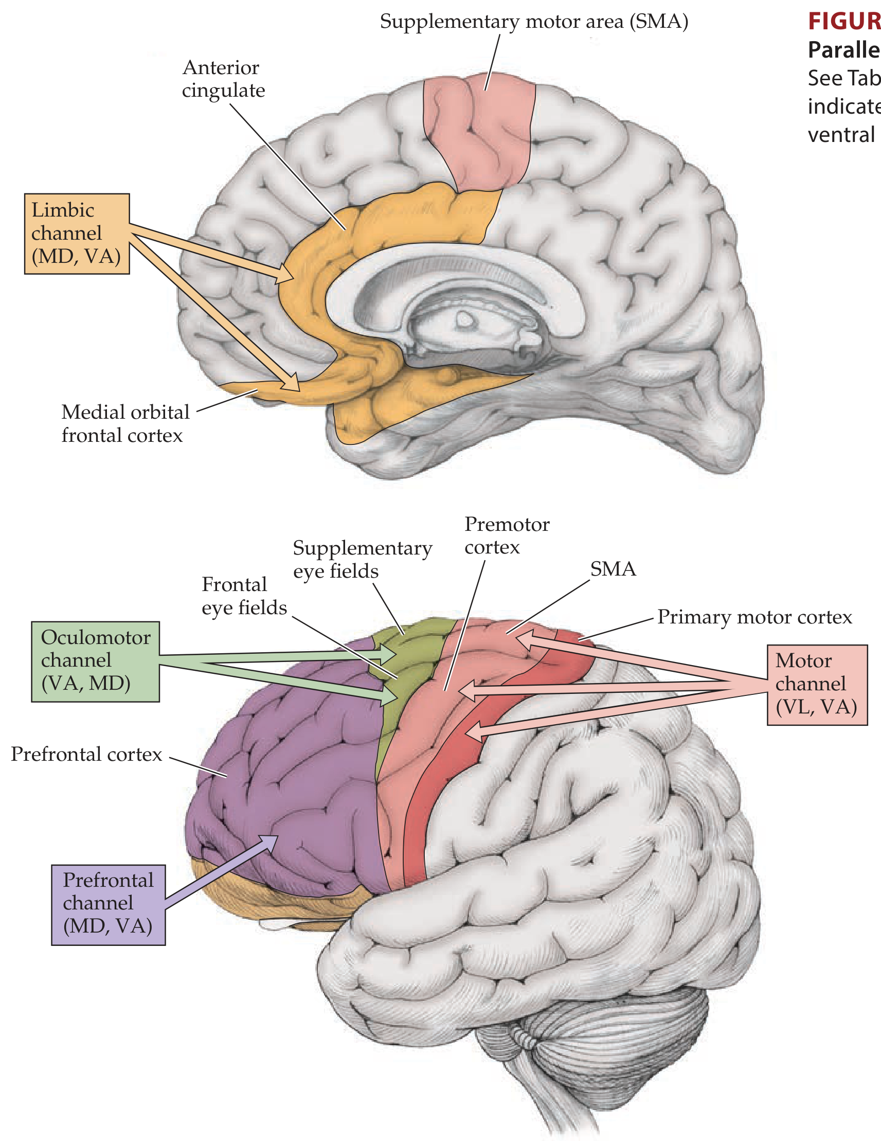
The limbic channel runs in parallel with the motor, oculomotor, and prefrontal channels:
| Channel | Cortical/Limbic Input | Striatal Input Nucleus | Output Nucleus | Thalamic Relay | Cortical Target |
|---|---|---|---|---|---|
| Limbic | Temporal cortex, hippocampus, amygdala | Nucleus accumbens; ventral caudate & putamen | Ventral pallidum; GPi; SNr | MD, VA | Anterior cingulate; orbitofrontal cortex |
| Motor | Somatosensory, motor, premotor cortex | Putamen | GPi; SNr | VL, VA | SMA; premotor; primary motor |
| Oculomotor | Posterior parietal; prefrontal | Caudate, body | GPi; SNr | VA, MD | Frontal/supplementary eye fields |
| Prefrontal | Posterior parietal; premotor | Caudate, head | GPi; SNr | VA, MD | Dorsolateral prefrontal cortex |
(Neuroanatomy through Clinical Cases, 3rd Ed.; Table 16.2)
The Limbic Circuit Through the Basal Ganglia
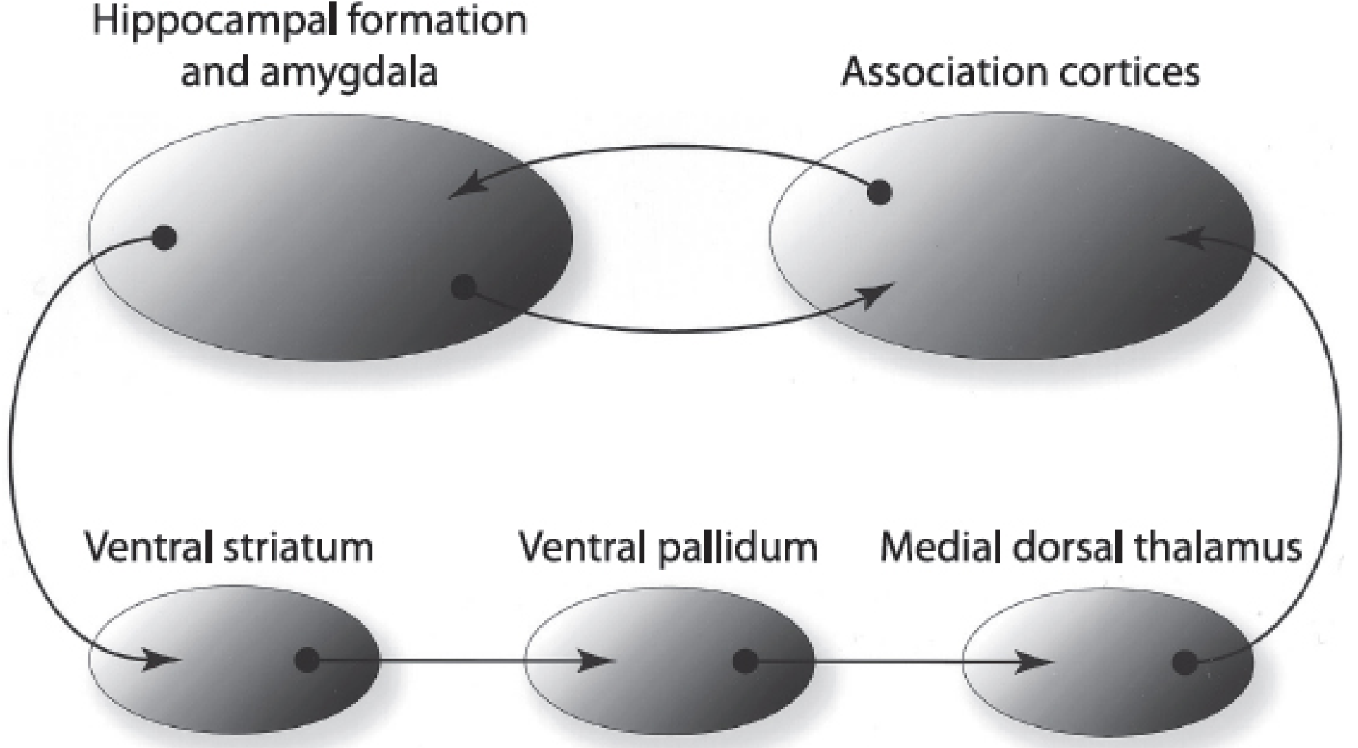
The pathway runs as follows:
Hippocampus + Amygdala (+ Association Cortices)
↓ (glutamatergic input via ventral amygdalofugal pathway and hippocampal projections)
Ventral Striatum / Nucleus Accumbens
↓ (GABAergic output)
Ventral Pallidum (= ventral extension of globus pallidus)
↓ (GABAergic output)
Mediodorsal Thalamus (MD)
↓ (glutamatergic output)
Orbitofrontal Cortex + Anterior Cingulate Cortex
↓ (back to striatum, closing the loop)
This illustrates that the functions of the basal ganglia extend far beyond motor control - they are essential for translating emotional and motivational states into goal-directed behaviors. (Kaplan & Sadock)
Key Anatomical Connections
1. Nucleus Accumbens - The "Limbic-Motor Interface"
The nucleus accumbens (NAcc) sits at the junction of the caudate and putamen in the ventral striatum. It is the most important anatomical interface between the limbic system and the basal ganglia.
Inputs to the Nucleus Accumbens:
- Amygdala (basolateral complex) - via the ventral amygdalofugal pathway - carries emotional valence and threat/reward signals
- Hippocampus - context and memory signals
- Prefrontal cortex (medial/orbitofrontal) - top-down cognitive and evaluative signals
- VTA dopamine neurons - reward prediction signals (mesolimbic pathway)
- Hypothalamus - homeostatic drives (hunger, thirst, sex)
Outputs from the Nucleus Accumbens:
- → Ventral pallidum → mediodorsal thalamus → anterior cingulate / orbitofrontal cortex
- → Hypothalamus (influencing autonomic/endocrine outputs)
- → Brainstem (motor output nuclei)
The NAcc functions as a modulation center between the limbic and motor systems: it receives emotional/motivational input from limbic structures and translates it into behavioral (motor) outputs through its connections with the motor basal ganglia. (Neuroanatomy through Clinical Cases)
2. Amygdala - Basal Ganglia Connections
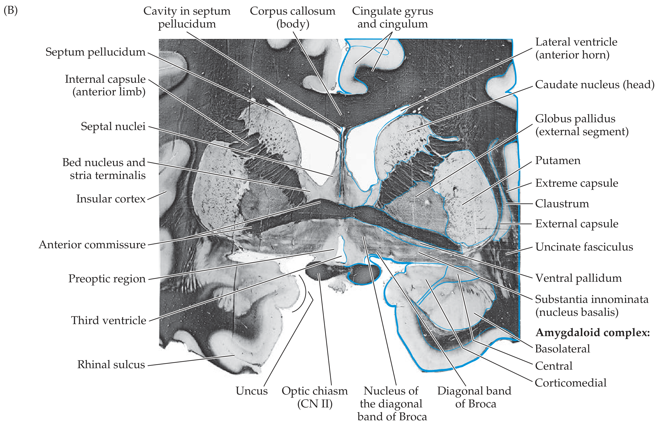
- The basolateral amygdala projects directly to the nucleus accumbens via the ventral amygdalofugal pathway - this is how fear, reward, and threat signals influence motivated behavior
- The central amygdala (part of the "extended amygdala") is continuous via the sublenticular substantia innominata with the bed nucleus of the stria terminalis, and is reciprocally connected with the lateral hypothalamus and brainstem
- The stria terminalis carries amygdaloid fibers to the hypothalamus and septal nuclei
3. Extended Amygdala
The extended amygdala is a continuous subcortical structure linking the central amygdaloid nucleus, the sublenticular substantia innominata, and the bed nucleus of the stria terminalis. This structure spans the border between the amygdala, the basal forebrain, and portions of the basal ganglia, and is intimately involved in anxiety, stress responses, and addiction.
4. Septal Nuclei - Basal Ganglia Relations
The septal area participates in both limbic circuits (hippocampal connections) and basal forebrain/basal ganglia circuits:
- Medial septal nucleus (cholinergic): projects to hippocampal formation (septohippocampal pathway = anxiety regulation)
- Lateral septal nucleus (GABAergic): receives hippocampal output via fornix
- Nucleus accumbens is sometimes classified within the broader septal/basal forebrain region
Dopamine: The Neurochemical Bridge
Dopamine is the key neuromodulator linking the limbic system to the basal ganglia. There are two critical dopaminergic pathways:
| Pathway | Origin | Target | Function |
|---|---|---|---|
| Nigrostriatal | Substantia nigra pars compacta (SNc) | Dorsal striatum (putamen/caudate) | Motor control (direct/indirect pathways) |
| Mesolimbic | Ventral tegmental area (VTA) | Nucleus accumbens + limbic structures | Reward, motivation, pleasure, addiction |
| Mesocortical | VTA | Prefrontal cortex (DLPFC, VMPFC) | Cognition, affective regulation |
| Tuberoinfundibular | Hypothalamus | Anterior pituitary | Prolactin inhibition |
The mesolimbic dopamine pathway (VTA → nucleus accumbens) is the biological substrate of:
- Pleasurable sensations and reward
- Powerful euphoria from drugs of abuse
- Reinforcement and habit learning
- In excess or dysregulation: delusions and hallucinations of psychosis (Stahl's Essential Psychopharmacology)
Medium spiny neurons in the nucleus accumbens receive convergent input from:
- Dopaminergic VTA neurons (reward signal)
- Glutamatergic hippocampal neurons (context)
- Glutamatergic amygdala neurons (emotional valence)
- Glutamatergic medial prefrontal cortex neurons (cognitive evaluation)
This convergence makes the NAcc a site where context + emotion + motivation + dopamine prediction error are integrated to generate approach or avoidance behavior.
Functional Significance: What This Integration Achieves
| Function | Limbic Input | Basal Ganglia Mechanism | Behavioral Output |
|---|---|---|---|
| Reward-motivated movement | Dopamine from VTA → NAcc | Limbic channel direct pathway activated | Approach behavior, seeking |
| Fear-motivated avoidance | Amygdala → NAcc | Indirect pathway activated | Withdrawal, avoidance |
| Habit formation | Hippocampus (context) + dopamine | Dorsal striatum reinforced | Automatic, stimulus-triggered actions |
| Decision making | OFC, anterior cingulate → ventral striatum | Prefrontal-limbic-BG loop | Choice based on expected value |
| Extinction of behavior | Prefrontal cortex → NAcc | Reduction of dopamine signal | Suppression of formerly rewarded behavior |
Clinical Disorders at the Limbic-Basal Ganglia Interface
| Disorder | Pathophysiology | Key Circuit |
|---|---|---|
| Drug addiction | Repeated dopamine surges in NAcc → sensitization and hijacking of limbic-BG reward circuits | VTA → NAcc (mesolimbic); OFC/ACC loops |
| Schizophrenia | Mesolimbic dopamine overactivity → positive symptoms (hallucinations, delusions); mesocortical dopamine underactivity → negative/cognitive symptoms | VTA → NAcc; VTA → PFC |
| Depression | Reduced dopamine and serotonin in limbic-BG circuits; hyperactivity of subgenual anterior cingulate | Limbic channel; NAcc; ACC-BA25 |
| OCD | Hyperactivity in orbitofrontal cortex - caudate - thalamus - OFC loop (CSTC loop); failed suppression of unwanted thoughts/actions | OFC → caudate → GPi/SNr → thalamus → OFC |
| Tourette syndrome | Disinhibition within limbic-motor BG circuitry; tics emerge from failure to gate motor and vocal programs | Striatum; limbic inputs to motor BG |
| PTSD/Anxiety | Hyperactive amygdala signals to NAcc and hypothalamus; failure of prefrontal top-down control | Amygdala → NAcc → ventral pallidum |
| Huntington's disease | Striatal degeneration disrupts both motor AND limbic/cognitive BG channels | Caudate (cognitive/limbic) + putamen (motor) |
Summary: The Limbic-Basal Ganglia Relationship in Three Sentences
The basal ganglia contain a dedicated limbic channel (ventral striatum/NAcc → ventral pallidum → mediodorsal thalamus → anterior cingulate/orbitofrontal cortex) that operates in parallel with their motor, oculomotor, and prefrontal channels. The nucleus accumbens is the anatomical interface where limbic signals from the hippocampus, amygdala, and prefrontal cortex converge with dopaminergic reward signals from the VTA to translate emotion and motivation into action. Dysregulation of this limbic-basal ganglia interface underlies a broad spectrum of neuropsychiatric disorders including addiction, depression, schizophrenia, and OCD.
Sources:
- Neuroanatomy through Clinical Cases, 3rd Edition (Blumenfeld) - Chapters 16 & 18
- Kaplan & Sadock's Comprehensive Textbook of Psychiatry
- Stahl's Essential Psychopharmacology, Neuroscientific Basis
Hippocampus role in Limbic system
The hippocampus is the central memory structure of the limbic system, critical for consolidating short-term memories into long-term storage, spatial navigation, and contextual learning. It is also part of the Papez circuit and modulates emotional responses via connections with the amygdala and hypothalamus.
Searching sources...
Finding Sources
Finding Sources
Searching the Web
hippocampus role limbic system memory consolidation spatial navigation
Excellent sources. Let me read from Kandel's Principles of Neural Science (most authoritative) and Guyton simultaneously.
Reading File
Reading File
Reading File
Excellent. Now let me get the subregion functions, LTP section, and clinical/Kaplan content in parallel.
Reading File
Reading File
Reading File
Excellent content. Now let me get the Kaplan & Sadock hippocampus section and fetch the key diagrams.
Reading File
Loading Image
Loading Image
Excellent material gathered. Let me get the hippocampal histology image and the internal circuit image.
Loading Image
Reading File
I now have comprehensive, authoritative material from Kandel's Principles of Neural Science, Guyton & Hall, and Kaplan & Sadock. Let me compile the complete answer.
The Hippocampus: Role in the Limbic System
Overview
The hippocampus is perhaps the most studied structure in neuroscience. Situated in the floor of the temporal horn of the lateral ventricle, it serves as the principal memory gateway of the limbic system - essential for converting immediate experience into lasting long-term memories. It also plays key roles in spatial navigation, contextual processing, emotional modulation, and future-oriented thinking. Its connections within the limbic system make it a hub where sensory, cognitive, and emotional information converge.
Anatomy: Location and Structure
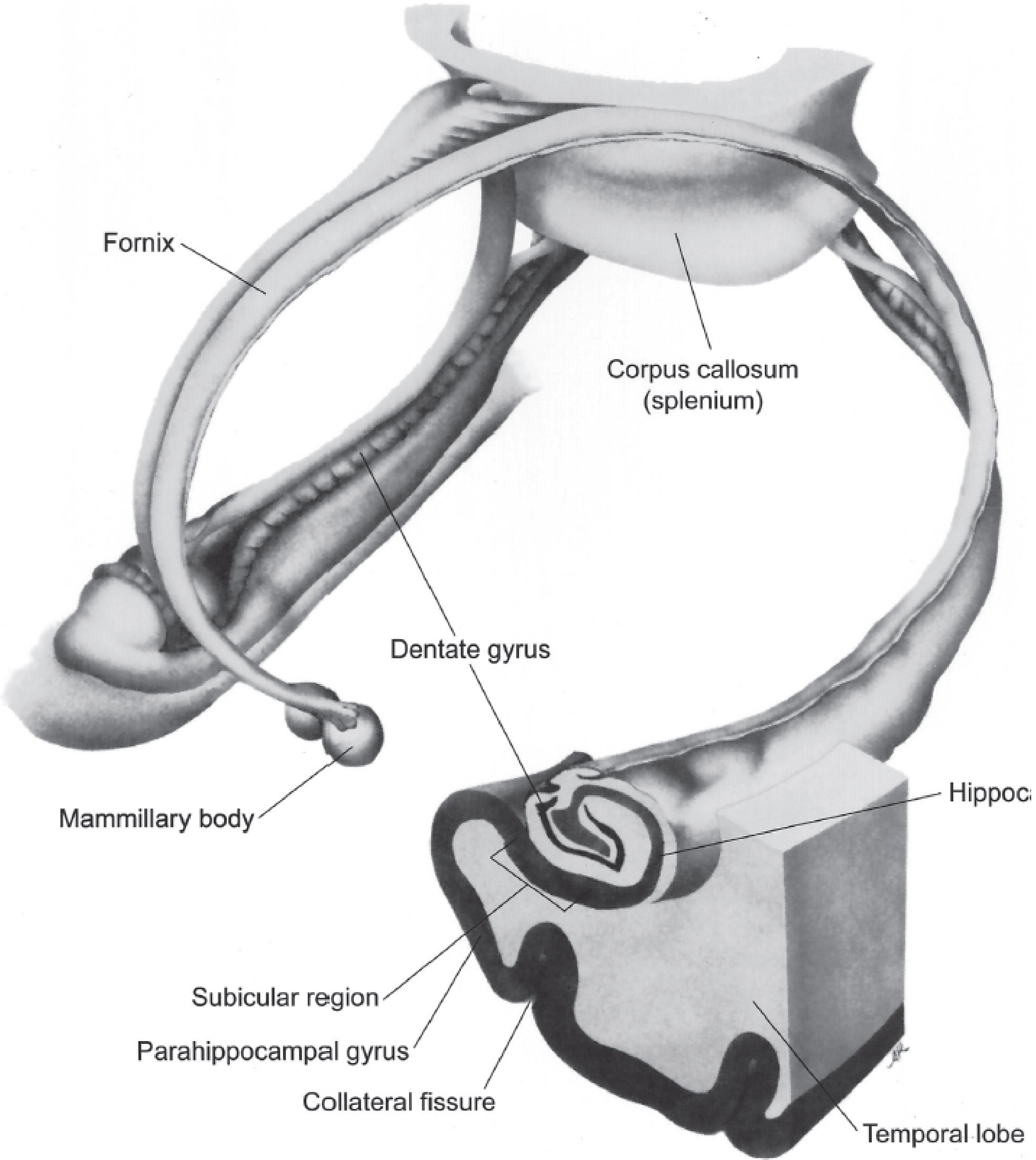
The hippocampal formation comprises three distinct adjacent zones folded together in a C-shape:
- Dentate gyrus - most medial; three-layered (molecular, granule cell, polymorphic layers)
- Hippocampus proper - divided into fields:
- CA3 (proximal, adjacent to dentate): mossy fiber input from dentate granule cells; autoassociative recurrent collaterals; important for pattern completion
- CA2: encodes social memory; relatively resistant to degeneration
- CA1 (distal): receives Schaffer collaterals from CA3; the major output zone; most vulnerable in Alzheimer's disease and hypoxia
- Hilus (sometimes called CA4): part of the dentate gyrus
- Subicular complex (presubiculum, parasubiculum, prosubiculum, subiculum, postsubiculum): transition zone between hippocampus and parahippocampal gyrus; major output to entorhinal cortex and fornix
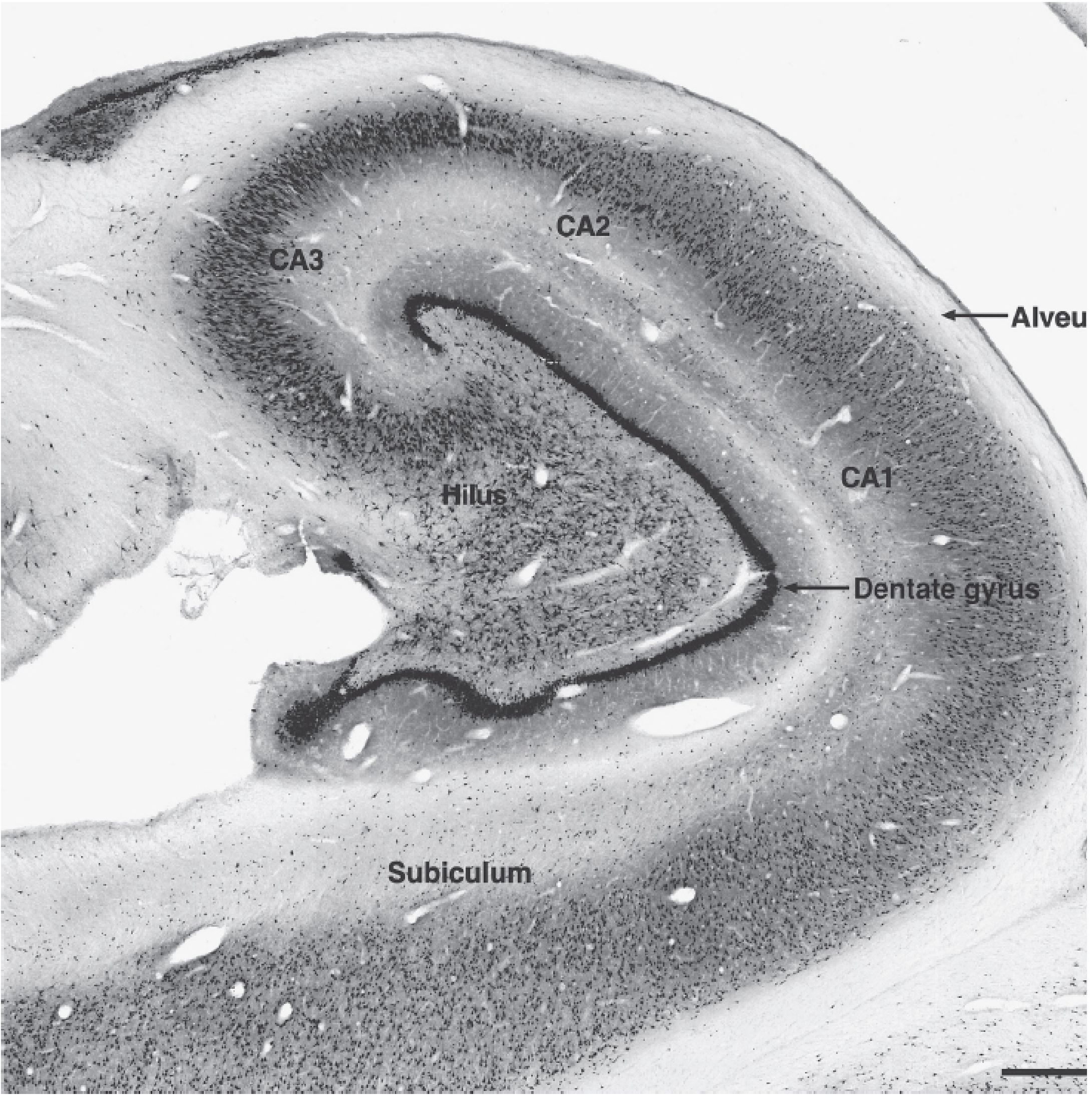
The white matter adjacent to the hippocampus is the alveus, which feeds into the fimbria, which becomes the crus of the fornix - the main hippocampal output tract running to the mammillary bodies and hypothalamus.
The Hippocampal Circuit: Information Flow
The hippocampus processes information through a largely unidirectional trisynaptic circuit:
Trisynaptic (Indirect) Pathway
Entorhinal Cortex (Layer II) → (perforant path) → Dentate Gyrus granule cells
↓ (mossy fiber pathway)
CA3 pyramidal cells
↓ (Schaffer collateral pathway)
CA1 pyramidal cells
↓ (via subiculum)
Deep layers of Entorhinal Cortex → (distributed to neocortex)
Direct Pathway
Entorhinal Cortex (Layer III) → (temporo-ammonic pathway) → CA1 (bypassing dentate and CA3)
This provides two routes - the indirect (trisynaptic) path for pattern-separated, transformed encoding, and the direct path for more faithful transmission of cortical information.
Output via Fornix
CA1 / Subiculum → Alveus → Fimbria → Fornix → Mammillary bodies + Septal nuclei + Hypothalamus
(Kaplan & Sadock; Kandel Principles of Neural Science)
The Hippocampus in the Papez Circuit
The hippocampus is the entry point and re-entry point of the Papez circuit - the classical circuit for emotional and memory processing:
Hippocampus → (fornix) → Mammillary bodies → (mammillothalamic tract) → Anterior thalamic nucleus → Cingulate gyrus → Parahippocampal gyrus → Entorhinal cortex → Hippocampus
This loop, originally proposed by James Papez in 1937 as the substrate of emotion, is now understood to be critical for episodic memory consolidation and spatial-contextual processing - the hippocampus both feeds information into this circuit and receives it back.
Roles of the Hippocampus in the Limbic System
1. Explicit (Declarative) Memory Consolidation
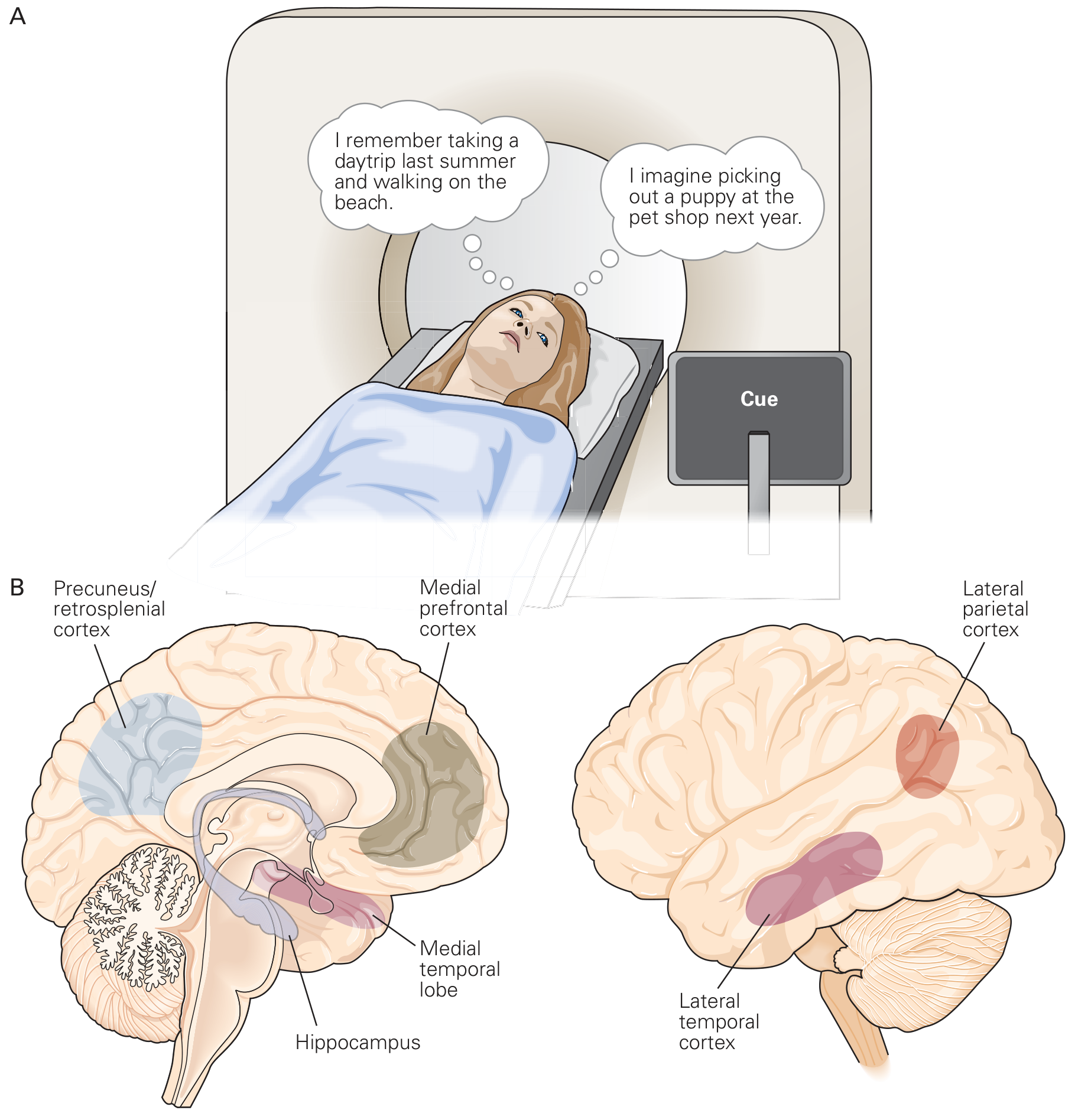
The hippocampus is the gateway for converting short-term memories into long-term explicit memories. It does not store memories permanently itself - the ultimate storage site for most declarative memory is the cerebral cortex - but the process of stabilizing those memories (memory consolidation) requires the hippocampus.
Types of explicit memory dependent on the hippocampus:
- Episodic memory: personally experienced events ("what happened, when, where")
- Semantic memory: factual knowledge about the world
- Spatial memory: knowledge of environments and navigation
Evidence: Patient H.M. (Henry Molaison), who had bilateral hippocampal removal for epilepsy, showed profound anterograde amnesia - he could not form any new declarative memories - while his procedural (skill) memory and remote memories remained intact. (Guyton & Hall; Kandel)
The hippocampus is also strongly linked to future imagination - the same network activated during recall of past events is activated when imagining future events, suggesting the hippocampus builds "relational maps" of experience usable for both memory and forward planning.
2. Gatekeeper of Memory Relevance
The hippocampus is one of the most important output pathways from the reward and punishment centers of the limbic system. It helps make the decision about which experiences are emotionally significant enough to be stored long term. Experiences that generate strong reward or punishment signals are preferentially consolidated. (Guyton & Hall)
3. Spatial Navigation and Cognitive Maps
The hippocampus contains place cells - neurons that fire specifically when an animal occupies a particular location in space. This was first discovered by O'Keefe and Dostrovsky (1971) in rodents and later confirmed in humans.
- Right hippocampus activity increases when spatial information is recalled
- Left hippocampus activity increases when words, objects, or people are recalled
Related cells:
- Place cells (hippocampus CA1): fire at specific locations; encode allocentric space
- Grid cells (entorhinal cortex): form a hexagonal coordinate system for navigation
- Head direction cells: encode facing direction
The hippocampus provides a general mechanism for forming complex multimodal associations - binding the separate spatial, temporal, sensory, and emotional elements of experience into a coherent relational map. (Kandel)
4. Subregion-Specific Functions
| Subregion | Key Function | Mechanism |
|---|---|---|
| Dentate gyrus | Pattern separation | Expansion recoding: sparse, orthogonalized representations from entorhinal input; prevents confusing similar memories; contains adult neurogenesis |
| CA3 | Pattern completion | Autoassociative recurrent collaterals allow partial cues to trigger complete memory retrieval |
| CA2 | Social memory | Encodes recognition of social identity; parvalbumin interneurons critical here |
| CA1 | Main output; context-sensitive encoding | Final integration point; projects to subiculum and entorhinal cortex |
| Subiculum | Output relay | Projects to entorhinal cortex, mammillary bodies, prefrontal cortex |
(Kandel, Principles of Neural Science)
5. Contextual Modulation of Fear and Emotion
The hippocampus provides contextual information to the amygdala - it tells the amygdala where a fearful event occurred. This is the basis of contextual fear conditioning:
- A tone (cued fear) → amygdala-dependent
- Fear in a specific context (room/environment) → hippocampus + amygdala dependent
Without the hippocampus, animals can still be conditioned to fear a tone (amygdala intact) but lose the context-specific fear response.
The hippocampus also sends projections to the nucleus accumbens (ventral striatum), translating contextual memory signals into motivational and reward-based behaviors via the limbic channel of the basal ganglia.
6. Stress and Neuroendocrine Regulation
The hippocampus expresses high levels of glucocorticoid receptors and provides inhibitory feedback to the hypothalamic-pituitary-adrenal (HPA) axis:
- Cortisol → binds hippocampal glucocorticoid receptors → inhibits further CRF release from hypothalamus (negative feedback)
- Chronic stress and high cortisol → hippocampal neuronal damage and atrophy
- This creates a vicious cycle: stress damages the hippocampus → impaired HPA feedback → more cortisol
Cellular Mechanism: Long-Term Potentiation (LTP)
The cellular substrate of hippocampal memory storage is long-term potentiation (LTP) - a persistent increase in synaptic strength following high-frequency stimulation.
NMDA Receptor as Coincidence Detector
At Schaffer collateral → CA1 synapses, LTP is mediated by NMDA receptors (N-methyl-D-aspartate type glutamate receptors). The NMDA receptor requires both:
- Glutamate release (presynaptic)
- Strong postsynaptic depolarization (to relieve Mg²⁺ block)
This makes it a Hebbian coincidence detector - "cells that fire together, wire together."
Three Key Properties of LTP
| Property | Meaning | Significance |
|---|---|---|
| Cooperativity | Near-simultaneous activation of many inputs needed | Prevents trivial stimuli from being stored |
| Associativity | Weak input paired with strong input → both potentiated | Allows context/association links (e.g., smell + memory) |
| Synapse specificity | Only active synapses are strengthened | Memory is precisely and selectively encoded |
LTP Phases
- Early LTP (E-LTP): Minutes to hours; AMPA receptor insertion/phosphorylation; no new protein synthesis needed
- Late LTP (L-LTP): Hours to days/weeks; requires gene transcription and new protein synthesis; PKA → CREB → immediate early genes; structural synaptic changes
(Kandel, Principles of Neural Science)
Adult Neurogenesis in the Dentate Gyrus
One of the most remarkable properties of the hippocampus is the continued birth of new neurons (neurogenesis) in the dentate gyrus throughout adult life - one of only two known sites of adult neurogenesis in mammals (the other is the olfactory bulb/subventricular zone).
New dentate granule cells are thought to:
- Contribute to pattern separation of closely related memories
- Be important for certain forms of hippocampal-dependent learning
- Be inhibited by chronic stress (reduced by glucocorticoids)
- Be promoted by exercise, enriched environments, and antidepressants
Clinical Correlates
| Condition | Hippocampal Involvement | Key Features |
|---|---|---|
| Bilateral hippocampal lesions (surgery, HSV encephalitis) | Complete loss of memory formation | Profound anterograde amnesia; intact procedural memory and remote memories |
| Alzheimer's disease | Earliest and most severe degeneration in entorhinal cortex → CA1 (via perforant path); Aβ plaques and tau tangles disrupt LTP | Short-term memory loss first; spatial disorientation; progressive global dementia |
| Temporal lobe epilepsy (TLE) | Hippocampal sclerosis (CA1 most vulnerable); seizures arise from medial temporal structures | Auras of fear, déjà vu, olfactory hallucinations; mesial TLE most common form |
| Korsakoff syndrome | Mammillary body damage + thalamic damage disrupts Papez circuit; hippocampal outflow blocked | Anterograde amnesia, confabulation; due to thiamine deficiency |
| PTSD | Hippocampal volume reduction (glucocorticoid toxicity from chronic stress); impaired context discrimination | Inability to distinguish "safe" from "dangerous" contexts; overgeneralized fear responses |
| Schizophrenia | Reduced hippocampus-prefrontal synchrony; loss of CA2 parvalbumin interneurons | Impaired working memory; context processing deficits |
| Depression | Reduced neurogenesis in dentate gyrus; hippocampal volume loss | Cognitive and memory impairment; some antidepressants restore neurogenesis |
| Right hippocampal lesions | Impaired spatial memory | Poor navigation, getting lost |
| Left hippocampal lesions | Impaired verbal memory | Difficulty learning new word lists, names, verbal information |
Summary: The Hippocampus as Limbic Hub
The hippocampus integrates where, when, what, and how significant into a unified memory trace. Its connections make it simultaneously:
- A memory consolidator (via entorhinal cortex ↔ neocortex)
- A spatial navigator (place cells + grid cell input)
- A Papez circuit node (via fornix → mammillary bodies → anterior thalamus → cingulate)
- A fear context provider (→ amygdala)
- A HPA axis regulator (inhibitory glucocorticoid feedback)
- A limbic-motor interface (→ nucleus accumbens)
- A stress vulnerability site (glucocorticoid receptor density; neurogenesis)
Sources:
- Kandel, Principles of Neural Science, 6th Edition - Chapters 52 & 54
- Guyton and Hall Textbook of Medical Physiology
- Kaplan & Sadock's Comprehensive Textbook of Psychiatry
This is a shared conversation. Sign in to Orris to start your own chat.