/teach me CNS Pharmacology

Reading File
Asking for Preferences
Finding Sources
Finding Sources
Finding Sources
Finding Sources
Finding Sources
Finding Sources
Finding Sources
Finding Sources
Finding Sources
Reading File
Reading File
Reading File
Reading File
Reading File
Reading File
Reading File
Reading File
Reading File
Reading File
Searching Images

benzodiazepine GABA receptor mechanism of action

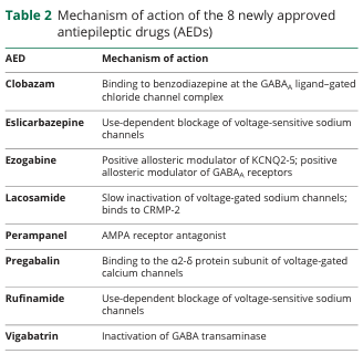
Table 2 Mechanism of action of the 8 newly approved antiepileptic drugs (AEDs)
<table><thead><tr><th>AED</th><th>Mechanism of action</th></tr></thead><tbody><tr><td>Clobazam</td><td>Binding to benzodiazepine at the GABA<sub>A</sub> ligand-gated chloride channel complex</td></tr><tr><td>Eslicarbazepine</td><td>Use-dependent blockage of voltage-sensitive sodium channels</td></tr><tr><td>Ezogabine</td><td>Positive allosteric modulator of KCNQ2-5; positive allosteric modulator of GABA<sub>A</sub> receptors</td></tr><tr><td>Lacosamide</td><td>Slow inactivation of voltage-gated sodium channels; binds to CRMP-2</td></tr><tr><td>Perampanel</td><td>AMPA receptor antagonist</td></tr><tr><td>Pregabalin</td><td>Binding to the α2-δ protein subunit of voltage-gated calcium channels</td></tr><tr><td>Rufinamide</td><td>Use-dependent blockage of voltage-sensitive sodium channels</td></tr><tr><td>Vigabatrin</td><td>Inactivation of GABA transaminase</td></tr></tbody></table>

Table 2 Mechanism of action of the 8 newly approved antiepileptic drugs (AEDs) <table><thead><tr><th>AED</th><th>Mechanism of action</th></tr></thead><tbody><tr><td>Clobazam</td><td>Binding to benzodiazepine at the GABA<sub>A</sub> ligand-gated chloride channel complex</td></tr><tr><td>Eslicarbazepine</td><td>Use-dependent blockage of voltage-sensitive sodium channels</td></tr><tr><td>Ezogabine</td><td>Positive allosteric modulator of KCNQ2-5; positive allosteric modulator of GABA<sub>A</sub> receptors</td></tr><tr><td>Lacosamide</td><td>Slow inactivation of voltage-gated sodium channels; binds to CRMP-2</td></tr><tr><td>Perampanel</td><td>AMPA receptor antagonist</td></tr><tr><td>Pregabalin</td><td>Binding to the α2-δ protein subunit of voltage-gated calcium channels</td></tr><tr><td>Rufinamide</td><td>Use-dependent blockage of voltage-sensitive sodium channels</td></tr><tr><td>Vigabatrin</td><td>Inactivation of GABA transaminase</td></tr></tbody></table>

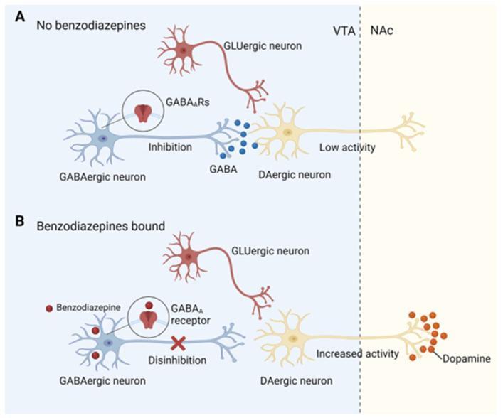
This pathophysiology diagram illustrates the neurobiological mechanism of benzodiazepine addiction through disinhibition of dopaminergic neurons in the mesolimbic reward system. The illustration is divided into two comparative panels. Panel A (No benzodiazepines) shows a baseline state in the Ventral Tegmental Area (VTA), where a blue GABAergic interneuron releases GABA molecules to inhibit a yellow dopaminergic (DAergic) neuron via GABAA receptors, resulting in 'Low activity' and minimal dopamine release in the Nucleus Accumbens (NAc). A GLUergic neuron is also shown nearby. Panel B (Benzodiazepines bound) depicts the introduction of red benzodiazepine molecules, which bind to GABAA receptors on the GABAergic interneuron. This binding induces 'Disinhibition' of the downstream DAergic neuron (indicated by an 'X' over the inhibitory synapse). Consequently, the DAergic neuron exhibits 'Increased activity,' leading to a significant rise in dopamine release (red molecules) within the NAc. The diagram highlights how benzodiazepines modulate neuronal signaling to trigger the brain's reward circuitry.

This pathophysiology diagram illustrates the neurobiological mechanism of benzodiazepine addiction through disinhibition of dopaminergic neurons in the mesolimbic reward system. The illustration is divided into two comparative panels. Panel A (No benzodiazepines) shows a baseline state in the Ventral Tegmental Area (VTA), where a blue GABAergic interneuron releases GABA molecules to inhibit a yellow dopaminergic (DAergic) neuron via GABAA receptors, resulting in 'Low activity' and minimal dopamine release in the Nucleus Accumbens (NAc). A GLUergic neuron is also shown nearby. Panel B (Benzodiazepines bound) depicts the introduction of red benzodiazepine molecules, which bind to GABAA receptors on the GABAergic interneuron. This binding induces 'Disinhibition' of the downstream DAergic neuron (indicated by an 'X' over the inhibitory synapse). Consequently, the DAergic neuron exhibits 'Increased activity,' leading to a significant rise in dopamine release (red molecules) within the NAc. The diagram highlights how benzodiazepines modulate neuronal signaling to trigger the brain's reward circuitry.

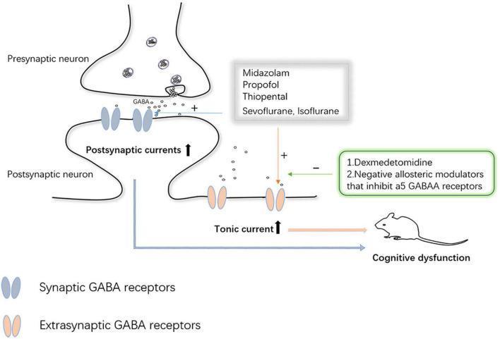
This pathophysiology diagram illustrates the mechanism of GABA receptor-mediated inhibition in neurons and the modulatory effects of anesthetic agents. The illustration depicts a synaptic cleft between a presynaptic neuron, which releases GABA from vesicles, and a postsynaptic neuron. GABA binds to two types of receptors: synaptic GABA receptors, which increase postsynaptic currents, and extrasynaptic GABA receptors, which increase tonic current. The diagram highlights how specific anesthetics—Midazolam, Propofol, Thiopental, Sevoflurane, and Isoflurane—act as positive modulators (+) on both receptor types, particularly enhancing tonic current. Conversely, Dexmedetomidine and negative allosteric modulators of α5 GABAA receptors exert inhibitory effects (-) on these extrasynaptic pathways. The net increase in GABAergic inhibition and tonic current is linked via arrows to a mouse model labeled 'Cognitive dysfunction,' suggesting a clinical correlation between excessive GABAergic tonic signaling and impaired cognitive outcomes. This diagram serves as an educational tool for neuropharmacology and anesthesiology, focusing on GABA receptor distribution and drug-induced neurotoxicity.

This pathophysiology diagram illustrates the mechanism of GABA receptor-mediated inhibition in neurons and the modulatory effects of anesthetic agents. The illustration depicts a synaptic cleft between a presynaptic neuron, which releases GABA from vesicles, and a postsynaptic neuron. GABA binds to two types of receptors: synaptic GABA receptors, which increase postsynaptic currents, and extrasynaptic GABA receptors, which increase tonic current. The diagram highlights how specific anesthetics—Midazolam, Propofol, Thiopental, Sevoflurane, and Isoflurane—act as positive modulators (+) on both receptor types, particularly enhancing tonic current. Conversely, Dexmedetomidine and negative allosteric modulators of α5 GABAA receptors exert inhibitory effects (-) on these extrasynaptic pathways. The net increase in GABAergic inhibition and tonic current is linked via arrows to a mouse model labeled 'Cognitive dysfunction,' suggesting a clinical correlation between excessive GABAergic tonic signaling and impaired cognitive outcomes. This diagram serves as an educational tool for neuropharmacology and anesthesiology, focusing on GABA receptor distribution and drug-induced neurotoxicity.

Searching Images

dopamine pathway antipsychotic mechanism schizophrenia

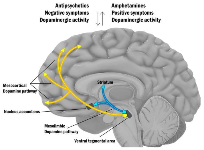
An anatomical diagram of the human brain in a midsagittal view illustrating the dopamine hypothesis of schizophrenia. The illustration highlights two primary dopaminergic pathways originating from the Ventral Tegmental Area (VTA) in the midbrain: the Mesocortical Dopamine Pathway, represented by yellow arrows projecting to the prefrontal cortex, and the Mesolimbic Dopamine Pathway, indicated by yellow arrows projecting to the Nucleus Accumbens. Additionally, blue arrows indicate projections toward the Striatum. At the top, a conceptual comparison lists clinical correlations: 'Antipsychotics' are associated with reduced 'Dopaminergic activity' and 'Negative symptoms,' while 'Amphetamines' are linked to increased 'Dopaminergic activity' and 'Positive symptoms.' Vertical arrows between these lists indicate an inverse relationship between the effects of these substances. This diagram serves as a neurobiological model for medical students and clinicians to visualize the pathophysiology of schizophrenia and the mechanism of action for antipsychotic medications and psychostimulants.

An anatomical diagram of the human brain in a midsagittal view illustrating the dopamine hypothesis of schizophrenia. The illustration highlights two primary dopaminergic pathways originating from the Ventral Tegmental Area (VTA) in the midbrain: the Mesocortical Dopamine Pathway, represented by yellow arrows projecting to the prefrontal cortex, and the Mesolimbic Dopamine Pathway, indicated by yellow arrows projecting to the Nucleus Accumbens. Additionally, blue arrows indicate projections toward the Striatum. At the top, a conceptual comparison lists clinical correlations: 'Antipsychotics' are associated with reduced 'Dopaminergic activity' and 'Negative symptoms,' while 'Amphetamines' are linked to increased 'Dopaminergic activity' and 'Positive symptoms.' Vertical arrows between these lists indicate an inverse relationship between the effects of these substances. This diagram serves as a neurobiological model for medical students and clinicians to visualize the pathophysiology of schizophrenia and the mechanism of action for antipsychotic medications and psychostimulants.

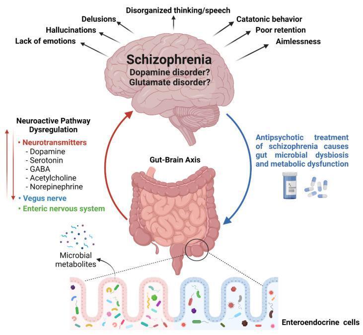
This medical pathophysiology diagram illustrates the bidirectional relationship of the gut-brain axis in the context of schizophrenia. At the top, a brain illustration represents schizophrenia, listing primary clinical symptoms (hallucinations, delusions, catatonic behavior, etc.) and highlighting the 'dopamine disorder' and 'glutamate disorder' hypotheses. A circular flow connects the brain to the gastrointestinal tract (large intestine). The ascending 'Neuroactive Pathway Dysregulation' (red arrow) demonstrates how the gut influences the brain via microbial metabolites, the vagus nerve, the enteric nervous system, and key neurotransmitters including dopamine, serotonin, GABA, acetylcholine, and norepinephrine. Conversely, the descending pathway (blue arrow) illustrates the impact of schizophrenia management, noting that antipsychotic treatments can cause gut microbial dysbiosis and metabolic dysfunction. A detailed inset at the bottom shows the gut barrier with enteroendocrine cells and diverse microbiota, emphasizing the role of microbial diversity in systemic homeostasis. This infographic is designed for educational use in psychiatry and gastroenterology to explain neurobiological mechanisms and treatment side effects.

This medical pathophysiology diagram illustrates the bidirectional relationship of the gut-brain axis in the context of schizophrenia. At the top, a brain illustration represents schizophrenia, listing primary clinical symptoms (hallucinations, delusions, catatonic behavior, etc.) and highlighting the 'dopamine disorder' and 'glutamate disorder' hypotheses. A circular flow connects the brain to the gastrointestinal tract (large intestine). The ascending 'Neuroactive Pathway Dysregulation' (red arrow) demonstrates how the gut influences the brain via microbial metabolites, the vagus nerve, the enteric nervous system, and key neurotransmitters including dopamine, serotonin, GABA, acetylcholine, and norepinephrine. Conversely, the descending pathway (blue arrow) illustrates the impact of schizophrenia management, noting that antipsychotic treatments can cause gut microbial dysbiosis and metabolic dysfunction. A detailed inset at the bottom shows the gut barrier with enteroendocrine cells and diverse microbiota, emphasizing the role of microbial diversity in systemic homeostasis. This infographic is designed for educational use in psychiatry and gastroenterology to explain neurobiological mechanisms and treatment side effects.

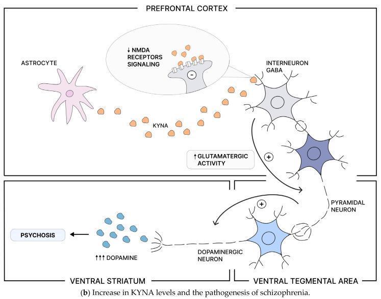
This pathophysiology diagram illustrates the neurobiological mechanism linking increased kynurenic acid (KYNA) levels to the pathogenesis of schizophrenia and psychosis across specific brain regions. In the Prefrontal Cortex (PFC), an astrocyte is shown producing KYNA, which acts as an antagonist at N-methyl-D-aspartate (NMDA) receptors. This interaction results in decreased NMDA receptor signaling on GABAergic interneurons. The subsequent reduction in inhibitory GABAergic output leads to disinhibition and increased glutamatergic activity of cortical pyramidal neurons (indicated by a '+' sign). This pyramidal neuron projects to the Ventral Tegmental Area (VTA), where it provides excitatory stimulation to dopaminergic neurons. This activation of the mesolimbic pathway causes excessive dopamine release in the Ventral Striatum (indicated by '↑↑↑ DOPAMINE'). The final outcome of this increased dopaminergic signaling is labeled as 'PSYCHOSIS'. The diagram highlights the complex interaction between the kynurenine pathway, glutamatergic signaling, and dopaminergic neurotransmission in psychiatric disorders.

This pathophysiology diagram illustrates the neurobiological mechanism linking increased kynurenic acid (KYNA) levels to the pathogenesis of schizophrenia and psychosis across specific brain regions. In the Prefrontal Cortex (PFC), an astrocyte is shown producing KYNA, which acts as an antagonist at N-methyl-D-aspartate (NMDA) receptors. This interaction results in decreased NMDA receptor signaling on GABAergic interneurons. The subsequent reduction in inhibitory GABAergic output leads to disinhibition and increased glutamatergic activity of cortical pyramidal neurons (indicated by a '+' sign). This pyramidal neuron projects to the Ventral Tegmental Area (VTA), where it provides excitatory stimulation to dopaminergic neurons. This activation of the mesolimbic pathway causes excessive dopamine release in the Ventral Striatum (indicated by '↑↑↑ DOPAMINE'). The final outcome of this increased dopaminergic signaling is labeled as 'PSYCHOSIS'. The diagram highlights the complex interaction between the kynurenine pathway, glutamatergic signaling, and dopaminergic neurotransmission in psychiatric disorders.

Searching Images

Parkinson disease levodopa dopamine basal ganglia

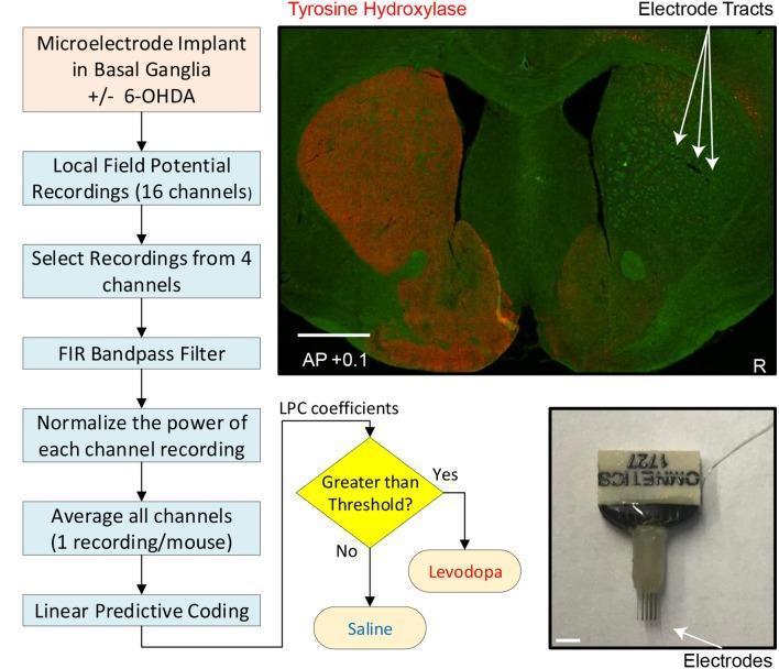
Educational composite image illustrating an experimental approach to modeling Parkinson's disease using a rodent model. Left: A diagnostic flowchart outlines the methodological sequence from microelectrode implantation in the basal ganglia to the classification of treatment (Saline vs. Levodopa) using Linear Predictive Coding (LPC) coefficients derived from Local Field Potential (LFP) recordings. Top Right: A coronal diagnostic image of a mouse striatum (AP +0.1) using Tyrosine Hydroxylase (TH) immunostaining (red fluorescence). The image demonstrates unilateral dopamine depletion in the right hemisphere following 6-OHDA neurotoxin injection, characterized by a significant loss of red staining compared to the intact left hemisphere. Arrows indicate the electrode tracts of the 16-channel microelectrode array within the dorsal striatum. Bottom Right: A clinical photograph of an Omnetics microelectrode array, showing the structural configuration of the fine wire electrodes used for localized neural recording. This visual summary integrates pathophysiology, neuroanatomical histology, and biomedical engineering concepts relevant to movement disorder research.

Educational composite image illustrating an experimental approach to modeling Parkinson's disease using a rodent model. Left: A diagnostic flowchart outlines the methodological sequence from microelectrode implantation in the basal ganglia to the classification of treatment (Saline vs. Levodopa) using Linear Predictive Coding (LPC) coefficients derived from Local Field Potential (LFP) recordings. Top Right: A coronal diagnostic image of a mouse striatum (AP +0.1) using Tyrosine Hydroxylase (TH) immunostaining (red fluorescence). The image demonstrates unilateral dopamine depletion in the right hemisphere following 6-OHDA neurotoxin injection, characterized by a significant loss of red staining compared to the intact left hemisphere. Arrows indicate the electrode tracts of the 16-channel microelectrode array within the dorsal striatum. Bottom Right: A clinical photograph of an Omnetics microelectrode array, showing the structural configuration of the fine wire electrodes used for localized neural recording. This visual summary integrates pathophysiology, neuroanatomical histology, and biomedical engineering concepts relevant to movement disorder research.

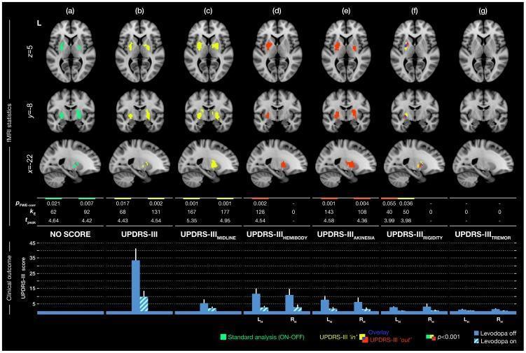
This composite diagnostic image presents fMRI statistical maps and clinical outcome data illustrating brain activity responses to levodopa in Parkinson’s disease (PD) patients. The top section displays seven columns (a–g) of axial (z=5), coronal (y=-8), and sagittal (x=-22) brain slices focusing on the basal ganglia. Columns compare different analytical approaches using UPDRS-III subscores: (a) standard ON-OFF medication analysis (green), (b) total UPDRS-III, (c) midline, (d) hemibody, (e) akinesia, (f) rigidity, and (g) tremor. Yellow clusters indicate a positive correlation with the score ('in' approach), red clusters represent treatment effects when accounting for the score as a nuisance variable ('out' approach), and blue indicates anatomical overlap. Notably, total UPDRS-III and midline subscores show significant correlations (yellow) in the basal ganglia, while akinesia and hemibody scores reveal strong treatment-related sensitivity (red). Rigidity shows a unique overlap of both approaches. The bottom section contains a bar plot demonstrating clinical outcomes, showing significant reduction in UPDRS-III scores across all subscores (except tremor) when comparing 'Levodopa off' (solid blue) to 'Levodopa on' (hatched blue) states.

This composite diagnostic image presents fMRI statistical maps and clinical outcome data illustrating brain activity responses to levodopa in Parkinson’s disease (PD) patients. The top section displays seven columns (a–g) of axial (z=5), coronal (y=-8), and sagittal (x=-22) brain slices focusing on the basal ganglia. Columns compare different analytical approaches using UPDRS-III subscores: (a) standard ON-OFF medication analysis (green), (b) total UPDRS-III, (c) midline, (d) hemibody, (e) akinesia, (f) rigidity, and (g) tremor. Yellow clusters indicate a positive correlation with the score ('in' approach), red clusters represent treatment effects when accounting for the score as a nuisance variable ('out' approach), and blue indicates anatomical overlap. Notably, total UPDRS-III and midline subscores show significant correlations (yellow) in the basal ganglia, while akinesia and hemibody scores reveal strong treatment-related sensitivity (red). Rigidity shows a unique overlap of both approaches. The bottom section contains a bar plot demonstrating clinical outcomes, showing significant reduction in UPDRS-III scores across all subscores (except tremor) when comparing 'Levodopa off' (solid blue) to 'Levodopa on' (hatched blue) states.

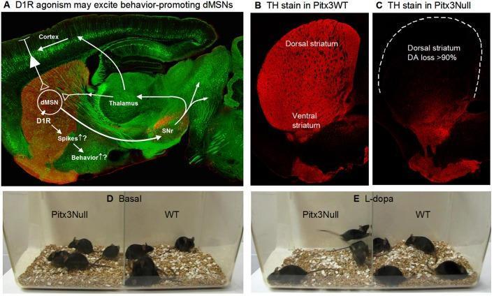
Educational figure illustrating dopamine (DA) denervation in the basal ganglia motor control circuit, using a mouse model (Pitx3Null) for Parkinson’s disease research. (A) Confocal sagittal brain section and overlay diagram: Red Tyrosine Hydroxylase (TH) stain indicates dopaminergic projections, while Green Fluorescent Protein (GFP) outlines basal ganglia structures. The diagram maps the direct pathway medium spiny neuron (dMSN) circuit from the cortex and thalamus to the substantia nigra reticulata (SNr), highlighting D1 receptor (D1R) involvement in behavior promotion. (B) High-magnification 3 μm confocal section of a wild-type (Pitx3WT) mouse striatum shows dense, uniform TH staining (red) across both dorsal and ventral regions, representing normal dopamine innervation. (C) Corresponding section from a Pitx3Null mouse shows a severe dorso-ventral gradient of DA loss; the dorsal striatum (outlined) exhibits >90% loss of TH staining, while some ventral innervation is preserved. (D, E) Clinical behavioral comparison photographs show Pitx3Null and WT mice in basal and L-dopa-treated states, correlating histological dopamine deficiency with motor performance and pharmacological response.

Educational figure illustrating dopamine (DA) denervation in the basal ganglia motor control circuit, using a mouse model (Pitx3Null) for Parkinson’s disease research. (A) Confocal sagittal brain section and overlay diagram: Red Tyrosine Hydroxylase (TH) stain indicates dopaminergic projections, while Green Fluorescent Protein (GFP) outlines basal ganglia structures. The diagram maps the direct pathway medium spiny neuron (dMSN) circuit from the cortex and thalamus to the substantia nigra reticulata (SNr), highlighting D1 receptor (D1R) involvement in behavior promotion. (B) High-magnification 3 μm confocal section of a wild-type (Pitx3WT) mouse striatum shows dense, uniform TH staining (red) across both dorsal and ventral regions, representing normal dopamine innervation. (C) Corresponding section from a Pitx3Null mouse shows a severe dorso-ventral gradient of DA loss; the dorsal striatum (outlined) exhibits >90% loss of TH staining, while some ventral innervation is preserved. (D, E) Clinical behavioral comparison photographs show Pitx3Null and WT mice in basal and L-dopa-treated states, correlating histological dopamine deficiency with motor performance and pharmacological response.

Finding Sources
Searching Images

opioid receptor mu delta kappa pain pathway analgesic

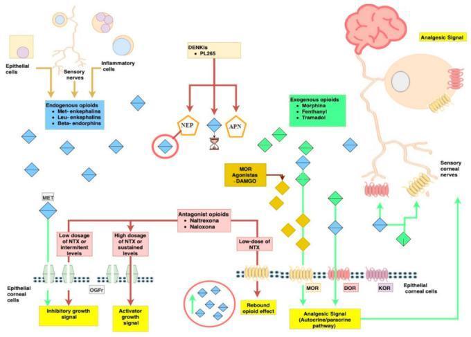
A pathophysiology diagram illustrating the opioid signaling pathways within corneal epithelial cells and sensory nerves. The diagram identifies endogenous opioids (Met-enkephalins, Leu-enkephalins, Beta-endorphins) produced by epithelial, inflammatory cells, and sensory nerves, alongside exogenous opioids like Morphine and Fentanyl. Key receptors shown on corneal cells include Mu (MOR), Delta (DOR), Kappa (KOR), and Opioid Growth Factor receptor (OGFr). Catabolic enzymes NEP and APN are depicted as mediators of opioid degradation, which can be inhibited by agents like PL265 (DENKIs). A critical educational focus is the dose-dependent effect of Naltrexone (NTX); low or intermittent dosages are linked to inhibitory growth signals and the 'rebound opioid effect' leading to analgesia, whereas high or sustained dosages trigger activator growth signals. The pathway concludes with the transmission of an analgesic signal from the corneal surface through sensory nerves to the brain, demonstrating the autocrine and paracrine mechanisms of ocular pain management and tissue homeostasis.

A pathophysiology diagram illustrating the opioid signaling pathways within corneal epithelial cells and sensory nerves. The diagram identifies endogenous opioids (Met-enkephalins, Leu-enkephalins, Beta-endorphins) produced by epithelial, inflammatory cells, and sensory nerves, alongside exogenous opioids like Morphine and Fentanyl. Key receptors shown on corneal cells include Mu (MOR), Delta (DOR), Kappa (KOR), and Opioid Growth Factor receptor (OGFr). Catabolic enzymes NEP and APN are depicted as mediators of opioid degradation, which can be inhibited by agents like PL265 (DENKIs). A critical educational focus is the dose-dependent effect of Naltrexone (NTX); low or intermittent dosages are linked to inhibitory growth signals and the 'rebound opioid effect' leading to analgesia, whereas high or sustained dosages trigger activator growth signals. The pathway concludes with the transmission of an analgesic signal from the corneal surface through sensory nerves to the brain, demonstrating the autocrine and paracrine mechanisms of ocular pain management and tissue homeostasis.

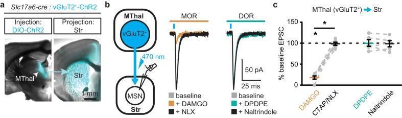
This composite educational image illustrates the selective opioid modulation of thalamic excitatory inputs to the dorsal medial striatum (DMS) in a mouse model. (a) Coronal brain section micrographs demonstrate Cre-dependent expression of ChR2-EYFP (cyan) in vGluT2-positive neurons of the medial thalamus (MThal) and their subsequent axonal projections into the striatum (Str). (b) A schematic diagram shows the experimental setup for optogenetic stimulation of MThal terminals while recording from medium spiny neurons (MSNs) in the DMS. Accompanying electrophysiological traces show that the mu-opioid receptor (MOR) agonist DAMGO significantly suppresses optically evoked excitatory postsynaptic currents (oEPSCs), an effect reversed by the antagonist naloxone (NLX). In contrast, the delta-opioid receptor (DOR) agonist DPDPE and its antagonist naltrindole show no significant effect on oEPSC amplitude. (c) A summary dot plot quantifies these findings, displaying a marked decrease in % baseline EPSC following DAMGO application, whereas DPDPE maintains levels near 100%. This illustrates that MORs, but not DORs, selectively gate the vGluT2+ thalamo-striatal pathway, providing insight into the neurocircuitry of pain and reward processing.

This composite educational image illustrates the selective opioid modulation of thalamic excitatory inputs to the dorsal medial striatum (DMS) in a mouse model. (a) Coronal brain section micrographs demonstrate Cre-dependent expression of ChR2-EYFP (cyan) in vGluT2-positive neurons of the medial thalamus (MThal) and their subsequent axonal projections into the striatum (Str). (b) A schematic diagram shows the experimental setup for optogenetic stimulation of MThal terminals while recording from medium spiny neurons (MSNs) in the DMS. Accompanying electrophysiological traces show that the mu-opioid receptor (MOR) agonist DAMGO significantly suppresses optically evoked excitatory postsynaptic currents (oEPSCs), an effect reversed by the antagonist naloxone (NLX). In contrast, the delta-opioid receptor (DOR) agonist DPDPE and its antagonist naltrindole show no significant effect on oEPSC amplitude. (c) A summary dot plot quantifies these findings, displaying a marked decrease in % baseline EPSC following DAMGO application, whereas DPDPE maintains levels near 100%. This illustrates that MORs, but not DORs, selectively gate the vGluT2+ thalamo-striatal pathway, providing insight into the neurocircuitry of pain and reward processing.

Searching Images

antiepileptic drug seizure sodium channel mechanism

Table 2 Mechanism of action of the 8 newly approved antiepileptic drugs (AEDs)
<table><thead><tr><th>AED</th><th>Mechanism of action</th></tr></thead><tbody><tr><td>Clobazam</td><td>Binding to benzodiazepine at the GABA<sub>A</sub> ligand-gated chloride channel complex</td></tr><tr><td>Eslicarbazepine</td><td>Use-dependent blockage of voltage-sensitive sodium channels</td></tr><tr><td>Ezogabine</td><td>Positive allosteric modulator of KCNQ2-5; positive allosteric modulator of GABA<sub>A</sub> receptors</td></tr><tr><td>Lacosamide</td><td>Slow inactivation of voltage-gated sodium channels; binds to CRMP-2</td></tr><tr><td>Perampanel</td><td>AMPA receptor antagonist</td></tr><tr><td>Pregabalin</td><td>Binding to the α2-δ protein subunit of voltage-gated calcium channels</td></tr><tr><td>Rufinamide</td><td>Use-dependent blockage of voltage-sensitive sodium channels</td></tr><tr><td>Vigabatrin</td><td>Inactivation of GABA transaminase</td></tr></tbody></table>

Table 2 Mechanism of action of the 8 newly approved antiepileptic drugs (AEDs) <table><thead><tr><th>AED</th><th>Mechanism of action</th></tr></thead><tbody><tr><td>Clobazam</td><td>Binding to benzodiazepine at the GABA<sub>A</sub> ligand-gated chloride channel complex</td></tr><tr><td>Eslicarbazepine</td><td>Use-dependent blockage of voltage-sensitive sodium channels</td></tr><tr><td>Ezogabine</td><td>Positive allosteric modulator of KCNQ2-5; positive allosteric modulator of GABA<sub>A</sub> receptors</td></tr><tr><td>Lacosamide</td><td>Slow inactivation of voltage-gated sodium channels; binds to CRMP-2</td></tr><tr><td>Perampanel</td><td>AMPA receptor antagonist</td></tr><tr><td>Pregabalin</td><td>Binding to the α2-δ protein subunit of voltage-gated calcium channels</td></tr><tr><td>Rufinamide</td><td>Use-dependent blockage of voltage-sensitive sodium channels</td></tr><tr><td>Vigabatrin</td><td>Inactivation of GABA transaminase</td></tr></tbody></table>

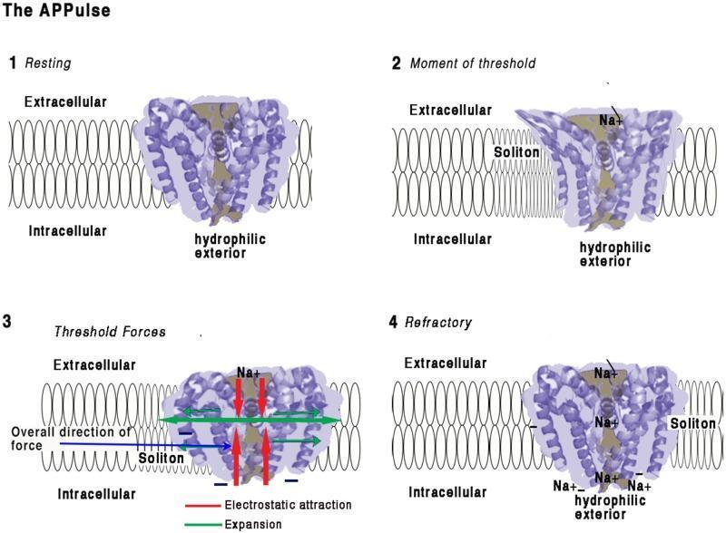
This pathophysiology diagram illustrates the four-stage mechanism of a voltage-gated sodium (Na+) ion channel embedded in a lipid bilayer, focusing on the mechanical and electrostatic changes during depolarization. (1) Resting: The channel's coiled helices are closed, maintaining an electrostatic insulating seal across the 10 nm membrane. (2) Moment of Threshold: An approaching membrane soliton (mechanical wave) exerts pressure, displacing helices and disrupting the electrical insulation. This allows Na+ ions to enter the pore and interact with hydrophilic, negatively charged interior surfaces. (3) Threshold Forces: Inward Na+ influx creates electrostatic attraction (red arrows) toward the intracellular space. Mechanical 'iris-like' opening and contraction occur, causing lateral membrane expansion (green arrows) and a net intracellular force vector (blue arrow). (4) Refractory: Positive charge equalization occurs on the intracellular side, leading to pore closure. The channel remains in a refractory state with localized Na+ accumulation until charges are cleared by diffusion, preventing immediate reactivation.

This pathophysiology diagram illustrates the four-stage mechanism of a voltage-gated sodium (Na+) ion channel embedded in a lipid bilayer, focusing on the mechanical and electrostatic changes during depolarization. (1) Resting: The channel's coiled helices are closed, maintaining an electrostatic insulating seal across the 10 nm membrane. (2) Moment of Threshold: An approaching membrane soliton (mechanical wave) exerts pressure, displacing helices and disrupting the electrical insulation. This allows Na+ ions to enter the pore and interact with hydrophilic, negatively charged interior surfaces. (3) Threshold Forces: Inward Na+ influx creates electrostatic attraction (red arrows) toward the intracellular space. Mechanical 'iris-like' opening and contraction occur, causing lateral membrane expansion (green arrows) and a net intracellular force vector (blue arrow). (4) Refractory: Positive charge equalization occurs on the intracellular side, leading to pore closure. The channel remains in a refractory state with localized Na+ accumulation until charges are cleared by diffusion, preventing immediate reactivation.

Reading File
Reading File
Reading File
Reading File
Here is your complete CNS Pharmacology teaching guide for MBBS/MD level, built from Katzung's Basic & Clinical Pharmacology (16th ed.) and Goodman & Gilman's Pharmacological Basis of Therapeutics.

CNS Pharmacology - Complete MBBS Study Notes


MODULE 1: SEDATIVES, HYPNOTICS & ANXIOLYTICS

Core Concept

These drugs depress CNS activity by enhancing GABAergic inhibition. The main drug classes differ in receptor specificity, duration, and safety profile.

GABA-A Receptor - The Master Target

The GABA-A receptor is a pentameric ligand-gated Cl⁻ channel (subunits: α, β, γ). When GABA binds → Cl⁻ influx → hyperpolarization → CNS depression.
Benzodiazepine disinhibition of dopaminergic neurons via GABA-A modulation
Drug ClassSite of ActionEffect on GABA-AKey Feature
BenzodiazepinesBenzodiazepine binding site (between α and γ subunits)↑ frequency of Cl⁻ channel openingRequire GABA to be present (allosteric modulators)
BarbituratesSeparate site on β subunit↑ duration of Cl⁻ channel opening; at high doses, open channel directlyCan act WITHOUT GABA at high doses - more dangerous
Z-drugs (Zolpidem, Zaleplon, Eszopiclone)Same as benzodiazepines↑ frequency of openingSelective for α1 subunit → hypnotic not anxiolytic
Buspirone5-HT1A partial agonistNo GABA effectAnxiolytic only, no sedation, no abuse potential
RamelteonMT1/MT2 melatonin receptorsNo GABA effectHypnotic for circadian rhythm disorders
Suvorexant / LemborexantOrexin (OX1R/OX2R) antagonistNo GABA effectNewer hypnotics; block wake-promoting orexin

Benzodiazepines - Key Drug Table

DrugHalf-lifeClinical UseKey Point
DiazepamLong (24-48 h)Anxiety, status epilepticus (IV/rectal), muscle relaxantActive metabolite: desmethyldiazepam
LorazepamIntermediate (10-20 h)Status epilepticus (preferred - longer CNS duration)No active metabolites; safe in liver disease
MidazolamShort (2-4 h)Pre-anesthesia, ICU sedation, out-of-hospital status epilepticus (IM)Fastest onset; water-soluble at acidic pH
AlprazolamIntermediatePanic disorder, GADHigh potency; significant dependence risk
ClonazepamLong (18-50 h)Seizures (absence, myoclonic, atonic), panic disorderMost potent antiseizure benzodiazepine
TriazolamVery short (2-5 h)Short-term insomniaRapid onset; rebound insomnia common
Oxazepam, Lorazepam, TemazepamIntermediateElderly, liver diseaseOnly undergo conjugation (glucuronidation) - no CYP
Memory tip: "LOT" drugs (Lorazepam, Oxazepam, Temazepam) = safe in liver disease - glucuronidation only.

Adverse Effects & Overdose

EffectNotes
CNS depressionSedation, anterograde amnesia, ataxia
Dependence & withdrawalIrritability, insomnia, seizures (7-10 days after stopping long-term use)
Paradoxical excitationEspecially in children and elderly
Respiratory depressionEspecially combined with alcohol or opioids
Overdose antidoteFlumazenil - competitive benzodiazepine antagonist (IV); short-acting - may need repeat dosing. Caution: can precipitate seizures in chronic users

Barbiturates (Less Used Now)

DrugUse
PhenobarbitalLong-term epilepsy, neonatal seizures
ThiopentalInduction of anesthesia (ultra-short acting)
PentobarbitalRefractory status epilepticus (ICU)
Key danger: Narrow therapeutic index, no antagonist, severe respiratory depression.

MODULE 2: ANTIEPILEPTIC DRUGS (AEDs)

Seizure Classification (ILAE)

  • Focal onset (previously "partial") - arise from one hemisphere
  • Generalized onset - involve both hemispheres from the start
  • Unknown onset

Mechanisms of AEDs

MechanismDrugsSeizure Types
Na⁺ channel blockade (prolong inactivated state)Phenytoin, Carbamazepine, Valproate, Lamotrigine, Oxcarbazepine, Lacosamide, EslicarbazepineFocal, GTC
T-type Ca²⁺ channel blockadeEthosuximide, ValproateAbsence seizures ONLY
GABA enhancement (↑ GABA-A)Benzodiazepines, Barbiturates, Vigabatrin (↑ GABA by blocking GABA-T), Tiagabine (blocks GABA reuptake)Broad spectrum
Glutamate (AMPA) blockadePerampanelBroad spectrum (adjunct)
SV2A vesicle proteinLevetiracetam, BrivaracetamBroad spectrum
α2δ Ca²⁺ channel subunitGabapentin, PregabalinFocal seizures, neuropathic pain
HCN channel (Ih)Ivermectin (not clinical), Lamotrigine (partial)-

Drug-Seizure Type Match (Exam Essential)

Seizure TypeFirst-LineSecond-Line/Adjunct
Focal (with/without awareness)Carbamazepine, Lamotrigine, OxcarbazepineLevetiracetam, Topiramate, Valproate
Generalized tonic-clonic (GTC)Valproate (broad spectrum), LamotrigineLevetiracetam, Topiramate
AbsenceEthosuximide (DRUG OF CHOICE), ValproateLamotrigine
Juvenile Myoclonic Epilepsy (JME)ValproateLevetiracetam, Lamotrigine
Status EpilepticusLorazepam/Diazepam IV (1st) → Phenytoin/Fosphenytoin IV (2nd) → Phenobarbital/Valproate IV (3rd) → Midazolam/Propofol (refractory)
Febrile seizuresDiazepam rectal/IV-
Neonatal seizuresPhenobarbital IV
Lennox-GastautValproate, Rufinamide, ClobazamLamotrigine
Dravet SyndromeValproate, Clobazam, StiripentolAvoid Na-channel blockers

Key Drug Profiles

DrugMechanismUnique Features / Adverse Effects
PhenytoinNa⁺ channelZero-order kinetics at therapeutic doses; gingival hyperplasia, hirsutism, megaloblastic anemia, teratogenic (fetal hydantoin syndrome), nystagmus, ataxia, CYP inducer
CarbamazepineNa⁺ channelDrug of choice for trigeminal neuralgia; induces its own metabolism (autoinduction); SJS/TEN (HLA-B*1502 in Asians); hyponatremia (SIADH); CYP3A4 inducer
ValproateNa⁺ + T-Ca²⁺ + ↑ GABABroad spectrum - works for all generalized seizures; teratogenic (neural tube defects - spina bifida, 1-2%); hepatotoxicity; weight gain; thrombocytopenia; polycystic ovary syndrome; pancreatitis
EthosuximideT-Ca²⁺ blockOnly for absence seizures; GI upset, headache; no effect on GTC - use valproate if both absence + GTC
LamotrigineNa⁺ channelBroad spectrum; risk of SJS (especially if dose escalated rapidly, or combined with valproate); good in pregnancy (relatively); needs slow titration
LevetiracetamSV2A bindingBroad spectrum; behavioral side effects (irritability, aggression); no drug interactions; renal excretion
Gabapentin / Pregabalinα2δ Ca²⁺ subunitNeuropathic pain, anxiety (pregabalin), focal seizures; renal excretion; no protein binding; no drug interactions
TopiramateNa⁺ + AMPA block + CA inhibitionBroad spectrum; kidney stones, cognitive slowing ("Dopamax"), metabolic acidosis, weight loss, teratogenic (cleft lip)
PhenobarbitalGABA-A (↑ Cl⁻ duration)Oldest; neonatal seizures; enzyme inducer; sedation, tolerance
Critical exam point: Valproate in women of childbearing age - always counsel about neural tube defect risk and folate supplementation.

MODULE 3: OPIOIDS & PAIN PHARMACOLOGY

Opioid Receptor Types

ReceptorLocationAgonist EffectEndogenous Ligand
μ (mu, MOR)Brain (PAG, VTA, dorsal horn), gutAnalgesia, euphoria, respiratory depression, constipation, miosis, physical dependenceβ-endorphins
κ (kappa, KOR)Brain, spinal cordAnalgesia, dysphoria, sedation, miosisDynorphins
δ (delta, DOR)Brain, spinal cordAnalgesia, modulates μ-receptorEnkephalins
All opioid receptors are Gi-protein coupled → inhibit adenylyl cyclase → ↓ cAMP → ↑ K⁺ efflux (hyperpolarization) + ↓ Ca²⁺ influx → reduced neurotransmitter release.
Key insight: μ-opioids cause euphoria because they inhibit GABAergic interneurons in the VTA, which disinhibits dopamine neurons projecting to the nucleus accumbens. κ-agonists directly inhibit these dopamine neurons → dysphoria.

Opioid Drug Classification

ClassDrugsReceptorClinical Notes
Full μ-agonistsMorphine, Codeine, Oxycodone, Hydrocodone, Fentanyl, Meperidine (Pethidine), Methadoneμ (full)Gold standard for severe pain
Partial μ-agonistBuprenorphineμ (partial), κ (antagonist)Ceiling effect for respiratory depression; used in opioid use disorder (OUD)
Mixed agonist-antagonistPentazocine, Butorphanol, Nalbuphineκ agonist / μ antagonistCan precipitate withdrawal in μ-opioid-dependent patients
AntagonistsNaloxone, Naltrexone, Methylnaltrexoneμ antagonistReversal of overdose; OUD treatment

Key Drug Profiles

DrugKey Facts
MorphineStandard; active metabolite morphine-6-glucuronide (M6G) - accumulates in renal failure; histamine release
CodeineProdrug → converted to morphine by CYP2D6; ultra-rapid metabolizers at risk of toxicity; poor metabolizers get no effect
Fentanyl100× more potent than morphine; rapid onset; transdermal patch, IV, nasal; responsible for most opioid overdose deaths
Meperidine (Pethidine)Metabolite normeperidine → seizures (avoid in renal failure); serotonin syndrome risk with MAOIs; not for chronic use
MethadoneLong half-life (24-36 h); QTc prolongation; used for OUD maintenance; complex pharmacokinetics
BuprenorphinePartial agonist; "ceiling effect" on respiratory depression - safer than full agonists; high receptor affinity - blocks other opioids
NaloxoneIV/IM/intranasal; reverses overdose within minutes; half-life 1 h (shorter than most opioids - repeat dosing may be needed)
NaltrexoneOral, long-acting; used for OUD and alcohol dependence; liver toxicity at high doses
MethylnaltrexonePeripheral-acting; treats opioid-induced constipation without reversing CNS analgesia

Opioid Adverse Effects

EffectMechanismNotes
Respiratory depressionμ receptors in brainstemMost dangerous; ↓ response to CO₂
Constipationμ receptors in GI tractDoes NOT develop tolerance; use peripherally-restricted antagonists
Miosis (pinpoint pupils)μ/κ on Edinger-Westphal nucleusDiagnostic sign of opioid toxicity
Nausea/vomitingStimulation of chemoreceptor trigger zoneTolerance develops
Urinary retention↑ sphincter tone
Histamine release(Morphine, codeine)Itching, hypotension
Tolerance & DependenceReceptor downregulation, uncouplingμ-receptor changes

Opioid Overdose Triad

Pinpoint pupils + Coma + Respiratory depression → Give Naloxone

Opioid Withdrawal (NOT life-threatening, unlike alcohol withdrawal)

Rhinorrhea, lacrimation, piloerection, mydriasis, muscle aches, yawning, nausea, vomiting, diarrhea, tachycardia, hypertension - peaks at 36-72 h for short-acting agents.

MODULE 4: ANTIDEPRESSANTS & ANTIPSYCHOTICS

ANTIDEPRESSANTS

Neurotransmitter Hypothesis of Depression

↓ Monoamines (5-HT, NE, DA) in synaptic cleft. All antidepressants (except newer agents) ultimately increase monoamine availability.

Classification and Mechanisms

ClassDrugsMechanismKey Adverse Effects
SSRIsFluoxetine, Sertraline, Paroxetine, Escitalopram, Citalopram, FluvoxamineBlock SERT → ↑ synaptic 5-HTNausea, sexual dysfunction, insomnia, serotonin syndrome, ↑ suicidality in young adults, SIADH
SNRIsVenlafaxine, Desvenlafaxine, Duloxetine, LevomilnacipranBlock SERT + NET → ↑ 5-HT + NEHypertension (especially venlafaxine at high doses), nausea, sexual dysfunction
TCAsAmitriptyline, Imipramine, Clomipramine, Nortriptyline, DesipramineBlock SERT + NET; also block H1, M1, α1Anticholinergic (dry mouth, urinary retention, constipation, blurred vision), sedation, orthostatic hypotension, QTc prolongation, lethal in overdose (cardiac arrhythmias, seizures)
MAOIsPhenelzine, Tranylcypromine, Selegiline (transdermal)Inhibit MAO-A and/or MAO-B → ↑ 5-HT, NE, DAHypertensive crisis with tyramine-rich foods (cheese, wine, cured meats); many drug interactions; serotonin syndrome
NaSSAMirtazapineα2 antagonist (↑ NE + 5-HT release) + H1 blockSedation, weight gain; good for depression with insomnia/weight loss
NDRIBupropionBlock NET + DATSeizures at high doses; no sexual dysfunction; useful for smoking cessation; contraindicated in eating disorders and seizure history
SARITrazodone5-HT2 antagonist + weak SERT inhibitorPriapism; sedation; used for insomnia
MelatonergicAgomelatineMT1/MT2 agonist + 5-HT2C antagonistHepatotoxicity; monitor LFTs
MultimodalVortioxetineSERT inhibitor + 5-HT modulatorCognitive improvement; GI side effects

Important Drug Interactions

  • SSRIs + MAOIsSerotonin Syndrome (hyperthermia, agitation, clonus, hyperreflexia, autonomic instability) - washout period required (fluoxetine: 5 weeks due to long half-life; others: 2 weeks)
  • TCAs + MAOIs → Serotonin syndrome, hypertensive crisis
  • Fluoxetine/Paroxetine (strong CYP2D6 inhibitors) → ↑ levels of many drugs (codeine, tamoxifen, TCAs)

Antidepressant Use Beyond MDD

ConditionPreferred Agent
OCDSSRIs (high dose), Clomipramine
Panic disorder, GAD, PTSDSSRIs, SNRIs
Neuropathic pain, fibromyalgiaSNRIs (duloxetine), TCAs
Smoking cessationBupropion
Insomnia (co-morbid depression)Mirtazapine, Trazodone
PMDDSSRIs (cyclically or continuously)

ANTIPSYCHOTICS

Dopamine Hypothesis of Schizophrenia

  • Positive symptoms (hallucinations, delusions, disorganized speech) = ↑ mesolimbic DA (D2 receptors)
  • Negative symptoms (flat affect, social withdrawal, avolition) = ↓ mesocortical DA
  • All effective antipsychotics block D2 receptors
Dopamine pathways - mesocortical and mesolimbic - and their relation to antipsychotic effects

Four Key Dopamine Pathways

PathwayFunctionEffect of D2 Blockade
Mesolimbic (VTA → nucleus accumbens)Reward, motivation; ↑ in schizophrenia↓ Positive symptoms ✓
Mesocortical (VTA → PFC)Cognition, negative symptoms; ↓ in schizophreniaWorsens negative symptoms ✗
Nigrostriatal (SNc → striatum)Motor controlEPS: dystonia, parkinsonism, akathisia, tardive dyskinesia ✗
Tuberoinfundibular (hypothalamus → pituitary)Inhibits prolactin releaseHyperprolactinemia → galactorrhea, amenorrhea ✗

First-Generation (Typical) vs Second-Generation (Atypical)

FeatureFGA (Typical)SGA (Atypical)
MechanismD2 blockadeD2 + 5-HT2A blockade
Positive symptoms++++
Negative symptoms+ (may worsen)++ (better)
EPSHighLow (except risperidone, haloperidol in some doses)
Tardive dyskinesiaHigh riskLower risk
Hyperprolactinemia++Less (except risperidone, amisulpride)
Metabolic syndromeLessHigh (especially clozapine, olanzapine)

Key Drug Profiles

First-Generation (Typical) Antipsychotics:
DrugPotencyKey Notes
HaloperidolHighHigh EPS risk; available IV/IM for acute psychosis; useful for delirium
ChlorpromazineLowSedating; antiemetic; photosensitivity; pigmentary retinopathy
Fluphenazine, FlupentixolHighLong-acting depot injections
ThioridazineLowQTc prolongation (withdrawn in many countries)
Second-Generation (Atypical) Antipsychotics:
DrugSpecial FeaturesKey Adverse Effects
ClozapineGold standard for treatment-resistant schizophrenia; reduces suicidalityAgranulocytosis (1-2%) - mandatory WBC monitoring; seizures; metabolic syndrome; myocarditis; excessive sedation
OlanzapineBroad efficacy; widely usedMetabolic syndrome (↑ weight, ↑ glucose, dyslipidemia)
RisperidoneMost D2 selective among atypicalsEPS at higher doses; highest hyperprolactinemia among SGAs; available as long-acting injectable
QuetiapineLow EPS; sedatingMetabolic effects; used for bipolar depression
AripiprazoleD2 partial agonist + 5-HT1A partial agonist + 5-HT2A antagonistWeight neutral; akathisia; used in bipolar, MDD augmentation
ZiprasidoneLow metabolic effectsQTc prolongation; must be taken with food
LurasidoneLow metabolic effectsAkathisia; must be taken with food
PaliperidoneActive metabolite of risperidoneLong-acting injectable available (monthly/3-monthly)

Extrapyramidal Side Effects (EPS)

EPS TypeTime of OnsetTreatment
Acute dystoniaHours to daysAnticholinergics (benztropine, diphenhydramine IV)
AkathisiaDays to weeksDose reduction, propranolol, benzodiazepines
ParkinsonismWeeksReduce dose; add anticholinergic; switch to SGA
Tardive dyskinesiaMonths to yearsDose reduction; switch to clozapine; Valbenazine or deutetrabenazine (VMAT2 inhibitors)
Neuroleptic Malignant Syndrome (NMS)Any timeSTOP drug; supportive care; dantrolene, bromocriptine
NMS vs Serotonin Syndrome:
  • NMS = bradykinesia, lead-pipe rigidity, hyperthermia, autonomic instability - SLOW onset (days)
  • Serotonin syndrome = tremor, clonus, hyperreflexia, agitation - FAST onset (hours)

MODULE 5: DRUGS FOR PARKINSON'S DISEASE & ALZHEIMER'S DISEASE

PARKINSON'S DISEASE

Pathophysiology

  • Progressive loss of dopaminergic neurons in substantia nigra pars compacta (SNpc)
  • Lewy bodies (α-synuclein aggregates) are pathological hallmark
  • Motor features appear when ~60-80% of dopaminergic neurons are lost
  • Braak staging: pathology spreads from olfactory bulb/medulla → midbrain → cortex
Cardinal features: Bradykinesia (required) + Tremor (resting, "pill-rolling") + Rigidity (lead-pipe/cogwheel) + Postural instability

Drug Treatment Strategies

StrategyDrugsMechanism
Dopamine replacementLevodopa + CarbidopaLevodopa → DA in CNS; Carbidopa blocks peripheral decarboxylation
Dopamine agonistsPramipexole, Ropinirole (oral); Rotigotine (patch); Apomorphine (SC)Direct D2/D3 receptor stimulation
MAO-B inhibitorsSelegiline, RasagilineBlock MAO-B → ↓ dopamine breakdown
COMT inhibitorsEntacapone, Tolcapone (avoid - hepatotoxic)Block COMT → ↓ levodopa peripheral breakdown → extend "on" time
AnticholinergicsBenztropine, TrihexyphenidylRestore DA/ACh balance; useful for tremor, especially in young
NMDA antagonistAmantadine↓ glutamate excitotoxicity; reduces dyskinesias; early mild benefit

Levodopa - The Cornerstone

  • Combined with Carbidopa (3:1 to 4:1 ratio, e.g., Sinemet) - carbidopa is a peripheral DOPA decarboxylase inhibitor that cannot cross the BBB
  • This combination reduces levodopa dose needed by ~75% and reduces peripheral side effects (nausea, cardiac effects)
  • Adverse effects:
    • Nausea, vomiting (stimulate CTZ)
    • Dyskinesias (involuntary movements) - long-term, dose-related
    • Wearing off - reduced duration of action after years of use
    • "On-off" phenomenon - sudden unpredictable fluctuations between mobility and immobility
    • Orthostatic hypotension
    • Hallucinations, confusion (especially in elderly)
    • Not effective in drug-induced parkinsonism (antipsychotics)

Dopamine Agonists vs Levodopa Comparison

FeatureLevodopaDopamine Agonists
EfficacyHigherModerate
DyskinesiasCommon (long-term)Less
Psychiatric effectsLessMore (impulse control disorders - gambling, hypersexuality)
UseAdvanced disease, any ageYoung patients (<70), early disease

Surgical Treatment: Deep Brain Stimulation (DBS)

High-frequency stimulation of subthalamic nucleus (STN) or globus pallidus interna (GPi) - for advanced PD with motor fluctuations and dyskinesias not controlled by medication.

ALZHEIMER'S DISEASE

Pathophysiology

  • Cholinergic hypothesis: Loss of basal forebrain cholinergic neurons (nucleus basalis of Meynert)
  • Amyloid plaques (Aβ peptide aggregates) + Neurofibrillary tangles (hyperphosphorylated tau protein)
  • Genetic risk: ApoE ε4 allele (most common); PSEN1/PSEN2 mutations (familial early-onset)

Drug Treatment Table

DrugClassMechanismClinical UseAdverse Effects
DonepezilAChEIReversible acetylcholinesterase inhibitor (piperidine)Mild to severe AD; also Lewy body dementia, PD dementiaNausea, diarrhea, bradycardia, insomnia
RivastigmineAChEIPseudo-irreversible AChEI + BuChEIMild-moderate AD; PD dementia; available as patchGI side effects (less with patch)
GalantamineAChEIReversible AChEI + nicotinic ACh receptor modulatorMild-moderate ADGI side effects
MemantineNMDA antagonistBlocks excessive glutamate (excitotoxicity) - uncompetitive NMDA-R antagonistModerate to severe AD (alone or + AChEI)Dizziness, constipation, confusion
LecanemabAnti-amyloid mAbRemoves Aβ protofibrils from brainEarly AD (FDA-approved 2023)Amyloid-related imaging abnormalities (ARIA - edema/microhemorrhage)
Key: AChEIs work for mild-moderate; Donepezil is also approved for severe AD. Memantine is added in moderate-severe disease.

Comparison of AChEIs

DrugReversibilityAdditional Action
DonepezilReversibleNone
RivastigminePseudo-irreversible (carbamylates enzyme)Also inhibits butyrylcholinesterase
GalantamineReversibleNicotinic receptor positive allosteric modulator

QUICK RECALL TABLES

"One Drug, One Disease" Unique Associations

DrugUnique Feature
EthosuximideOnly for absence seizures
ClozapineTreatment-resistant schizophrenia + agranulocytosis risk
BupropionAntidepressant + smoking cessation; avoid in seizure disorder/eating disorders
MemantineNMDA antagonist for moderate-severe Alzheimer's
LecanemabFirst anti-amyloid antibody for early AD
NaloxoneOpioid overdose antidote
FlumazenilBenzodiazepine antidote (caution in chronic users)
ValproateBroadest spectrum AED (works for all generalized seizures)
CarbidopaPeripheral DOPA decarboxylase inhibitor (given with levodopa)
SelegilineMAO-B inhibitor (low doses - safe with tyramine; high doses - MAO-A also inhibited)

Teratogenic CNS Drugs - HIGH YIELD

DrugTeratogenic Risk
ValproateNeural tube defects (spina bifida) - highest risk among AEDs (~1-2%)
PhenytoinFetal hydantoin syndrome (cleft lip, digit hypoplasia, mental retardation)
TopiramateCleft lip/palate
CarbamazepineNeural tube defects (lower risk than valproate)
LithiumEbstein's anomaly (cardiac)
BenzodiazepinesNeonatal withdrawal; possible cleft palate (weak evidence)

CNS Drug - Receptor Summary

ReceptorDrugs That ActivateDrugs That Block
GABA-ABenzodiazepines, barbiturates, Z-drugs, alcoholFlumazenil (BZD site antagonist)
D2Levodopa (indirect), dopamine agonistsAll antipsychotics
μ-opioidMorphine, fentanyl, codeine, buprenorphine (partial)Naloxone, naltrexone
NMDA-Memantine, Ketamine, PCP
5-HT2ALSD, psilocybinSGAs (key mechanism)
AChE (inhibit = ↑ACh)Donepezil, rivastigmine, galantamine-
MAO-B (inhibit = ↑DA)Selegiline, rasagiline-
MT1/MT2Ramelteon, agomelatine-
Orexin OX1/OX2-Suvorexant, lemborexant

HIGH-YIELD EXAM MNEMONICS

  • LOT = Safe in Liver: Lorazepam, Oxazepam, Temazepam (only glucuronidation, no CYP)
  • CAVE (valproate SEs): Congenital defects, Alopecia, Vomiting/hepatotoxicity, Elevated weight
  • SHAKES (opioid withdrawal): Sweating, Hyperalgesia/Hyperthermia, Agitation, Kicking/cramps, Eyewatering/rhinorrhea, Stomach cramps
  • DETER for Parkinson's drugs: Dopamine precursor (levodopa), Enzyme inhibitors (MAO-B, COMT), Transporter blockers, Esterase support, Receptor agonists
  • SSRI SSRI starts with S and ends with nothing dangerous in overdose (unlike TCAs which are lethal in overdose)

Sources: Katzung's Basic & Clinical Pharmacology, 16th Ed. | Goodman & Gilman's Pharmacological Basis of Therapeutics | Stahl's Essential Psychopharmacology

This covers all 5 modules you selected. Let me know which topic you'd like to go deeper on - we can do detailed mechanisms, clinical cases, or a Q&A session on any section.
This is a shared conversation. Sign in to Orris to start your own chat.