
Mechanishm of asphyxia bata de fmt n
I'll look up the mechanism of asphyxia from the forensic medicine textbooks right away.
Searching sources...
Reading File
Finding Sources
Finding Sources
Good - found the key FMT books. Let me read the main asphyxia sections from both Dikshit and The Essentials (36th ed).
Finding Sources
Finding Sources
Reading File
Reading File
Excellent content retrieved. Let me get the stages/phases of asphyxia and the postmortem signs from The Essentials (2026 edition).
Reading File
I have all the key content. Let me now read the stages of asphyxia section.
Reading File
I now have comprehensive content from both FMT textbooks. Let me also fetch the vicious cycle diagram image.
Loading Image
I now have everything needed for a comprehensive answer. Here is the full breakdown:
Mechanism of Asphyxia - FMT Notes
(Sources: The Essentials of Forensic Medicine & Toxicology, 36th ed. 2026; P.C. Dikshit Textbook of FMT)
Definition
Asphyxia is a condition caused by interference with respiration, or due to lack of oxygen in the respired air, by which the organs and tissues are deprived of oxygen (together with failure to eliminate CO2), causing unconsciousness or death. The term asphyxia indicates a mode of dying, rather than a cause of death.
Core Mechanism - Physiological Basis
| Parameter | Normal (Young/Middle-aged) | Fatal Asphyxia |
|---|---|---|
| Arterial O2 tension | 90-100 mmHg | Falls to 20-40 mmHg |
| Brain uses | 20% of total O2 | First organ affected |
| Cortical function ceases | - | 8-15 seconds |
| Brainstem ganglia cease | - | 25-30 seconds |
| Irreparable cortex damage | - | ~3 minutes |
| Basal ganglia damage | - | 6-7 minutes |
| Vagal center damage | - | 9-10 minutes |
Thumb rule: "Breathing stops within 20 seconds of cardiac arrest; heart stops within 20 minutes of stopping of breathing."
The Vicious Cycle of Asphyxia
This is the central pathophysiological mechanism (especially in mechanical/neck compression asphyxia):
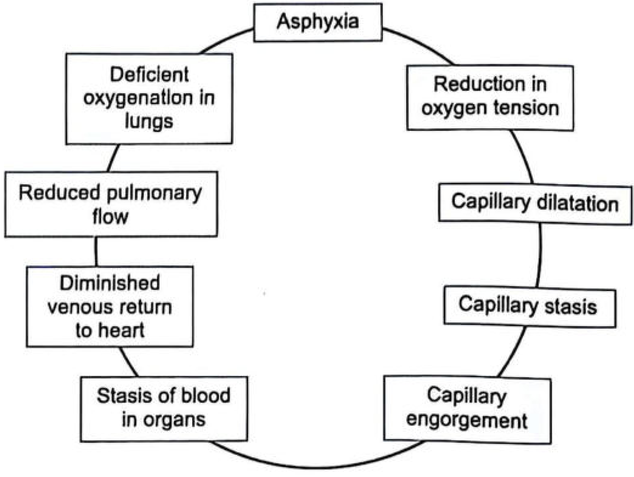
Fig. 6.1: Vicious cycle of asphyxia (The Essentials of FMT, 36th ed.)
Step-by-step:
- Asphyxia (any cause) --> Reduction in oxygen tension
- --> Capillary dilatation
- --> Capillary stasis
- --> Capillary engorgement (capillo-venous engorgement)
- --> Stasis of blood in organs (visceral congestion)
- --> Diminished venous return to heart (anoxia)
- --> Reduced pulmonary flow
- --> Deficient oxygenation in lungs
- --> Worsening asphyxia (cycle repeats and escalates to death)
In neck compression: jugular veins are occluded (preventing venous drainage from head) but carotid/vertebral arteries continue supplying blood --> raises venous pressure --> leads to petechiae + congestion.
Mechanical Asphyxia - 3 Core Mechanisms (Dikshit)
Mechanical asphyxia occurs by:
- Impeding access of air to lungs - airway obstruction (smothering, choking, drowning)
- Reducing blood supply to the head and neck - carotid/jugular compression (hanging, strangulation, throttling)
- Sudden cardiac arrest due to stimulation of carotid sinus - triggering vagal reflex mechanism (vagal inhibition)
Stages/Symptoms of Mechanical Asphyxia (3 Stages)
Stage 1 - Dyspnea (Stimulation Phase)
- Excess CO2 in blood stimulates the respiratory centre
- Respiratory movements increase in rate and amplitude
- Blood pressure rises, pulse rate increases
- Slight cyanosis appears
Stage 2 - Convulsions (Excitation Phase)
- Effort to breathe is mostly expiratory
- Face is deeply congested, blood pressure still elevated
- Neck veins become swollen
- Convulsions occur
- Victim becomes insensible; reflexes are abolished
Stage 3 - Exhaustion (Terminal Phase)
- Respiratory centre is paralyzed
- Muscles become flaccid; complete insensibility
- Reflexes lost, pupils widely dilated
- Breathing becomes gasping and inspiratory with long intervals
- Blood pressure falls; respiration ceases --> death
- Heart may continue to beat for some minutes after respiration has ceased
All three stages together last approximately 3-5 minutes in complete mechanical asphyxia.
Classical Signs of Asphyxia (Post-mortem)
All three form the classic triad - caused by raised venous pressure:
1. Cyanosis
- Requires at least 5 g% of reduced hemoglobin to become visible
- In neck constriction: venous blood pools, blood becomes bluer as it accumulates
- Significant only if seen within a few hours of death (after 24 hrs, may be purely post-mortem change)
2. Congestion (Capillo-venous Congestion)
- Due to hypoxia-induced capillary dilatation --> stasis in dilated capillaries and venules
- Leads to systemic and pulmonary congestion, and right heart dilatation
- Blood fluid state is maintained by fibrinolysin released from endothelium (defense mechanism against clotting in stagnant blood)
3. Petechial Hemorrhages (Most Specific Cardinal Sign)
- Caused by raised venous pressure --> overdistension and rupture of venules
- NOT due to hypoxia of vessel walls
- Minimum 15-30 seconds required to produce congestion and petechiae
- Size: 0.1 to 2 mm (larger = ecchymoses)
- Seen in: skin, sclerae, conjunctivae, eyelid surfaces, mucosal surfaces of mouth, serous membranes (subpleural, subepicardial), and brain substance
- Tardieu spots = subpleural and subepicardial petechiae
Other PM findings:
- Lungs heavier (450-500 g or more) = heart continued beating after respiration stopped
- Histology: Disruption of alveolar septa, intra-alveolar hemorrhage/oedema, brick-red discoloration of cortical nerve cells, vacuolar degeneration of Purkinje cells (cerebellum) and liver cells (prolonged suffocation)
Asphyxial Stigmata - Important Exam Point
The triad of (1) cyanosis, (2) petechiae, (3) visceral congestion are:
- Consistent with asphyxia but NOT diagnostic of it alone
- They are non-specific and can appear in deaths from other causes
- To confirm mechanical asphyxia, reliable local indications of fatal obstructing trauma must be demonstrated
Sources:
- The Essentials of Forensic Medicine and Toxicology, 36th ed. (2026), Chapter 6 - Asphyxia
- P.C. Dikshit Textbook of Forensic Medicine and Toxicology, Chapter on Mechanical Asphyxia & Classical Signs of Asphyxia
This is a shared conversation. Sign in to Orris to start your own chat.