
Diabetes in pregnancy
"diabetes mellitus" AND "pregnancy" AND "gestational"
gestational diabetes mellitus pregnancy complications management
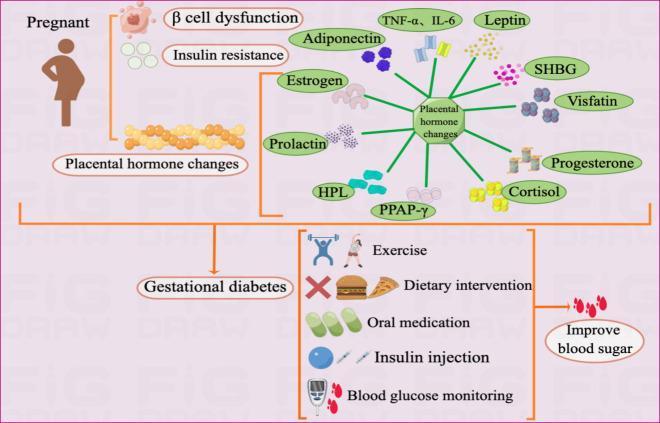
Educational infographic illustrating the pathogenesis and management of Gestational Diabetes Mellitus (GDM). The diagram is divided into sections showing contributing factors, hormonal changes, and therapeutic interventions. Pathogenesis factors listed include ̢ cell dysfunction, insulin resistance, and placental hormone changes. A central hub detailing placental dysfunction connects to various hormonal and inflammatory mediators such as TNF-̑, IL-6, Leptin, SHBG, Visfatin, Progesterone, Cortisol, PPAR-̣, HPL, Prolactin, Estrogen, and Adiponectin. The lower section of the diagram outlines management strategies categorized into non-pharmacological interventions (exercise, dietary intervention, and self-monitoring of blood glucose) and pharmacological treatments (oral medication and insulin injection). The visual workflow culminates in the educational goal of improving blood sugar levels. This schematic serves as a comprehensive overview of endocrine dysfunction during pregnancy and the multidisciplinary approach required for GDM glycemic control.
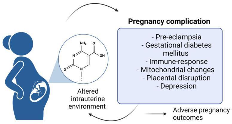
A pathophysiology diagram illustrating the relationship between molecular changes in gestation and clinical complications. On the left, a silhouette of a pregnant woman contains a circular inset magnification of the 'Altered intrauterine environment.' This inset displays a chemical structure (cytosine-like pyrimidine ring with nitrogen, oxygen, and hydrogen atoms), suggesting epigenetic or metabolic modifications at the molecular level. A bidirectional arrow system connects this environment to a primary box titled 'Pregnancy complication.' The list of complications includes pre-eclampsia, gestational diabetes mellitus, immune-response alterations, mitochondrial changes, placental disruption, and depression. A final arrow leads from these complications toward 'Adverse pregnancy outcomes,' which then feeds back into the intrauterine environment loop. The diagram serves as an educational summary of how maternal-fetal health is influenced by biochemical signaling pathways and physiological stressors, highlighting the cyclic nature of obstetric pathology.
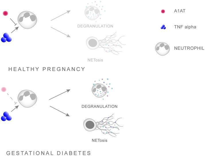
This pathophysiology diagram illustrates the regulatory role of Alpha-1 Antitrypsin (A1AT) in neutrophil priming during healthy pregnancy versus Gestational Diabetes Mellitus (GDM). The schematic identifies three primary components: A1AT (red circle), TNF-alpha (blue spheres), and Neutrophils (gray cells with multi-lobed nuclei). In a 'Healthy Pregnancy' scenario, A1AT acts as an inhibitory mediator, blocking the interaction between TNF-alpha and the neutrophil, thereby limiting inflammatory activation. In contrast, the 'Gestational Diabetes' section depicts a reduced or weakened presence of A1AT (represented by a faded red circle and dashed inhibitory line), allowing TNF-alpha to unimpededly prime the neutrophil. Both pathways culminate in two downstream effector processes: 'DEGRANULATION,' shown as the release of cytoplasmic granules, and 'NETosis,' visualized as the ejection of extracellular chromatin traps (stringy extensions). The diagram highlights how a shift in the A1AT/TNF-alpha balance in GDM promotes hyper-inflammation through enhanced neutrophil activation, making it a critical educational resource for understanding inflammatory signaling and obstetric complications.
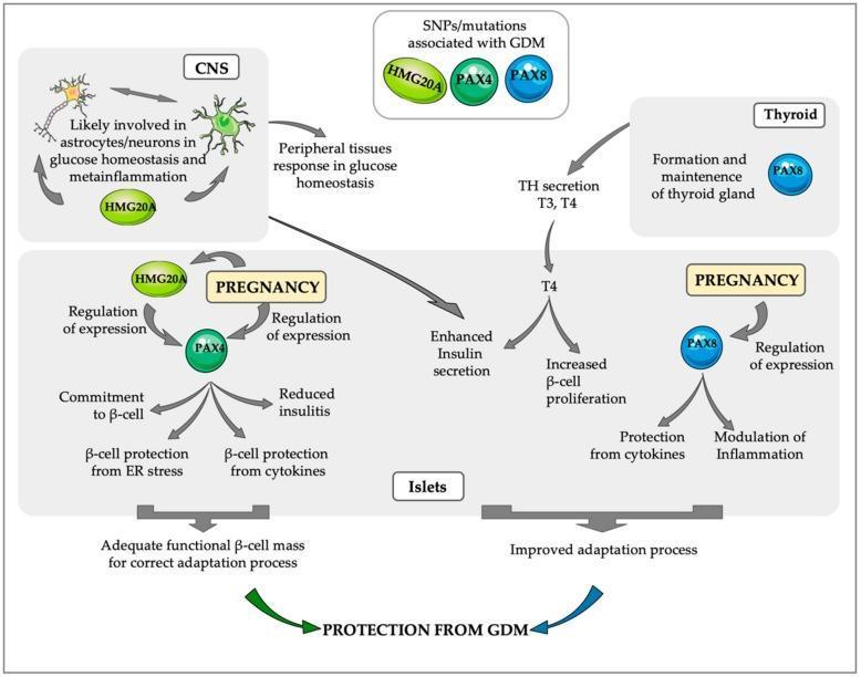
This pathophysiology diagram illustrates the systemic and molecular pathways involving PAX4, HMG20A, and PAX8 in maintaining glucose homeostasis during pregnancy and providing protection against gestational diabetes mellitus (GDM). The diagram is divided into anatomical compartments: Central Nervous System (CNS), Thyroid, and Pancreatic Islets. In the CNS, HMG20A is depicted modulating astrocytes and neurons to regulate metainflammation and peripheral glucose responses. In the Thyroid, PAX8 is essential for gland maintenance and secretion of thyroid hormones (T3, T4), which enhance insulin secretion and beta-cell proliferation. Within the Islets during pregnancy, a complex regulatory network is shown: HMG20A and PAX4 co-regulate expression, leading to beta-cell commitment, protection from endoplasmic reticulum (ER) stress/cytokines, and reduced insulitis. Simultaneously, PAX8 expression in islets modulates inflammation and offers cytokine protection. These pathways converge to ensure an adequate functional beta-cell mass and an improved adaptation process, ultimately conferring 'Protection from GDM'. The diagram also highlights that SNPs or mutations in HMG20A, PAX4, and PAX8 are clinically associated with GDM development.
Diabetes in Pregnancy
Overview
- Pregestational diabetes mellitus (pgDM) - pre-existing Type 1 (T1DM) or Type 2 (T2DM) diabetes before conception
- Gestational diabetes mellitus (GDM) - impaired glucose tolerance that first develops or is recognized during pregnancy
- Robbins & Cotran Pathologic Basis of Disease, p. 2123
Why Pregnancy Is Diabetogenic
- Human placental lactogen (HPL)
- Progesterone and cortisol
- TNF-alpha, IL-6, leptin, visfatin
- Reduced adiponectin
- Creasy & Resnik's Maternal-Fetal Medicine, p. 1429
Gestational Diabetes (GDM)
Risk Factors
- Previous GDM
- Obesity / BMI >30
- Family history of T2DM
- Prior macrosomic infant (birth weight >4 kg)
- Polycystic ovary syndrome (PCOS)
- Advanced maternal age
- Certain ethnicities (South Asian, East Asian, Hispanic, Indigenous)
Screening and Diagnosis
| Step | Test | Cutoff |
|---|---|---|
| Step 1 | 50 g oral glucose, 1-hour challenge (no fasting required) | ≥ 140 mg/dL = abnormal |
| Step 2 | 100 g OGTT, 3-hour (fasting required), if Step 1 abnormal | See below |
| Timepoint | NDDG Criteria | Carpenter-Coustan Criteria |
|---|---|---|
| Fasting | 105 mg/dL | 95 mg/dL |
| 1 hour | 190 mg/dL | 180 mg/dL |
| 2 hour | 165 mg/dL | 155 mg/dL |
| 3 hour | 145 mg/dL | 140 mg/dL |
- 75 g OGTT at 24-28 weeks; GDM if fasting ≥ 92, 1-hour ≥ 180, or 2-hour ≥ 153 mg/dL (any single value sufficient)
- Textbook of Family Medicine 9e, p. 491; Creasy & Resnik's, Goldman-Cecil Medicine
Pregestational Diabetes - Preconception Care
-
Target HbA1c < 6% before conception to minimize fetal congenital malformation risk (organogenesis occurs in the first 8 weeks, often before the woman presents for prenatal care)
-
Assess for end-organ complications: retinopathy, nephropathy, neuropathy, cardiovascular disease
-
Insulin is the preferred agent for pgDM in pregnancy - it does not cross the placenta, has a strong safety profile, and lower treatment failure rates vs. oral agents
-
Low-dose aspirin (60-162 mg/day) should be initiated before 16 weeks in women with T1DM or T2DM to reduce preeclampsia risk by approximately 50% and IUGR by 20% (SMFM/ACOG/ADA recommendation)
-
Harrison's Principles of Internal Medicine 22E, p. 394
Glycemic Targets During Pregnancy
| Parameter | Target |
|---|---|
| Fasting blood glucose | < 95 mg/dL (< 5.3 mmol/L) |
| 1-hour postprandial | < 140 mg/dL (< 7.8 mmol/L) |
| 2-hour postprandial | < 120 mg/dL (< 6.7 mmol/L) |
- 1st trimester: 0.7-0.8 units/kg/day
- 2nd trimester: 0.8-1.0 units/kg/day
- 3rd trimester: 0.9-1.2 units/kg/day (insulin sensitivity decreases as placental hormones peak)
- Harrison's, p. 396
Management of GDM
- Medical nutrition therapy: 30-35 kcal/kg lean body weight/day, carbohydrate restriction
- Regular moderate exercise (e.g., walking)
- SMBG: fasting + postprandial glucose
- Fetal growth monitoring by ultrasound every 4-6 weeks
- Effective in the majority of women
-
Insulin is first-line (preferred)
-
Metformin - alternative; associated with lower birth weight, gestational weight gain, and preeclampsia rates vs. glyburide/insulin. However, metformin crosses the placenta and long-term neonatal metabolic effects (including higher childhood adiposity) remain uncertain
-
Glyburide - used less frequently; associated with higher rates of neonatal hypoglycemia and macrosomia vs. insulin
-
Harrison's, p. 406-408; Textbook of Family Medicine, p. 491
Complications
Maternal Complications
| Complication | Notes |
|---|---|
| Preeclampsia | 2-4x more common in pgDM; >1/3 of women diabetic for >20 years develop it |
| Hypertension | OR = 14.2 in pregestational DM |
| Cesarean delivery | OR = 11.3 |
| Preterm birth | OR = 4.4 |
| Diabetic ketoacidosis (DKA) | Occurs at lower glucose thresholds than non-pregnant state (<1% incidence now); fetal mortality from DKA has dropped from 35% to <5% with modern management |
| Retinopathy progression | Risk proportional to duration of DM and prepregnancy HbA1c; worsened by rapid improvement in glycemic control; CSII (insulin pump) reduces risk |
| Nephropathy | Proteinuria increases during pregnancy but reverts postpartum; pregnancy does not appear to accelerate progression to ESRD |
| Heart disease | Rare but maternal mortality 50%+ if significant atherosclerotic CAD |
Fetal and Neonatal Complications
| Complication | Notes |
|---|---|
| Macrosomia | Most common in GDM; birth weight >4 kg; increases risk of shoulder dystocia, birth trauma |
| Congenital malformations | Risk doubled or more (cardiac, neural tube, caudal regression syndrome); related to glycemic control during organogenesis (first 8 weeks) |
| Spontaneous abortion / stillbirth | Increased in poorly controlled pgDM |
| Neonatal hypoglycemia | From fetal hyperinsulinemia in response to maternal hyperglycemia |
| Polycythemia, hypocalcemia, hyperbilirubinemia | Metabolic complications of the neonate |
| Respiratory distress syndrome | Hyperinsulinemia delays fetal lung maturation |
| Long-term risks | Child has increased risk of obesity and T2DM ("Barker hypothesis" / in utero programming) |
- Creasy & Resnik's, pp. 1431-1432; Goldman-Cecil Medicine, p. 2591
Obstetric Management
- Antenatal fetal surveillance begins in the third trimester: non-stress tests (NST), biophysical profiles
- Delivery timing:
- Well-controlled GDM on diet alone: delivery by 40-41 weeks
- GDM on insulin: induction at 39-40 weeks
- pgDM: induction at 37-39 weeks is common
- Cesarean section considered if estimated fetal weight ≥ 4500 g to prevent shoulder dystocia
- Intrapartum glucose control: target 70-120 mg/dL to prevent neonatal hypoglycemia from fetal hyperinsulinemia
Postpartum Follow-Up
-
GDM resolves in most women after delivery
-
All women with GDM should have a 75 g, 2-hour OGTT at 4-12 weeks postpartum to screen for overt DM or impaired glucose tolerance
-
7-10 fold increased risk of developing T2DM in later life; 10% risk within 5 years of delivery
-
30-60% lifetime risk of T2DM after GDM (O'Sullivan data)
-
Lifestyle interventions (weight loss, exercise) and metformin reduce the risk of progression to T2DM
-
Annual screening recommended thereafter
-
Harrison's, p. 410; Textbook of Family Medicine, p. 491; Creasy & Resnik's
Key Evidence (Recent)
Summary: GDM vs. pgDM at a Glance
| Feature | GDM | pgDM (T1/T2DM) |
|---|---|---|
| Onset | During pregnancy | Pre-existing |
| Key risk | Macrosomia, T2DM later | Congenital anomalies, macrosomia, preeclampsia |
| Management | Diet → insulin/metformin | Insulin (preferred); CGM in T1DM |
| HbA1c target pre-pregnancy | N/A | < 6% |
| Postpartum | Resolves; screen at 4-12 weeks | Continues |
Sources: Creasy & Resnik's Maternal-Fetal Medicine, Harrison's Principles of Internal Medicine 22E, Robbins & Cotran Pathologic Basis of Disease, Goldman-Cecil Medicine, Textbook of Family Medicine 9e