

What can be the diagnosis
dermatofibroma skin lesion hyperpigmented firm nodule abdomen
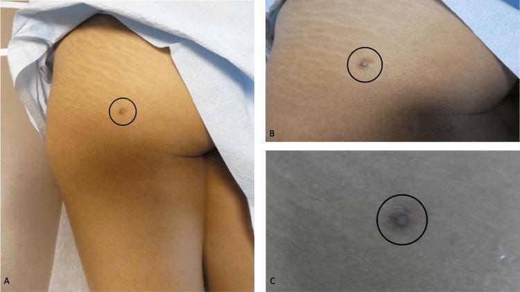
Clinical photograph series (distant, medium, and close-up views) depicting a dermatofibroma on the left buttock of a 42-year-old female. The lesion consists of a central, well-circumscribed, brown-colored dermal nodule measuring approximately 1.2 x 1.2 cm. This nodule is protuberant and elevated above the skin surface. Surrounding the central nodule is a larger, ill-defined, hyperpigmented patch measuring approximately 3 x 2 cm, which displays irregular borders and a diffuse, tan-to-brown discoloration compared to the adjacent normal skin. The surrounding skin exhibits normal anatomical features, including visible striae distensae in the superior and lateral aspects. The central nodule is marked with a black circle across all three panels (A, B, and C) for localization. This visual presentation is characteristic of a benign fibrous histiocytoma (dermatofibroma), highlighting the common morphological features of firm, raised dermal nodules with associated peripheral hyperpigmentation, frequently found on the extremities or trunk.
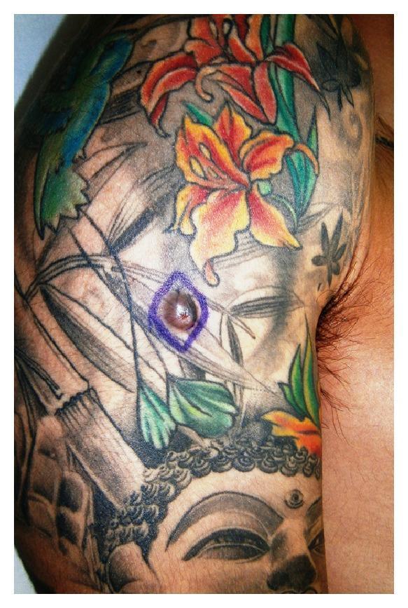
This clinical photograph displays a 10 mm dermatofibroma appearing as a firm, raised, light-red nodular lesion on the right arm of a 21-year-old male. The nodule is specifically localized within the black-pigmented area of an extensive multi-colored tattoo featuring floral, bamboo, and religious iconography. Visually, the lesion is circular with a smooth surface texture and lacks a central punctum or visible ulceration. The surrounding skin shows no acute inflammatory changes or distinct abnormalities apart from the tattoo ink. This image illustrates a potential dermatologic complication arising within tattooed skin, specifically a benign fibrohistiocytic proliferation. Clinically, such lesions are relevant for differential diagnosis against other tattoo-related reactions or malignancies, requiring histopathological confirmation for definitive diagnosis of dermatofibroma.
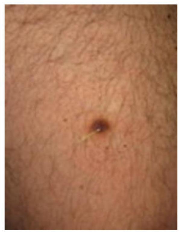
Clinical photograph of a solitary dermatofibroma (DF) presenting as a dark brown to black, well-circumscribed, firm nodule on the skin. A key diagnostic feature illustrated is the 'Meyerson phenomenon', characterized by an inflammatory, eczematous-like reaction or halo surrounding the fibrous lesion, often visible as a lighter or erythematous ring. The primary lesion is centrally located within the image and features a terminal hair protruding from the nodule, a common finding in benign skin lesions. The surrounding skin exhibits a light brown tone with visible hair follicles and several smaller, non-specific melanocytic nevi scattered in the periphery. This image serves as an educational example of clinical variations of dermatofibroma and the associated Meyerson phenomenon, relevant for dermatology residents and medical students studying pigmented skin tumors.
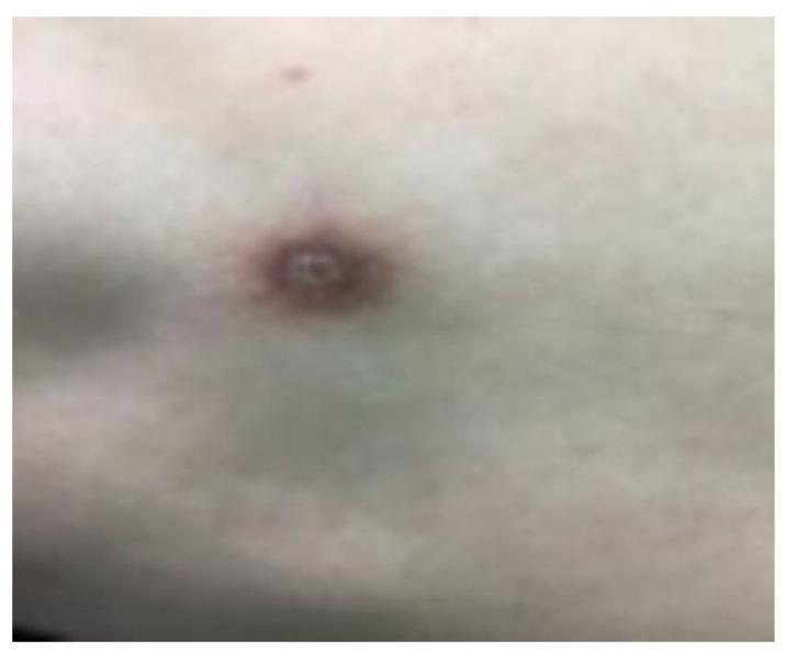
This clinical photograph displays a solitary skin lesion consistent with a dermatofibroma (DF) on a patient's skin. The lesion is a well-defined, circular, hyperkeratotic nodule measuring approximately 1 cm in diameter. It exhibits a distinct yellow-brown surface coloration and is clearly demarcated from the surrounding integument. The visual presentation highlights the characteristic firm, slow-growing nature of these benign fibrous histiocytomas, which frequently present as solitary papules or nodules. This image serves as an educational reference for dermatology students and clinicians to recognize the common clinical morphology of dermatofibromas, distinguishing them from other pigmented or nodular skin conditions through their specific texture, color, and border definition.
Image Findings
- A solitary, oval-to-linear, slightly depressed macule/flat plaque
- Dark brown to grayish-brown hyperpigmentation
- Faint whitish/lighter central area
- ~5-10 mm in size
- Moderately defined borders blending into surrounding normal skin
- Surrounding skin is unremarkable
Most Likely Diagnosis: Dermatofibroma (Benign Fibrous Histiocytoma)
| Feature | This Lesion | Dermatofibroma |
|---|---|---|
| Color | Dark brown/grey-brown | Reddish-brown, sometimes yellowish hue |
| Shape | Oval, slightly depressed | Round/ovoid papule, may be depressed |
| Size | ~5-10 mm | Typically 4-20 mm |
| Location | Trunk/flank | Lower extremities, sides of trunk (classic) |
| Surrounding skin | Normal | Normal |
Differential Diagnoses to Consider
- Post-inflammatory hyperpigmentation (PIH) - flat, macular, follows a preceding inflammatory or traumatic event; no induration
- Melanocytic nevus (mole) - usually more uniformly pigmented, not depressed
- Scar/keloid (hypertrophic) - history of prior trauma/wound at site; scars tend to be linear
- Blue nevus - blue-grey, more deeply pigmented, dome-shaped
- Dermatofibrosarcoma protuberans (DFSP) - if the lesion is >2-3 cm or progressively enlarging, malignancy must be excluded by biopsy
Key Clinical Points (from Andrews' Diseases of the Skin)
- Dermatofibromas are common, benign lesions mostly in middle-aged adults
- Thought to be triggered by prior skin injury (insect bites, blunt trauma)
- Principal locations: lower extremities, above the elbows, or sides of the trunk (matches this case)
- Usually asymptomatic - no treatment needed
- Multiple dermatofibromas can be associated with: SLE, immunosuppressive therapy, HIV, chronic myelogenous leukemia
- Biopsy is indicated if the lesion grows beyond 2-3 cm or changes in appearance
Recommendation
This is a clinical assessment only. A definitive diagnosis requires physical examination with palpation (checking for the dimple sign and firmness), dermoscopy, and if uncertain, a skin biopsy. Please consult a dermatologist for formal evaluation.
It was red before then it became black as the photo and it pains me on touchits now for 3/4 months
nodular melanoma black pigmented painful skin lesion trunk
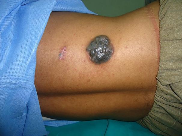
Clinical photograph of a 33-year-old male's trunk displaying a large, exophytic nodular melanoma located on the right dorsal aspect. The primary lesion is a heavily pigmented, multi-lobulated mass that appears charcoal-black to dark gray with a distinctive 'bunch of grapes' morphology. Its surface is irregular and demonstrates a glistening sheen. Adjacent to this primary mass is a smaller, horizontal, erythematous linear scar, possibly indicating a prior biopsy or minor surgical site. The background skin shows mild perifollicular erythema and a few scattered benign-appearing nevi. This image serves as a textbook example of advanced nodular melanoma, emphasizing the importance of identifying rapidly growing, pigmented, and elevated skin lesions in dermatological oncology. The presentation is contextually linked to high-stage (III or IV) disease management in adult patients.
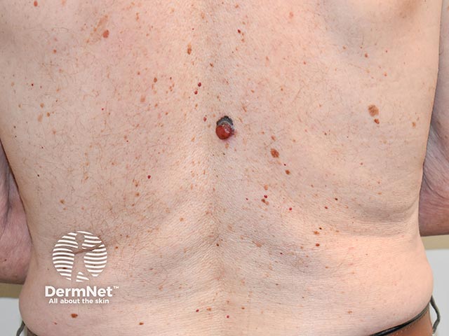
A dorsal trunk clinical skin photograph showing a nodular melanoma arising within a pre-existing superficial spreading melanoma (SSM) on the back. Modality and technique: clinical photography; non-contact, color image captured with standard white balance and diffuse daylight illumination; focal framing centers the lesion within an approximately 5–9 mm pigmented nodule embedded in a larger flat, irregular pigmented patch. The primary subject is the skin lesion on the back, with dorsal trunk anatomy and posterior view. Anatomical context: integumentary system; skin of the back (thoracic/dorsal region); epidermal/dermal skin structures; laterality not specified; is in the midline dorsum area. Lesion characteristics: solitary, dark brown to black pigmented lesion with a distinct nodular component; raised, rounded surface within a broader flat lesion; color variegation and irregular borders are present; surface may show minimal scaling or crusting consistent with malignant transformation. Clinical significance: appearance is highly suspicious for melanoma with nodular growth phase within a superficial spreading melanoma; requires prompt biopsy and histopathology; prognosis can be influenced by vertical growth and depth of invasion. Differential diagnoses include pigmented nevus with nodular change, blue nevus, seborrheic keratosis; typical educational use: melanoma recognition, lesion evolution, and clinical management discussion.
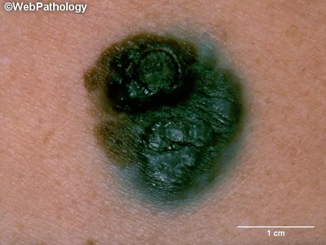
Clinical photography of a cutaneous pigmented lesion consistent with superficial spreading melanoma in a vertical growth phase. The image captures a dark, irregular, macro-nodular area on the skin surface with two elevated blue-black nodules surrounded by a blue-brown macular component. The lesion demonstrates classic melanoma hallmarks: asymmetry, irregular borders with indentations, variable pigmentation from tan to black, and surface elevation with nodularity indicating invasion. The surrounding skin shows a slightly lighter, mottled halo reflecting partial regression or desmoplastic reaction. The nodules are firm to palpation, suggesting vertical growth depth into the dermis, which portends poorer prognosis compared with radial growth alone. The macular component likely corresponds to in-situ or superficial spreading melanoma areas with pigment production by malignant melanocytes. The pigmentation range includes blue-black melanin-rich regions and lighter tan or pink foci often associated with regression or reduced melanin production. On clinical evaluation, the combination of nodular invasion and irregular, scalloped margins supports a diagnosis of malignant melanoma rather than a benign nevus or scar. This photograph is valuable for teaching melanoma morphology, recognizing vertical growth phase, and supporting differential diagnosis including pigmented nevus, seborrheic keratosis, and amelanotic melanoma. Practical utility includes dermoscopic correlation, lesion monitoring, and surgical planning today.
pyogenic granuloma black dark evolved red bleed trunk
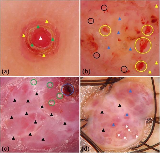
Dermoscopic images of pyogenic granuloma (PG) presented in a four-panel figure (a-d) demonstrating characteristic vascular and architectural features. Panel (a) shows a central red homogeneous area with surrounding yellow scabs and a peripheral white collar pattern. Panel (b) reveals a mixed red-white homogeneous background with distinct vascular structures (black circles), areas of hemorrhage (yellow circles), and a white collarette. Panel (c) highlights 'white rail' lines—whitish streaks intersecting the lesion—along with dark red scabs and superficial white scales. Panel (d) demonstrates a red-white background with prominent 'white rail' lines and serpentine (serpiginous) vessels marked by white arrows. These clinical photographs illustrate the diagnostic dermoscopic criteria for PG, including the white collarette, homogeneous red/white areas, and specific vascular morphologies like serpentine vessels, which are critical for differentiating PG from other vascular or melanocytic skin lesions in dermatology.
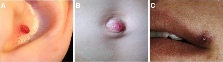
This composite clinical photograph displays three distinct presentations of pyogenic granuloma (lobular capillary hemangioma) in different anatomical locations. (A) Auricle: A small, well-defined, bright red oval nodule is situated within the ear's concha. (B) Umbilicus: A more prominent, dome-shaped red nodule protrudes from the umbilical base, demonstrating a fleshy, vascular appearance. (C) Lip: A smaller, slightly more violaceous or dark red papule is located on the cutaneous surface near the vermilion border. Across all sites, the lesions are characterized as solitary, exophytic, and hypervascular in nature with a typically smooth surface texture. The surrounding skin in each case shows no significant signs of inflammation or secondary changes. These benign vascular tumors commonly arise following minor trauma and are clinically significant for their tendency to bleed easily. This educational visual aids in identifying typical morphologies of pyogenic granulomas for dermatological and surgical classification.
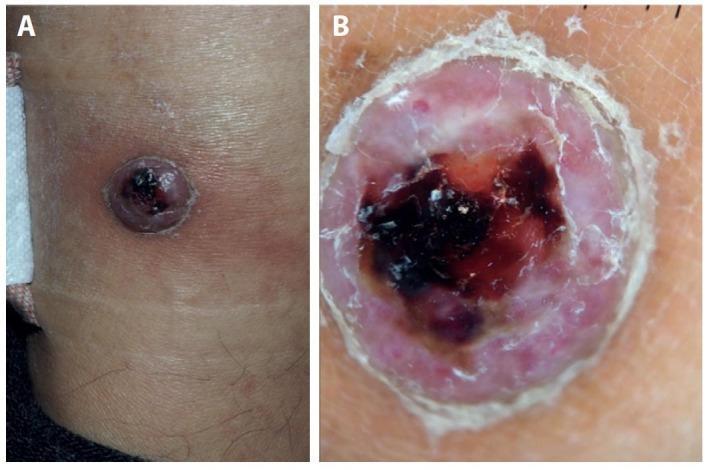
This composite image consists of a clinical photograph (A) and a dermoscopic image (B) of a solitary skin lesion on the ankle, demonstrating clinical features that mimic pyogenic granuloma but histopathologically confirmed as myopericytoma. Panel A shows a well-circumscribed, raised, dome-shaped nodule with a multi-colored surface. The lesion exhibits a central dark, violaceous to black crust indicative of hemorrhage or necrosis, surrounded by an erythematous, fleshy peripheral zone. Panel B provides a dermoscopic view revealing a characteristic pattern of homogeneous white-red areas. The center contains dark red to black blood lakes. A prominent, peripheral whitish collarette of scales is visible at the edge of the lesion, which is a key diagnostic feature for vascular tumors like pyogenic granuloma. The surrounding skin shows mild peri-lesional erythema. This comparison illustrates the visual overlap between benign vascular proliferations and rare perivascular tumors like myopericytoma, emphasizing the importance of histopathological correlation for definitive diagnosis.
Updated Clinical Picture
| Feature | Significance |
|---|---|
| Started red | Vascular/inflammatory origin |
| Turned dark black over time | Thrombosis, old blood/hemosiderin, or malignant pigmentation |
| Painful on touch (tender) | Active inflammation, nerve involvement, or rapid growth |
| Duration 3-4 months | Subacute - not healing as expected |
| Location: trunk/flank | Common site for several conditions |
Revised Top Differential Diagnoses
1. 🔴 Thrombosed / Involuting Pyogenic Granuloma (Most likely)
- Common on trunk, especially near waistband friction areas
- Tenderness from vascular congestion and thrombosis
- Can persist for months without treatment
2. ⚠️ Nodular Melanoma (Must be excluded urgently)
- Can appear initially as a red/pink/amelanotic lesion that darkens over weeks
- Grows rapidly (3-4 months fits the aggressive timeline)
- Can be tender or painful
- Does not follow a benign course
- Trunk is a common location, especially in adults
Andrews' Diseases of the Skin notes: "On the sole of the foot or nail bed, [pyogenic granuloma] may be mistaken for a melanoma" - and this distinction is critical because treatment of a bleeding amelanotic melanoma as pyogenic granuloma is dangerous.
3. Thrombosed Hemangioma / Angiokeratoma
4. Inflamed / Irritated Dermatofibroma
5. Blue Rubber Bleb Nevus / Glomus Tumor
⚠️ RED FLAGS in This Lesion
- Color change (red → black) over months
- Tenderness / pain on palpation
- Persistence > 3 months without resolution
- Darkening rather than fading
- Dermoscopy of the lesion
- Possible excision biopsy - this is the only way to definitively rule out melanoma
Important disclaimer: This is not a substitute for in-person medical evaluation. A changing, painful, darkening lesion that has persisted for 3-4 months requires biopsy to exclude malignancy. Please see a dermatologist or surgeon as soon as possible - do not delay.
What can be the treatment of keloid
keloid scar treatment intralesional injection
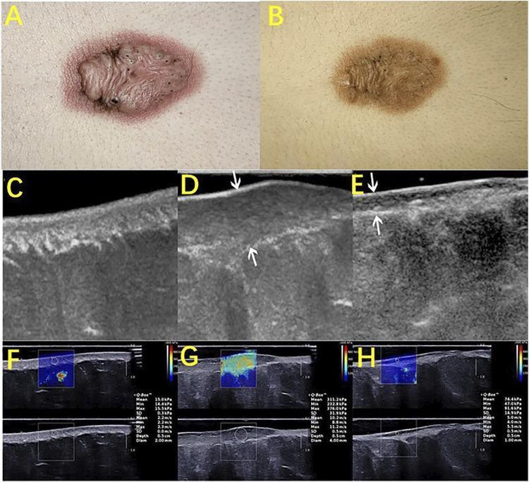
This composite figure illustrates the application of High-Frequency Ultrasound (HFUS) and Shear Wave Elastography (SWE) in monitoring keloid treatment efficacy. Panels A and B provide clinical photographs of a keloid before and after intralesional steroid injection, showing a reduction in erythema and lesion height. Panels C, D, and E display longitudinal B-mode ultrasound images comparing normal skin (C), a pretreated keloid (D), and a post-treated keloid (E). In panel D, the pretreated keloid appears as a thickened, hypoechoic elliptical area in the dermis. Panel E shows a significant reduction in scar thickness and a shift in echo texture toward normal dermal patterns following treatment. Panels F, G, and H present corresponding elastography maps. Normal skin (F) and post-treated tissue (H) exhibit relatively uniform blue-to-green color distributions, indicating lower stiffness. In contrast, the pretreated keloid (G) shows a heterogeneous color map with prominent yellow-red areas, signaling increased Young’s modulus and tissue stiffness. The educational focus is on using non-invasive imaging to objectively quantify changes in scar morphology and mechanical properties (elasticity) during therapy.
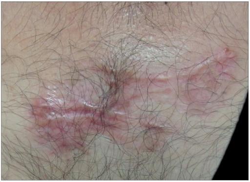
This clinical photograph displays a close-up view of a treated keloid on hair-bearing skin, one year after CO2 laser ablation and adjuvant intralesional triamcinolone acetonide therapy. The resulting scar is predominantly flat with a supple, parchment-like texture and visible skin lines, indicating successful resolution of the prior bulk. The scar exhibits a complex pigmentary pattern, including areas of hypopigmentation (depigmentation) and mild erythema with visible telangiectasias, suggesting chronic vascular changes or residual inflammation. The scar morphology is linear with minor branching patterns. Hair follicles are present in the immediate vicinity and partially overlap the edges of the scar tissue. This image serves as a clinical reference for the expected long-term outcome and potential local side effects, such as atrophy and vascular abnormalities, following combined surgical and corticosteroid treatment for hypertrophic or keloid scarring.
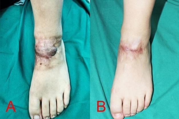
Clinical photograph comparison of a traumatic keloid scar on the dorsum of the left foot before (A) and after (B) intralesional diode laser treatment. Image A shows a highly elevated, irregularly shaped, hyperpigmented keloid mass crossing the anterior ankle joint. The lesion exhibits nodular texture with dark purplish and reddish-brown coloration, extending distally toward the midfoot. Image B demonstrates the clinical outcome after two sessions of 1470 nm bare-fiber diode laser therapy. There is a significant reduction in scar volume and height, with the mass appearing flattened. The hyperpigmentation has transitioned to a more uniform, light pinkish tone, indicating reduced vascularity and inflammation. The surrounding skin shows improved texture and blending, illustrating the efficacy of intralesional laser ablation in managing large keloid scars in areas of high tension like the ankle.
Treatment of Keloids
1. Intralesional Corticosteroid Injection (First-Line)
- Concentration: 40 mg/mL for initial treatment; reduce to 10-20 mg/mL as the lesion softens
- Technique: 30-gauge needle on a 1 mL tuberculin syringe, injected directly into the keloid
- Frequency: Every 6-8 weeks as needed
- Results: Reliably flattens the lesion and stops itching; does not narrow the scar
- Side effects: Hypopigmentation, skin atrophy (especially if steroid spreads via lymphatics)
2. Combination with 5-Fluorouracil (5-FU)
- Verapamil (calcium channel blocker - decreases IL-6 and VEGF in keloid fibroblasts)
- Methotrexate
- Intralesional IFN alfa-2b (post-excision)
3. Surgical Excision
- Post-operative intralesional triamcinolone injections
- Radiation therapy (adjuvant irradiation within 24-48 hours of excision)
- Topical imiquimod
- Useful when narrowing the keloid is a priority (not achievable with injections alone)
4. Radiation Therapy
- Used adjuvantly after surgical excision - significantly reduces recurrence
- Especially useful for large, recurrent, or surgically inaccessible keloids
- Requires careful planning to avoid radiation to sensitive underlying structures
5. Cryotherapy (Cryosurgery)
- Techniques: contact, intralesional needle cryoprobe, or spray methods
- Has demonstrated efficacy, especially for smaller lesions
- Works in part by suppressing TGF-β1 expression (a key driver of keloid fibrosis)
- Treatment times may need to be longer (up to 60 seconds) due to the dense, fibrotic nature
6. Laser Therapy
| Laser Type | Mechanism |
|---|---|
| Pulsed dye laser (PDL) | Reduces TGF-β1 expression, flattens and softens |
| Nd:YAG laser (long-pulsed) | Often combined with triamcinolone |
| CO2 laser | Used for excision, especially ear keloids |
| Diode laser (intralesional) | Reduces scar volume significantly |
7. Silicone Gel / Silicone Sheeting
- Applied topically as sheets or gel over the keloid
- Reduces mast cell numbers in the lesion and decreases itching
- Evidence base is modest (a Cochrane review found generally poor quality evidence)
- Used mainly as an adjunct to prevent recurrence after other treatments
- Andrews' Diseases of the Skin notes: "Results with these modalities have been mixed"
8. Pressure Therapy
- Compression garments or pressure earrings (for earlobe keloids)
- Useful for prevention and as adjunct, particularly after burns
9. Special Case: Ear Lobe Keloids (Piercing-Related)
- Young/new keloid: Intralesional triamcinolone alone is often sufficient
- Old/mature keloid: Excision under lidocaine + triamcinolone, followed by injections at 2-week intervals; CO2 laser excision also effective
Treatment Algorithm Summary
Small/early keloid
    → Intralesional triamcinolone ± 5-FU (repeat q6-8 weeks)

Refractory or large keloid
    → Add verapamil / methotrexate
    → Consider laser (PDL or Nd:YAG)
    → Consider cryotherapy

Narrowing is required / large lesion
    → Surgical excision + adjuvant radiotherapy + triamcinolone injections

Prevention of recurrence
    → Silicone sheeting + pressure therapy
Key point: Keloids must be distinguished from hypertrophic scars before starting aggressive treatment (especially radiation). Hypertrophic scars often improve spontaneously over months - keloids do not. If in doubt, a biopsy confirms the diagnosis. - Andrews' Diseases of the Skin & Dermatology 5e