
Explain tonsillectomy in ent mbbs oriented dhingra textbook
tonsillectomy dissection technique anatomy diagram
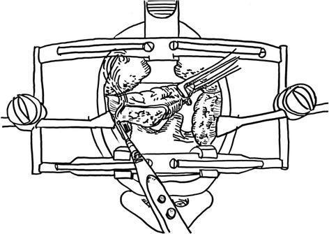
A surgical illustration depicting a tonsillectomy procedure using a monopolar electrocautery technique. The diagram shows an oral cavity held open by a McIvor or Dingman mouth gag retractor, providing optimal visualization of the oropharynx. A palatine tonsil is being retracted medially using curved Kelly clamps, which provide the necessary tension to expose the extracapsular dissection plane. The tip of a monopolar electrocautery pencil is positioned at the junction between the tonsillar capsule and the surrounding pharyngeal musculature, demonstrating the process of surgical dissection. This educational diagram illustrates the key principles of cold and hot dissection tonsillectomy, including proper instrumentation, the importance of medial traction, and the precise identification of the peritonsillar space to minimize bleeding and protect the tonsillar pillars.
This composite educational graphic illustrates the surgical technique of pneumatic dissection of the anterior hyaloid membrane (AHM) during ophthalmic surgery. It consists of three panels: a cross-sectional diagram, a top-down microscopic diagram, and a real-time intraoperative surgical photograph. The cross-sectional view details the ocular anatomy, including the cornea, anterior chamber, iris, crystalline lens, and the posterior segment layers (sclera and choroid/retina). A 30-gauge needle is shown entering through the pars plana, with the tip positioned posterior to the lens. The top-down diagram illustrates the needle orientation relative to the iris and limbus. The clinical photograph demonstrates the actual surgical field, showing an eye with multiple sclerotomy cannulas (trocars) in place and a needle-mounted syringe performing the air injection near the pupillary margin. The primary educational focus is on the safe entry and positioning of the needle tip behind the lens within the vitreous cavity to facilitate AHM dissection while avoiding iatrogenic damage to the lens or retina.
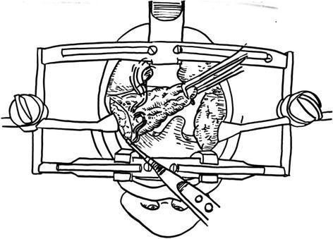
This surgical illustration depicts an intraoperative view of a tonsillectomy procedure, emphasizing the dissection technique. A specialized mouth retractor (such as a Dingman or McIvor system) is positioned to provide wide exposure of the oropharynx. The medial portion of the palatine tonsil is firmly grasped with right-angled Kelly clamps, applying medial traction to create necessary tension on the surgical plane. A monopolar electrocautery device is shown performing a precise incision along the base of the tonsil, targeting the extracapsular plane. The drawing illustrates the anatomical relationship between the tonsillar tissue, the anterior/posterior pillars, and the surrounding pharyngeal mucosa. This visual serves as an educational guide for Otorhinolaryngology trainees on the surgical steps of cold or hot dissection tonsillectomy, highlighting the importance of instrument placement, tissue traction, and the use of thermal energy for simultaneous cutting and hemostasis.
Tonsillectomy - ENT (Dhingra-Oriented MBBS Notes)
Note: The Dhingra ENT textbook is not in the library's digital collection, but the content below is compiled from Scott-Brown's Otorhinolaryngology, Cummings Otolaryngology, and Bailey & Love's Surgery - all covering the same core MBBS syllabus topics on tonsillectomy that Dhingra covers. The clinical facts, indications, technique, and complications are consistent across all standard ENT textbooks.
Definition
Indications
Absolute Indications
- Obstructive sleep apnoea (OSA) / chronic upper airway obstruction
- Cor pulmonale from chronic airway obstruction
- Suspected tonsillar malignancy (asymmetric enlargement, ulceration, fixity)
Relative Indications
- Recurrent acute tonsillitis - the Paradise criteria:
- 7 or more well-documented episodes in 1 year, OR
- 5 or more episodes per year for 2 consecutive years, OR
- 3 or more episodes per year for 3 consecutive years
- (Episodes must be disabling and prevent normal functioning)
- Chronic tonsillitis (persistent sore throat, halitosis, tonsillar debris)
- Peritonsillar abscess (quinsy) - especially recurrent
- Tonsillar asymmetry (to rule out malignancy)
- Tonsillitis causing febrile convulsions
- Diphtheria carriers unresponsive to antibiotics
- Systemic disease caused by beta-haemolytic Streptococcus (rheumatic fever, nephritis)
- Surgical access - to reach the parapharyngeal space or elongated styloid process
In children, adenotonsillectomy (combined removal of adenoids and tonsils) is the treatment of choice for obstructive sleep apnoea, with improvement in ~90% of cases.
Contraindications
- Acute tonsillitis (surgery deferred until infection subsides - ideally 3-4 weeks)
- Bleeding disorders (haemophilia, von Willebrand disease - must be corrected preoperatively)
- Severe systemic illness
- Cleft palate (tonsillectomy may worsen velopharyngeal insufficiency and cause hypernasal speech)
- Blood dyscrasias
Preoperative Assessment
- Exclude active infection - surgery only when tonsils are quiescent
- Assess for bleeding tendency (history and basic coagulation screen)
- Blood grouping and screening (cross-match for children < 15 kg)
- Consent must include discussion of risk of haemorrhage, pain, and referred otalgia
Technique
Position
- Patient supine under general anaesthesia
- Head extended (Rose position / Boyle-Davis mouth gag position)
- Boyle-Davis mouth gag opens the mouth and depresses the tongue, giving good surgical exposure
Dissection Tonsillectomy (Gold Standard - Cold Steel)
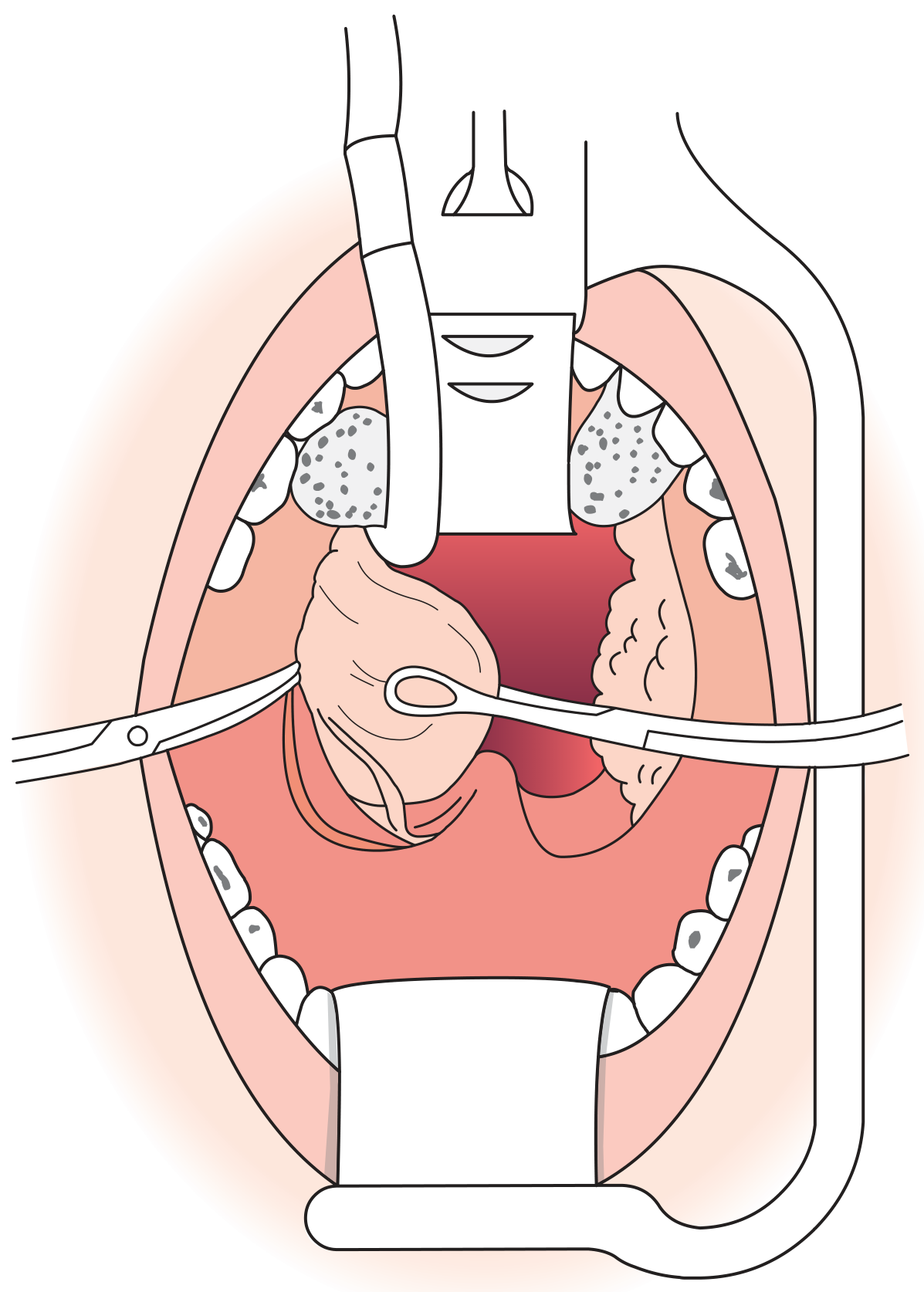
- Incision - Mucosa of the anterior faucial pillar is incised with a scalpel
- Capsule identification - The tonsil capsule is identified
- Blunt dissection - The tonsil is separated from its bed in the plane of loose areolar tissue between the capsule and the superior constrictor muscle, using a Negus dissector or gauze swab
- Inferior pedicle - Dissection proceeds superiorly to inferiorly until only a small inferior pedicle (attached to the lingual tonsil) remains
- Division - The pedicle is snared or divided with scissors
- Haemostasis - A tonsil swab is packed into the tonsillar fossa with pressure applied for several minutes; bleeding points are controlled with ligatures or bipolar diathermy
Guillotine Technique (historical)
- A specially designed guillotine amputates the tonsil
- Less commonly used today due to higher bleeding rates and incomplete removal
Other Techniques (with comparative haemorrhage data from the National Prospective Tonsillectomy Audit, n = 33,921):
| Technique | Primary Haemorrhage | Secondary Haemorrhage |
|---|---|---|
| Cold steel + ties/packs | 0.8% | 1.0% |
| Diathermy (monopolar/bipolar) | Higher than cold steel | Higher than cold steel |
| Coblation | Intermediate | Intermediate |
| Laser | Variable | Variable |
Postoperative Care
- Patient nursed in tonsillar position (semi-prone / lateral, head-down) immediately after surgery to prevent blood/secretions from entering the airway
- Regular monitoring: pulse, blood pressure, and observation for excessive swallowing (a sign of concealed haemorrhage)
- Encourage normal diet early (promotes healing of tonsillar fossa by mechanical debridement)
- Regular oral analgesics (NSAIDs + paracetamol)
- Discharge same day or next day
- Warn the patient about:
- Referred otalgia - pain in the ear due to referred pain via the glossopharyngeal nerve (Jacobson's nerve) - common, does not indicate ear pathology
- Secondary haemorrhage - can occur up to 10 days postoperatively (typically day 5-10 when the slough separates)
- White slough on tonsillar fossa is normal healing (not infection)
Complications
Haemorrhage (most important complication)
| Type | Timing | Cause | Management |
|---|---|---|---|
| Reactionary (primary) | First 24 hours | Slipped ligature, missed bleeder | Return to theatre under GA, suture/diathermy the bed |
| Secondary | Day 5-10 | Infection causing slough separation | IV antibiotics, remove clot, hydrogen peroxide gargles; may need GA for haemostasis |
- Secondary haemorrhage is life-threatening, especially in young children - never underestimate it
- In theatre: if a focal bleeder is found, ligate/diathermy; if generalised ooze, suture the tonsillar fossa with haemostatic gauze + bipolar diathermy
Other Complications
- Pain / otalgia (referred via glossopharyngeal nerve) - near-universal
- Infection - secondary tonsillitis/pharyngitis of the raw tonsillar bed
- Postoperative airway obstruction - especially in children with OSA (relative narrowing as oedema resolves can temporarily worsen before improving)
- Velopharyngeal insufficiency - rare; nasal regurgitation and hypernasal speech if too much soft palate tissue is removed; risk is higher in children with submucous cleft palate
- Nasopharyngeal stenosis - very rare; scarring between soft palate and posterior pharyngeal wall
- Dental/lip/tongue injury - from the mouth gag
- Temporomandibular joint injury - from prolonged mouth opening
- Taste disturbance - rare
Key Points for MBBS/MCQs
- Most common indication: Recurrent acute tonsillitis (using Paradise criteria)
- Absolute indication: OSA / suspected malignancy
- Standard technique: Dissection tonsillectomy under GA
- Most common complication: Haemorrhage
- Reactionary haemorrhage: Within 24 hours - return to theatre
- Secondary haemorrhage: Day 5-10 - usually infective
- Referred otalgia post-tonsillectomy: via glossopharyngeal nerve (not ear disease)
- Contraindication: Acute active tonsillitis, cleft palate, bleeding disorder
- Cold steel dissection has the lowest haemorrhage rate of all techniques
- Adenotonsillectomy is effective in ~90% of children with OSA