

Explanation please if something missing in this paragraph also you can add but explanation has to be good and understandable
B Lymphocytes - Full Explanation
1. Origin and Maturation
"These are formed and mature in the bone marrow."
2. Role in Specific (Adaptive) Immunity
"They confer upon us the most important aspect - the antibodies for our specific immunity."
What the paragraph doesn't mention: There are 5 classes of antibodies - IgM, IgG, IgA, IgE, and IgD - each with different roles. For example, IgM is the first antibody produced in any infection; IgG is the most abundant and crosses the placenta to protect the fetus; IgA is found in mucus and saliva; IgE is involved in allergic responses.
3. B Cell Activation - Two Pathways
"B Cells activation into effector cells called Plasma cell occurs either T-dependent mechanism or through T-Independent mechanisms."
A) T-Dependent (TD) Mechanism
"The former occurs when an activated T_H cell displays peptide antigen along with its MHC-II. Thus activated T_H cell releases the alert signals to naïve B-cell..."
-
Antigen Presentation: A B cell binds its specific antigen using its BCR and pulls the antigen inside. It chops the antigen into peptide fragments and displays them on MHC Class II molecules on its surface.
-
T Helper (T_H) Cell Recognition: A CD4+ T Helper cell (specifically a T Follicular Helper / Tfh cell) recognizes this peptide-MHC II complex using its T Cell Receptor (TCR).
-
Activation Signals: The activated T_H cell then delivers two critical activation signals to the B cell:
- Contact signal: The T_H cell expresses a surface protein called CD40L (CD40 Ligand), which binds to CD40 on the B cell surface - this is a direct contact "hand-shake."
- Chemical signals (cytokines): The T_H cell releases cytokines (chemical messengers like IL-4, IL-5, IL-21) that act as "alert signals," telling the B cell to grow, divide, and produce antibodies.
-
The B cell then grows in size, proliferates (divides rapidly), and starts actively producing and releasing antibodies.
Key feature of T-Dependent responses:
- Produces high-affinity antibodies
- Allows isotype/class switching (the antibody class can switch from IgM to IgG, IgA, or IgE)
- Generates memory B cells (critical for long-term immunity and vaccination)
- Undergoes affinity maturation - the antibodies produced get progressively "better" at binding their antigen over time
B) T-Independent (TI) Mechanism
Limitations of T-Independent responses (missing from the paragraph):
- Produces lower affinity antibodies (mainly IgM)
- No memory B cells are generated, so it cannot be boosted by vaccination
- Poorly developed in children under 2 years of age - this is why polysaccharide vaccines (like early pneumococcal vaccines) did not work in infants. Modern vaccines conjugate the polysaccharide to a protein carrier to convert the response to T-dependent and make it effective in young children.
4. Plasma Cells
"Such antibody secreting B-cells are termed as Plasma cells."
5. B Memory Cells (B_M)
"Some of the B-cells meanwhile turn into B Memory cells or (B_M). The latter, records all the data of the antigen for controlling future reinfection. They do play very important role as far as the immunization is concerned."
- Persist for years or even a lifetime in the body
- Have a lower activation threshold - they respond faster and more powerfully upon re-exposure to the same antigen
- Upon re-exposure, they rapidly expand and differentiate into new plasma cells, generating a faster and stronger antibody response - this is called the Secondary (Anamnestic) Immune Response
What the paragraph doesn't mention: Memory B cells also undergo somatic hypermutation in germinal centers - microscopic structures within lymph nodes and the spleen where B cells refine and improve their antibodies. This process, combined with clonal selection (only the B cells with the best-fitting antibodies survive), is why secondary responses produce better antibodies than primary responses.
Summary Table
| Feature | T-Dependent | T-Independent |
|---|---|---|
| Antigen type | Proteins, haptens | Polysaccharides, lipids |
| T_H cell required? | Yes | No |
| Memory cells? | Yes | No |
| Antibody class | IgG, IgA, IgE (class switching) | Mainly IgM |
| Antibody quality | High affinity (affinity maturation) | Lower affinity |
| Vaccination boost? | Yes | No |
| Effective in infants? | Yes | No (under 2 years) |
The Diagram at the Bottom (Cytotoxic T Cell Killing)
- Cytotoxic T cell binds to infected cell via the self-nonself complex (TCR recognizes foreign antigen displayed on MHC-I of the infected cell)
- Perforin makes holes in the infected cell's membrane - perforin is a protein released by the CTL that polymerizes and punches pores into the target cell membrane
- Infected cell is destroyed - through the pores, enzymes called granzymes enter and trigger apoptosis (programmed cell death) inside the infected cell, destroying it along with any viruses replicating within it
Create a visual flowchart showing B cell differentiation: antigen → activation → plasma cells vs memory cells
A detailed educational flowchart showing B cell differentiation pathway. Clean white background, scientific textbook style. Top: A blue circular "Naive B Cell" with BCR receptors shown on surface. Arrow down labeled "Antigen binds to BCR (B Cell Receptor)" Next box: "Antigen Internalized & Presented on MHC-II" Then the pathway splits into TWO branches side by side: LEFT BRANCH (labeled "T-Dependent Pathway" in green): - Box: "T Helper Cell (CD4+) recognizes peptide-MHC II complex" - Arrow: "CD40L binds CD40 + Cytokines released (IL-4, IL-5, IL-21)" - Box: "B Cell Activation & Proliferation (Clonal Expansion)" - Then splits into two sub-branches: LEFT: Arrow to "PLASMA CELLS" (large oval, purple color) with description: "Short-lived antibody factories | Secrete 1000s of antibodies/sec | High RER content | IgG, IgA, IgE (class switching)" RIGHT: Arrow to "B MEMORY CELLS (BM)" (star-shaped, golden yellow) with description: "Long-lived | Lower activation threshold | Rapid secondary response | Basis of vaccination" RIGHT BRANCH (labeled "T-Independent Pathway" in orange): - Box: "Repetitive antigen (polysaccharides) cross-links many BCRs" - Arrow down to "Plasma Cells only (mainly IgM)" (smaller purple oval) - Red X mark labeled "No Memory Cells formed" At the bottom: A comparison box with two columns: T-Dependent: High affinity | Class switching | Memory cells | Boosted by vaccine T-Independent: Low affinity | Mainly IgM | No memory | Not boostable Use clear arrows, color coding (blue=B cells, green=T-dependent, orange=T-independent, purple=plasma cells, gold=memory cells), bold labels, clean modern scientific design
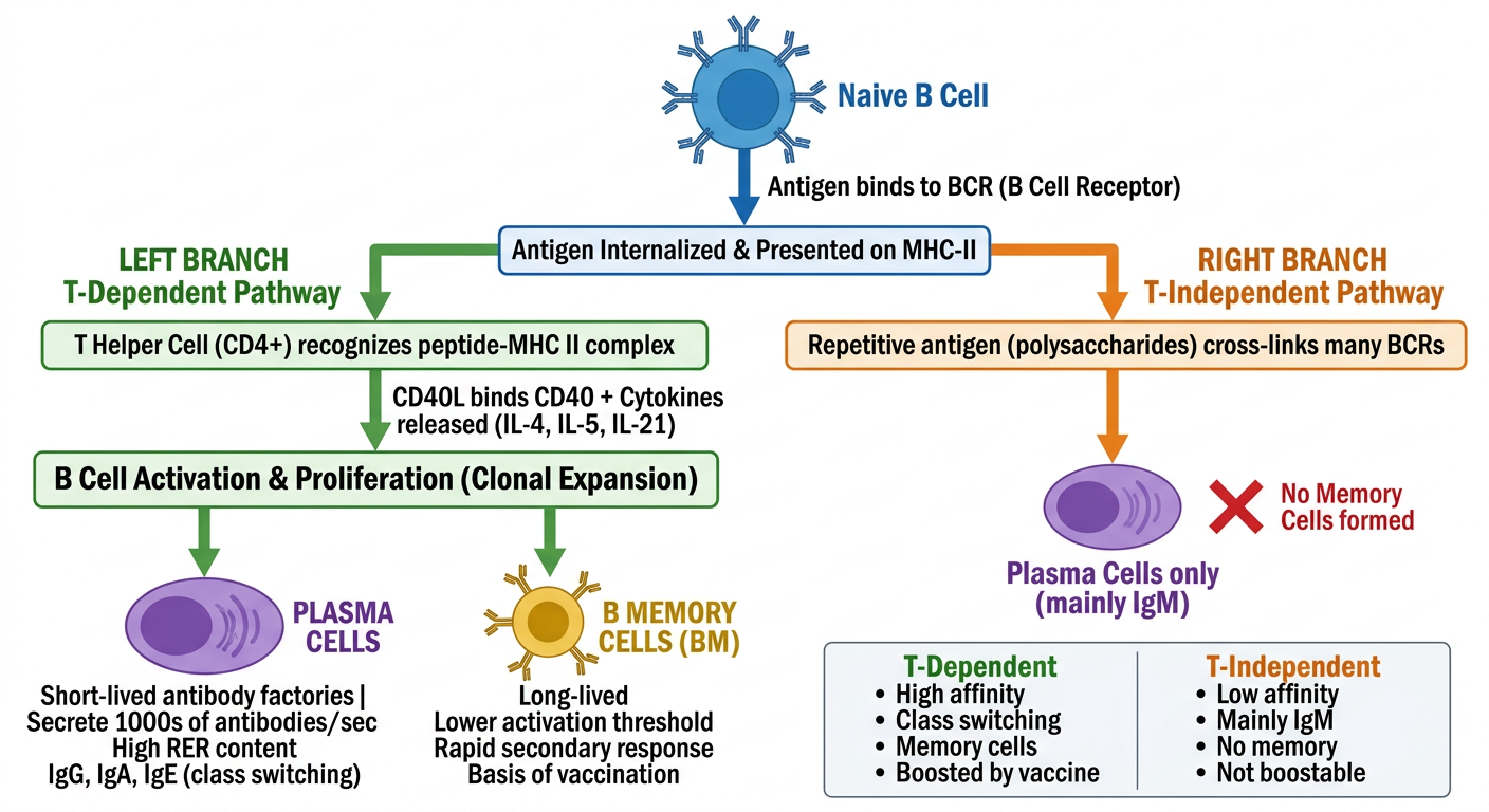
- Naive B Cell → Antigen binds BCR → Antigen presented on MHC-II
- Then splits into the two activation pathways:
- Plasma Cells (purple) - antibody factories, IgG/IgA/IgE, class switching
- Memory B Cells (gold) - long-lived, basis of vaccination