
1. Erythropoietin.[3][2010] PAGE 77 2. ESR.[3][2012] PAGE 86-87 3. Erythroblastosis Fetalis.[3][2016] PAGE 105-106 4. Hemoglobinopathies.[3][2017] PAGE 81,82,75 5. ESR[3][2018] PAGE 86-87 6. Rh Incompatibility.[2019]37 7. Law of Landsteiner. [2014 supple.] 8. IgG[2013 supple.] PAGE 110-111 9. Humoralimmunity.[2015 supple.] PAGE 107,108 10. B and T lymphocytes. [2016 supple.] PAGE 108 11.Tissue macrophage system. [2017 supple.] 12.Megaloblastic anaemia. [2019 supple.] PAGE 78-79 13. Starling forces.[2024]
Reading File
These are physiology exam questions (likely from a medical university question paper). Let me search the textbook library for all these topics simultaneously.
Finding Sources
Finding Sources
Good - I found relevant sections. Now let me read the key physiology textbook sections in parallel for the most clinically relevant content. I'll focus on Costanzo Physiology and Ganong's (the main physiology textbooks for these exam topics).
Reading File
Finding Sources
Finding Sources
Finding Sources
Now let me read the key sections in parallel:
Reading File
Reading File
Reading File
Now let me read more content on the remaining topics - Starling forces basics, hemoglobinopathies, and the immunology topics:
Reading File
Finding Sources
Reading File
Finding Sources
Reading File
Finding Sources
Now I have enough from the textbooks. Let me also read the IgG section and do a quick web search for Landsteiner's law and Rh incompatibility (for comprehensive coverage):
Reading File
Reading File
I now have comprehensive content from all key textbooks. Let me read a bit more on Starling forces (the Pc and other components) and get content on hemoglobinopathies/Rh incompatibility:
Reading File
Reading File
I now have rich content from authoritative textbooks for all 13 topics. Let me compile the comprehensive answer.Here is a complete, exam-ready write-up for all 13 topics from your question paper list, drawn from authoritative physiology and pathology textbooks.
Physiology Exam Answer Bank
1. Erythropoietin (EPO) [3 marks - 2010]
Definition: Erythropoietin is a glycoprotein growth factor synthesized primarily in the kidneys (and to a lesser extent in the liver) that serves as the major stimulus for erythropoiesis by promoting differentiation of pro-erythroblasts into red blood cells.
Mechanism of synthesis (steps):
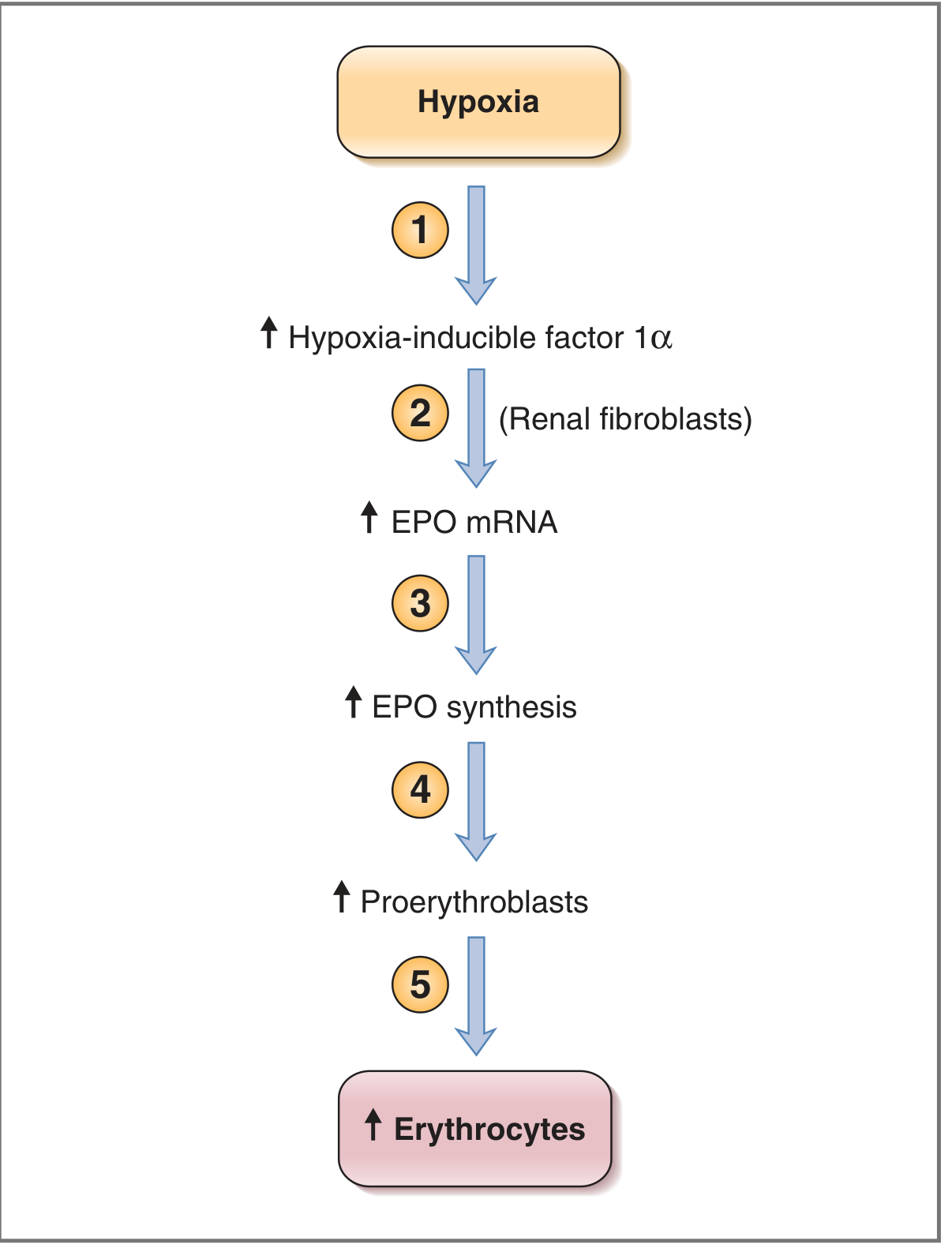
- Decreased O₂ delivery to the kidneys (due to low hemoglobin or low PaO₂) causes increased production of hypoxia-inducible factor 1α (HIF-1α).
- HIF-1α acts on fibroblasts in the renal cortex and medulla → stimulates synthesis of EPO mRNA.
- EPO mRNA directs increased synthesis of EPO.
- EPO causes differentiation of proerythroblasts. Further maturation steps (to mature RBCs) do not require EPO.
Why kidneys are the ideal site: The kidney can distinguish between decreased blood flow (which lowers both O₂ delivery and consumption equally - no alarm) and decreased O₂ content of arterial blood (which lowers delivery without reducing consumption - alarm triggered).
Clinical note: In chronic renal failure, decreased renal mass → decreased EPO synthesis → normochromic normocytic anemia. Recombinant EPO is used therapeutically.
(Source: Costanzo Physiology 7th Edition)
2 & 5. ESR (Erythrocyte Sedimentation Rate) [3 marks - 2012, 2018]
Definition: ESR is the rate at which red blood cells settle (sediment) in a vertical glass tube of anticoagulated blood over 1 hour, expressed in mm/hour.
Normal values: ≤15 mm/hr (varies slightly by method: Westergren vs Wintrobe).
Mechanism:
- Any acute stress (trauma, infection, inflammation) triggers the acute-phase response → the liver rapidly synthesizes and secretes inflammatory proteins, most importantly fibrinogen.
- Fibrinogen (a large asymmetric protein) causes RBCs to aggregate into rouleaux (coin stacks) → increases effective particle density.
- Rouleaux sediment faster due to reduced surface area-to-volume ratio and increased downward gravitational force relative to upward plasma viscosity resistance.
Three stages of ESR:
- Aggregation phase (first ~10 min): rouleaux formation
- Sedimentation phase (next ~40 min): rapid, constant settling
- Packing phase (last ~10 min): RBCs pack at bottom
Factors increasing ESR: Elevated fibrinogen, globulins, paraproteins, anemia (fewer RBCs, less ionic cloud repulsion), macrocytosis, female sex, pregnancy, age.
Factors decreasing ESR: Polycythemia, sickle cell disease (abnormal shape prevents rouleaux), microcytosis, spherocytosis, hyperviscosity.
Clinical utility: Although nonspecific, ESR is widely used to assess the presence and severity of inflammation. Example: in lupus erythematosus, an elevated ESR with nonspecific complaints (fatigue, weakness) suggests disease reactivation rather than depression or poor sleep.
(Source: Medical Physiology - Boron & Boulpaep, Box 18-1)
3. Erythroblastosis Fetalis [3 marks - 2016]
Definition: A hemolytic disease of the fetus/newborn caused by maternal antibody-mediated destruction of fetal red blood cells, most commonly due to Rh incompatibility (also ABO).
Pathogenesis:
- Rh-negative mother sensitized by Rh-positive fetal blood (fetomaternal hemorrhage) → produces anti-D IgG antibodies.
- In subsequent pregnancies, maternal IgG (the only Ig class to cross the placenta) enters fetal circulation and destroys Rh-positive fetal RBCs → hemolytic anemia.
Consequences:
- Fetal anemia → bone marrow and extramedullary hematopoiesis → normoblastic hyperplasia (erythroblastosis).
- Hemoglobin catabolism → unconjugated bilirubin; placenta normally removes this, so jaundice is not seen in utero, but amniotic fluid bilirubin rises.
- Severe cases: hydrops fetalis (congestive heart failure, massive edema, intrauterine death).
- After birth: bilirubin rises rapidly → kernicterus (bilirubin encephalopathy).
Diagnosis:
- Kleihauer-Betke test or flow cytometry to detect fetal cells in maternal blood.
- Amniotic fluid spectrophotometry: measurement of ΔOD₄₅₀ (net bilirubin absorbance at 450 nm) - the Liley test - to assess severity.
- Now largely replaced by MCA Doppler (middle cerebral artery peak systolic velocity) as a non-invasive measure of fetal anemia.
Management: Anti-D immunoglobulin (RhoGAM) given prophylactically to Rh-negative mothers at 28 weeks and within 72 hours of delivery.
(Source: Henry's Clinical Diagnosis and Management by Laboratory Methods)
4 & part of 6. Hemoglobinopathies [3 marks - 2017]
Definition: A group of genetic disorders affecting the structure or production of hemoglobin chains.
Classification:
| Type | Mechanism | Examples |
|---|---|---|
| Structural hemoglobinopathies | Abnormal globin chain sequence (qualitative) | Sickle cell disease (HbS), HbC, HbE |
| Thalassemias | Reduced/absent synthesis of normal globin chains (quantitative) | α-thalassemia, β-thalassemia |
Sickle Cell Disease (HbS):
- Mutation: Glu→Val at position 6 of β-globin chain.
- Deoxygenated HbS polymerizes → sickling of RBCs → hemolysis, vaso-occlusion, painful crises.
- Protects against falciparum malaria (heterozygous advantage).
β-Thalassemia:
- Decreased (β⁺) or absent (β⁰) β-globin chain synthesis.
- Excess α-chains precipitate in RBCs → ineffective erythropoiesis, hemolysis, bone marrow expansion.
- Thalassemia major (Cooley's anemia): severe, transfusion-dependent; skeletal deformities ("crew-cut" skull X-ray, chipmunk facies).
HbF: Fetal hemoglobin (α₂γ₂) - high O₂ affinity, present until ~6 months of age. Its persistence (HPFH) ameliorates sickle cell and β-thalassemia.
Lab findings (common): Microcytic hypochromic anemia, target cells, elevated HbA₂ (β-thalassemia trait), Hb electrophoresis diagnostic.
(Sources: Murray & Nadel's Respiratory Medicine; Robbins Pathologic Basis of Disease)
6. Rh Incompatibility [2019]
Rh System Basics:
- The Rh (Rhesus) antigen, specifically the D antigen, is the most clinically important. ~85% of people are Rh-positive (carry D antigen).
- Unlike ABO antibodies, anti-D antibodies are not naturally occurring - they develop only after exposure (transfusion or pregnancy).
Mechanism of sensitization:
- Rh-negative (D-negative) mother carries Rh-positive fetus.
- Fetomaternal hemorrhage (especially at delivery) → fetal Rh-positive RBCs enter maternal circulation.
- Maternal immune system responds → produces anti-D IgG (primary response, usually too late to affect first pregnancy).
- In subsequent Rh-positive pregnancies: maternal IgG crosses placenta → erythroblastosis fetalis (see topic 3 above).
ABO incompatibility is usually milder because anti-A and anti-B are IgM (cannot cross placenta) and because ABO antigens are expressed on many tissues.
Prevention: Anti-D immunoglobulin (passive immunization) given within 72 hours of delivery, miscarriage, or amniocentesis - destroys fetal cells before sensitization can occur.
7. Law of Landsteiner [2014 Supple]
Landsteiner's Law (1900-1901): States that if a blood group antigen is absent from the red blood cell surface, the corresponding antibody (agglutinin/isoantibody) will be present in the serum.
ABO Blood Group System:
| Blood Group | RBC Antigen | Serum Antibody | Genotype |
|---|---|---|---|
| A | A | Anti-B | AA or AO |
| B | B | Anti-A | BB or BO |
| AB | A and B | Neither | AB |
| O | Neither | Anti-A and Anti-B | OO |
Key points:
- Anti-A and anti-B are IgM antibodies (naturally occurring, do not require prior sensitization, do not cross placenta).
- ABO antigens are also present on most body tissues (not just RBCs).
- This law governs blood transfusion compatibility: mismatched transfusion causes acute hemolytic transfusion reaction (intravascular hemolysis, DIC, renal failure).
- Universal donor: Group O (no antigens, safe RBCs for all).
- Universal recipient: Group AB (no antibodies, accepts all).
8. IgG [2013 Supple]
Structure: IgG is a monomer with two heavy (γ) chains and two light chains joined by disulfide bonds. MW ~150 kDa. Has an Fc region and two Fab regions (antigen-binding).
Unique properties of IgG:
| Feature | Detail |
|---|---|
| Most abundant immunoglobulin | ~80% of total serum immunoglobulins |
| Secondary immune response | Copiously produced; long-lasting memory |
| Complement activation | Via classical pathway (needs ≥2 IgG molecules); IgG3 > IgG1 > IgG2 most effective |
| Opsonization | Fc region binds Fc receptors on macrophages and neutrophils → enhanced phagocytosis |
| ADCC | NK cells bearing Fc receptors kill IgG-coated target cells |
| Only Ig to cross placenta | Fc receptor-mediated transcytosis through syncytiotrophoblast; provides passive immunity to neonate until ~6 months |
| High diffusion coefficient | Distributes into extravascular spaces; major defense in tissues |
4 subclasses: IgG1 (most abundant), IgG2, IgG3 (most effective complement activator), IgG4 (cannot activate complement by classical pathway). All except IgG2 cross the placenta.
Clinical importance: IgG deficiency → recurrent bacterial infections. In autoimmune disease (e.g., Graves', myasthenia gravis), pathogenic IgG autoantibodies cross the placenta → transient neonatal disease.
(Source: Henry's Clinical Diagnosis and Management by Laboratory Methods)
9. Humoral Immunity [2015 Supple]
Definition: The arm of adaptive immunity mediated by antibodies (immunoglobulins) produced by B lymphocytes/plasma cells. Effective primarily against extracellular pathogens (bacteria, free viruses, toxins).
Key steps:
- Antigen recognition: B cells recognize antigen via surface immunoglobulin (B cell receptor).
- T cell help: For most antigens (T-dependent antigens), activated CD4+ T helper cells (Th2) provide costimulatory signals (CD40L-CD40 interaction) and cytokines (IL-4, IL-5, IL-6).
- B cell activation and proliferation.
- Class switching (isotype switching): Cytokines direct which Ig class is produced (IgM → IgG, IgA, or IgE).
- Somatic hypermutation and affinity maturation in germinal centres → higher-affinity antibodies.
- Differentiation into:
- Plasma cells: Effector cells secreting large amounts of antibody.
- Memory B cells: Long-lived; rapid secondary response on re-exposure.
Effector mechanisms of antibodies:
- Neutralization: Block pathogen binding to host cells (viruses, toxins).
- Opsonization: IgG/IgM coat pathogens → enhanced phagocytosis (Fc receptor and C3b receptor-mediated).
- Complement activation (classical pathway): IgM and IgG → lysis, opsonization, inflammation.
- ADCC: IgG-coated cells killed by NK cells.
- Mast cell degranulation: IgE cross-linking on mast cells → immediate hypersensitivity.
Deficiency: Hypogammaglobulinemia (e.g., Bruton's XLA) → recurrent pyogenic infections beginning after maternal IgG wanes (~6 months).
(Sources: Cellular and Molecular Immunology; Medical Microbiology 9e)
10. B and T Lymphocytes [2016 Supple]
B Lymphocytes
- Origin & maturation: Bone marrow (both origin and maturation). Express surface immunoglobulin (sIgM, sIgD).
- Function: Precursors of antibody-secreting plasma cells. Mediate humoral immunity.
- Markers: CD19, CD20, CD21, MHC Class II, surface Ig.
- Subsets: Follicular B cells (classical adaptive), Marginal zone B cells (rapid response), B1 cells (produce natural IgM against bacterial polysaccharides).
- On activation with T cell help → class switching, affinity maturation, memory formation.
T Lymphocytes
- Origin: Bone marrow; maturation in thymus (positive and negative selection).
- Markers: CD3 (all T cells), CD4 (helper), CD8 (cytotoxic).
- Recognize antigen only when presented by MHC molecules (MHC restriction).
- CD4+ Helper T cells (Th):
- Th1: activate macrophages, CD8+ T cells → cell-mediated immunity (intracellular pathogens, TB, viral infections). Cytokines: IFN-γ, TNF.
- Th2: help B cells, activate eosinophils → humoral immunity, allergy. Cytokines: IL-4, IL-5, IL-13.
- Th17: IL-17, IL-22 → mucosal immunity, autoimmunity.
- T-regulatory (Treg): FoxP3+, suppress immune responses, prevent autoimmunity.
- CD8+ Cytotoxic T cells (CTL): Kill virus-infected cells and tumor cells via perforin/granzyme and Fas-FasL pathway. Restricted to MHC Class I (expressed on all nucleated cells).
Comparison:
| Feature | B Cells | T Cells |
|---|---|---|
| Maturation site | Bone marrow | Thymus |
| Receptor | Surface Ig (BCR) | T cell receptor (TCR) |
| Antigen recognition | Free/soluble antigen | MHC-presented peptides only |
| Effector | Plasma cells → antibodies | Cytokines, cytotoxicity |
| Immunity type | Humoral | Cell-mediated |
(Sources: Robbins Basic Pathology; Junqueira's Basic Histology)
11. Tissue Macrophage System [2017 Supple]
Also called the Mononuclear Phagocyte System (MPS) (formerly reticuloendothelial system).
Definition: A network of phagocytic cells derived from bone marrow monocytes that mature into tissue-resident macrophages in virtually every organ.
Components and tissue locations:
| Tissue Macrophage Name | Location |
|---|---|
| Kupffer cells | Hepatic sinusoids (liver) |
| Alveolar macrophages | Lung alveoli |
| Microglia | Central nervous system |
| Osteoclasts | Bone |
| Langerhans cells | Skin epidermis |
| Mesangial cells | Kidney glomerulus |
| Splenic macrophages | Splenic red pulp (remove senescent RBCs) |
| Peritoneal/pleural macrophages | Body cavities |
Functions:
- Phagocytosis and killing of microorganisms (bacteria, fungi, parasites).
- Antigen presentation via MHC Class II → activate CD4+ T cells (bridge innate and adaptive immunity).
- Removal of senescent/damaged cells (e.g., aged RBCs cleared by splenic and hepatic macrophages).
- Blood cleansing: Kupffer cells phagocytose gut-derived bacteria from portal blood in <0.01 seconds - <1% of intestinal bacteria reach systemic circulation.
- Secretion of cytokines: TNF-α, IL-1, IL-6, IL-12 → inflammation, fever.
- Wound healing and tissue remodeling.
- Lipid metabolism: Foam cell formation (lipid-laden macrophages in atherosclerotic plaques).
Activation states:
- M1 (classical activation): IFN-γ + LPS → pro-inflammatory, microbicidal.
- M2 (alternative activation): IL-4, IL-13 → anti-inflammatory, tissue repair.
(Sources: Guyton and Hall Textbook of Medical Physiology; Medical Microbiology 9e; Junqueira's Basic Histology)
12. Megaloblastic Anaemia [2019 Supple]
Definition: A macrocytic anemia caused by impaired DNA synthesis (due to B₁₂ or folate deficiency), leading to nuclear-cytoplasmic asynchrony and ineffective erythropoiesis.
Causes:
Vitamin B₁₂ deficiency:
- Decreased intake: strict vegetarians/vegans
- Decreased intrinsic factor: pernicious anemia (autoimmune gastritis, anti-IF antibodies), gastrectomy
- Malabsorption: ileal disease/resection (terminal ileum is absorption site), bacterial overgrowth, fish tapeworm (Diphyllobothrium latum)
Folate deficiency:
- Decreased intake: alcoholism, poverty, infancy, pregnancy (↑ requirement)
- Malabsorption: celiac disease, tropical sprue
- Drugs: methotrexate (DHFR inhibitor), phenytoin, oral contraceptives
- Increased demand: hemolytic anemia, disseminated cancer
Pathophysiology: B₁₂ and folate are cofactors for synthesis of thymidine (a DNA base). Deficiency → impaired DNA synthesis → cells continue to grow (RNA/protein synthesis intact) but cannot divide → large cells with immature nuclei (nuclear-cytoplasmic asynchrony).
Morphology:
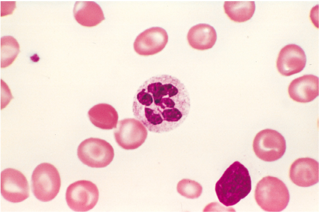
- Peripheral blood: Macro-ovalocytes, anisocytosis, poikilocytosis, low reticulocytes, hypersegmented neutrophils (≥5 lobes - pathognomonic), pancytopenia in severe cases.
- Bone marrow: Hypercellular; megaloblasts at all stages; giant metamyelocytes; nuclear chromatin remains fine (does not condense normally).
B₁₂ vs Folate deficiency - key distinguishing feature:
- B₁₂ deficiency ONLY causes subacute combined degeneration of spinal cord (posterior and lateral columns) due to demyelination → paresthesias, ataxia, spastic paraparesis.
- Serum methylmalonic acid (MMA) is elevated in B₁₂ deficiency only.
- Folate deficiency: no neurological involvement; serum homocysteine elevated (in both).
Treatment: IM hydroxocobalamin (B₁₂ deficiency); oral folic acid (folate deficiency). Note: giving folate to B₁₂-deficient patients may mask anemia but will not prevent neurological damage.
(Source: Robbins, Cotran & Kumar Pathologic Basis of Disease)
13. Starling Forces [2024]
The Starling Equation governs fluid movement across capillary walls:
Jv = Kf [ (Pc - Pi) - (πc - πi) ]
Where:
- Jv = Net fluid movement (mL/min); positive = filtration (out of capillary), negative = absorption (into capillary)
- Kf = Hydraulic conductance (water permeability of capillary wall)
- Pc = Capillary hydrostatic pressure (force favoring filtration)
- Pi = Interstitial hydrostatic pressure (force opposing filtration; normally ≈ 0 or slightly negative)
- πc = Capillary oncotic pressure (force opposing filtration; due to plasma proteins)
- πi = Interstitial oncotic pressure (force favoring filtration; normally very low)
The four Starling forces at a typical capillary:
| Force | Value | Direction |
|---|---|---|
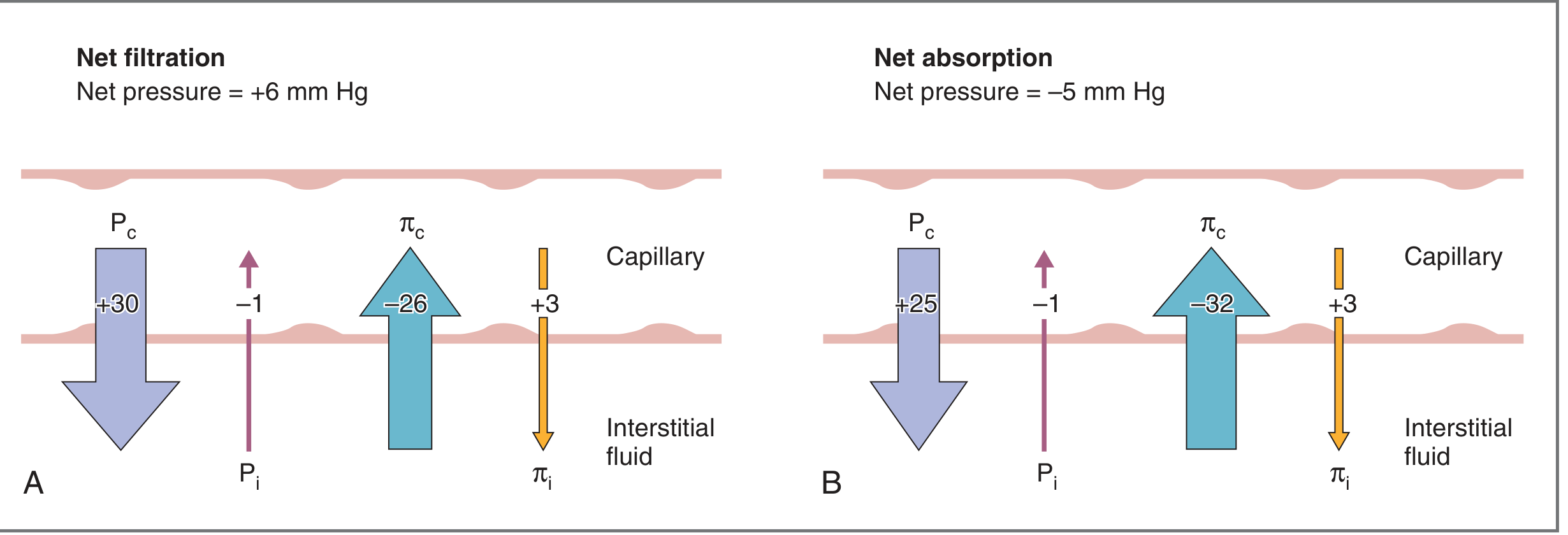
| Pc (capillary hydrostatic) | 30 mm Hg (arteriolar end) → 15 mm Hg (venous end) | Filtration ← |
| Pi (interstitial hydrostatic) | ~1 mm Hg | Absorption → |
| πc (plasma oncotic) | ~26 mm Hg | Absorption → |
| πi (interstitial oncotic) | ~3 mm Hg | Filtration ← |
- At the arteriolar end: Net pressure = +6 mm Hg → net filtration
- At the venous end: Net pressure = -5 mm Hg → net absorption
- Small net excess of filtration over absorption is drained by lymphatics back to circulation
Each parameter explained:
- Kf varies with capillary type (highest in glomerulus, lowest in brain). Increases in burns/toxin injury → edema.
- Pc is raised by increased venous pressure (more than arterial). It falls along capillary length due to filtration.
- πc falls with hypoproteinemia (e.g., nephrotic syndrome, liver disease) → increased filtration → edema.
- πi rises when protein leaks out of capillary (capillary injury) → further worsens edema.
Causes of edema (changes in Starling forces):
| Cause | Mechanism |
|---|---|
| Heart failure | ↑ venous/capillary hydrostatic pressure (Pc↑) |
| Hypoalbuminemia (nephrotic/cirrhosis) | ↓ capillary oncotic pressure (πc↓) |
| Burns/inflammation | ↑ Kf (capillary permeability ↑) + πi↑ |
| Lymphatic obstruction | Impaired return of excess filtrate |
| Filariasis | Lymphatic blockade → massive edema (elephantiasis) |
(Source: Costanzo Physiology 7th Edition)
Quick Summary Table
| # | Topic | Key Points to Remember |
|---|---|---|
| 1 | Erythropoietin | Kidney (HIF-1α) → EPO → proerythroblast differentiation; fails in CRF |
| 2/5 | ESR | Fibrinogen → rouleaux → settles faster; non-specific inflammation marker |
| 3 | Erythroblastosis Fetalis | IgG anti-D crosses placenta; ΔOD₄₅₀, MCA Doppler; prevent with RhoGAM |
| 4 | Hemoglobinopathies | Structural (HbS, HbC) vs quantitative (thalassemia); Hb electrophoresis diagnostic |
| 6 | Rh Incompatibility | D antigen; sensitization at delivery; affects subsequent pregnancies |
| 7 | Landsteiner's Law | Absent antigen → antibody present; ABO table; natural IgM antibodies |
| 8 | IgG | Most abundant; crosses placenta; opsonization; ADCC; complement; 4 subclasses |
| 9 | Humoral Immunity | B cell → plasma cell → antibodies; neutralization, opsonization, complement |
| 10 | B & T Lymphocytes | B: bone marrow maturation, antibodies; T: thymus maturation, CD4/CD8, MHC restriction |
| 11 | Tissue Macrophage System | MPS; Kupffer cells, microglia, alveolar macrophages; phagocytosis, APC function |
| 12 | Megaloblastic Anaemia | B₁₂/folate → impaired DNA; macro-ovalocytes, hypersegmented neutrophils; B₁₂ = neurological features |
| 13 | Starling Forces | Jv = Kf[(Pc-Pi)-(πc-πi)]; filtration at arteriolar end, absorption at venous end; edema mechanisms |
This is a shared conversation. Sign in to Orris to start your own chat.