
opthalmic neonatorum
ophthalmia neonatorum neonatal conjunctivitis
ophthalmia neonatorum neonatal conjunctivitis gonococcal

This clinical photograph shows an infant presenting with ophthalmia neonatorum, a form of neonatal conjunctivitis. The primary clinical signs include severe bilateral eyelid edema (swelling) and profuse, thick, purulent (pus-like) discharge emanating from the palpebral fissure. The discharge is thick and yellowish, characteristic of a hyperacute bacterial infection such as gonococcal conjunctivitis. An adult hand is shown gently retracting the upper eyelid to reveal the extent of the inflammation and ocular discharge. The infant is swaddled and using a pacifier, indicating a clinical examination setting. This image serves as a significant educational tool for recognizing the signs of neonatal eye infections, which require urgent diagnosis and treatment to prevent serious complications such as corneal ulceration, perforation, and permanent visual impairment. The anatomical focus is on the ocular and periorbital region, demonstrating the hallmark presentation of severe infectious conjunctivitis in the first weeks of life.
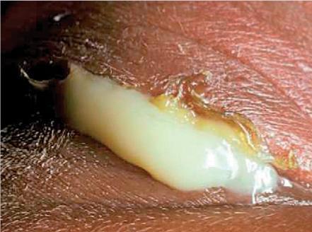
This clinical photograph displays a close-up view of a neonatal eye exhibiting severe ophthalmia neonatorum, likely caused by Neisseria gonorrhoeae infection. The primary visual finding is a massive, hyperacute purulent discharge characterized by a thick, creamy, yellowish-white consistency that is so copious it obscures the underlying globe. The superior and inferior eyelids are markedly edematous (swollen) and erythematous, indicating significant periorbital inflammation. The surrounding skin appears irritated and macerated from contact with the abundant exudate. This presentation is a medical emergency due to the risk of rapid corneal perforation and permanent vision loss. The image serves as a critical educational tool for recognizing the signs of neonatal gonococcal conjunctivitis, emphasizing the urgency of immediate systemic antibiotic treatment and ocular irrigation.
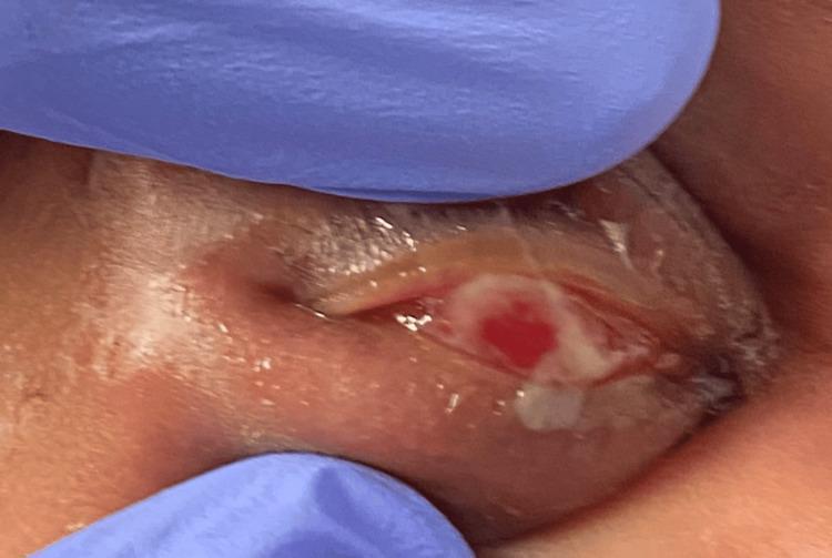
This clinical photograph displays a close-up, procedural view of the right eye of a neonate, with the upper eyelid everted by a clinician wearing blue nitrile gloves. The image demonstrates a primary clinical presentation of ophthalmia neonatorum (neonatal conjunctivitis). Significant findings include diffuse conjunctival injection (hyperemia) and a prominent accumulation of thick, yellowish mucopurulent discharge covering the palpebral and bulbar surfaces. The surrounding periorbital tissues and eyelids show marked erythema and edema. This visual illustrates the hallmark signs of infectious conjunctivitis in a newborn, highlighting the importance of assessing for N. gonorrhoeae or Chlamydia trachomatis. The photograph captures a key diagnostic step in the physical examination of a pediatric patient presenting with eye swelling and discharge, focusing on the character of the ocular secretion and the integrity of the ocular surface.
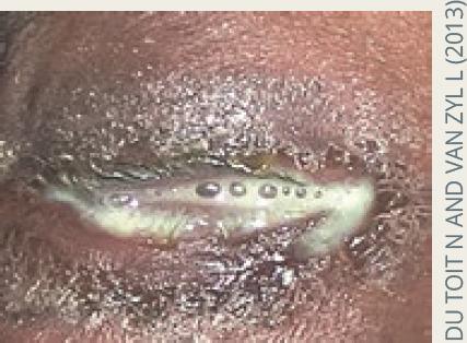
This clinical photograph displays a close-up view of an eye presenting with severe ophthalmia neonatorum, specifically hyperacute purulent conjunctivitis. The eyelids exhibit significant erythema and marked periorbital edema, causing them to be nearly swollen shut. A copious, thick, yellowish-white purulent discharge (pus) is seen oozing from the palpebral fissure and coating the eyelid margins and eyelashes. Small air bubbles or droplets are visible within the viscous exudate. The clinical appearance is highly characteristic of Neisseria gonorrhoeae infection, demonstrating the hallmark symptom of rapid-onset, profuse discharge that poses a high risk for corneal ulceration and perforation if not treated emergently. The image serves as an educational tool for identifying neonatal infectious emergencies and differentiating hyperacute bacterial conjunctivitis from milder forms of neonatal eye infections.
Ophthalmia Neonatorum (Neonatal Conjunctivitis)
Clinical Image
Classification by Etiology
| Type | Cause | Age of Onset | Key Findings | Treatment |
|---|---|---|---|---|
| Chemical | Erythromycin/silver nitrate prophylaxis | Within 24 h | Bilateral, watery discharge, negative Gram stain | Watchful waiting |
| Gonococcal | Neisseria gonorrhoeae | 2-7 days | Intense chemosis, copious purulent discharge, gram-negative diplococci | Admission, IV antibiotics |
| Chlamydial | Chlamydia trachomatis | 7-14 days | Intense palpebral erythema, mucopurulent discharge | Oral + topical erythromycin |
| Other bacterial | S. aureus, H. influenzae, S. epidermidis, E. coli, Pseudomonas | 7-14 days | Variable; identify by Gram stain | Topical antibiotics |
| Viral | HSV-1, HSV-2 | 14-28 days | Eyelid edema, corneal dendrites, mucocutaneous lesions | Admission, IV acyclovir |
1. Chemical Ophthalmia Neonatorum
- Occurs within 24 hours of birth following erythromycin ointment (or historically silver nitrate) prophylaxis
- Presents with bilateral conjunctivitis, inflamed eyelids, and watery discharge
- Gram stain: no pathologic bacteria, only a few WBCs
- Treatment: watchful waiting - resolves within 48 hours
- Note: the AAP recommends prophylaxis for all newborns; the Canadian Paediatric Society advises against routine use
2. Gonococcal Ophthalmia Neonatorum
- Presents at 2-7 days of life (sometimes as early as 3-4 days)
- Signs: intense bilateral bulbar conjunctival erythema, marked chemosis, and copious purulent discharge
- Risk: rapid corneal ulceration and perforation if untreated - this is the most dangerous form
- Diagnosis: Gram stain showing gram-negative intracellular diplococci; culture on chocolate agar (in CO2 atmosphere)
- Admit all infants; obtain ophthalmology consultation
- Rule out disseminated disease: blood, urine, CSF, and any other suspected sites
- Ceftriaxone 25-50 mg/kg IV/IM (max 125 mg) as a single dose
- If hyperbilirubinemia or disseminated infection suspected: cefotaxime 100 mg/kg IV/IM single dose
- Saline irrigation of conjunctiva and fornices until discharge clears (q.i.d.)
- Topical antibiotics alone are NOT sufficient
- Co-treat for chlamydia: erythromycin elixir 50 mg/kg/day in 4 divided doses for 14 days
- Treat mother and sexual partners for both gonorrhea and chlamydia
3. Chlamydial Ophthalmia Neonatorum
- Presents at 7-14 days of life (most common cause of neonatal conjunctivitis in developed countries)
- Signs: unilateral or bilateral purulent discharge, intense erythema of palpebral conjunctiva, possible pseudomembranes with bloody discharge
- Can progress to chlamydial pneumonia (evaluate for respiratory symptoms)
- Diagnosis:
- Giemsa stain: basophilic intracytoplasmic inclusion bodies in conjunctival epithelial cells
- PCR, ELISA, enzyme immunoassay, or DNA hybridization probe (most sensitive)
- Oral erythromycin 12.5 mg/kg PO every 6 hours for 14 days (50 mg/kg/d in 4 divided doses)
- PLUS erythromycin ophthalmic ointment q.i.d.
- Alternative: azithromycin 20 mg/kg orally for 3 days
- Topical therapy alone is NOT effective (systemic treatment is mandatory)
- Patients without respiratory symptoms may be discharged with 24-hour follow-up
- Treat mother and partners: doxycycline 100 mg b.i.d. x 7 days (if not pregnant/breastfeeding); azithromycin 1 g single dose or amoxicillin 500 mg t.i.d. x 7 days in pregnancy
4. Other Bacterial Ophthalmia Neonatorum
- Common organisms: S. aureus (including MRSA), streptococci, H. influenzae (nontypeable), S. epidermidis, E. coli, Pseudomonas
- Present within 2 weeks of birth; variable severity
- Diagnosis: Gram stain and culture
- Treatment: Topical antibiotics are sufficient for most cases
- Gram-positive organisms: bacitracin ointment q.i.d. for 2 weeks
- Gram-negative organisms: gentamicin, tobramycin, or ciprofloxacin ointment q.i.d.
- Exception: nontypeable H. influenzae requires admission, full septic workup, and parenteral antibiotics
5. Viral (Herpetic) Ophthalmia Neonatorum
- Caused by HSV-1 or HSV-2 (rare but potentially devastating)
- Presents at 14-28 days of life with bilateral lid edema and conjunctival erythema
- Signs: corneal dendrites on fluorescein examination, mucocutaneous vesicular lesions; maternal history of HSV not required for diagnosis
- Risk: keratitis and disseminated infection with CNS involvement
- Admit, full septic evaluation including lumbar puncture with HSV PCR of CSF
- IV acyclovir 60 mg/kg/day in 3 divided doses (20 mg/kg q8h)
- 14 days if limited to skin, eye, and mouth
- 21 days if disseminated or CNS involvement
- Topical antivirals: vidarabine 3% ointment, ganciclovir 0.15% gel, or trifluridine 1% drops 5-9x/day (optional when systemic therapy is given)
- Pediatric infectious disease consultation
Workup (General Approach)
- Maternal history: STIs during pregnancy, prenatal cervical cultures
- Ocular exam with fluorescein staining for corneal involvement
- Conjunctival scrapings: Gram stain + Giemsa stain (technique: irrigate fornices, topical anesthetic, then scrape palpebral conjunctiva of lower eyelid)
- Cultures: blood agar + chocolate agar (CO2 atmosphere for gonorrhea)
- Chlamydial immunofluorescent antibody test or PCR
- Viral culture if HSV suspected
- Systemic evaluation
Prophylaxis
- Erythromycin 0.5% ophthalmic ointment - currently the standard in the US
- Historically, silver nitrate 1% drops (now rarely used due to chemical irritation)
- Povidone-iodine 2.5% is used in resource-limited settings
Key Points to Remember
- Gonococcal = earliest onset, most severe, risk of corneal perforation - always admit
- Chlamydial = most common, requires systemic (not just topical) treatment
- HSV = latest onset, rare but life-threatening, needs IV acyclovir
- Always co-test and co-treat for chlamydia in confirmed gonococcal cases
- Always treat the mother and her sexual partners
- Inadequately treated chlamydial conjunctivitis can lead to chlamydial otitis or pneumonia