
What is pulmonary atelectasis?
pulmonary atelectasis chest X-ray or lung collapse diagram
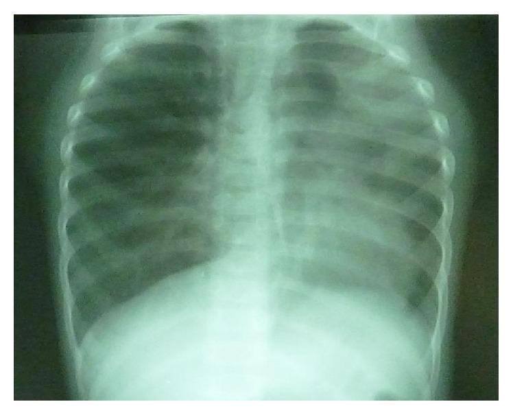
Anteroposterior (AP) chest X-ray of a pediatric patient demonstrating significant pulmonary asymmetry and mediastinal displacement. The left upper lung field shows increased opacity and volume loss, characteristic of a left upper lobe collapse (atelectasis). This is associated with an ipsilateral mediastinal shift, visible by the deviation of the tracheal air column and cardiac silhouette toward the left side of the thoracic cavity. In contrast, the right lung field appears markedly more radiolucent with increased volume, representing compensatory hyperinflation of the contralateral lung. These diagnostic findings are critical for identifying obstructive lung processes, such as follicular bronchiolitis or foreign body aspiration, which lead to differential ventilation and volume loss in the affected segments. The image serves as a clinical example of the secondary effects of focal lung collapse on thoracic anatomy and mediastinal positioning.
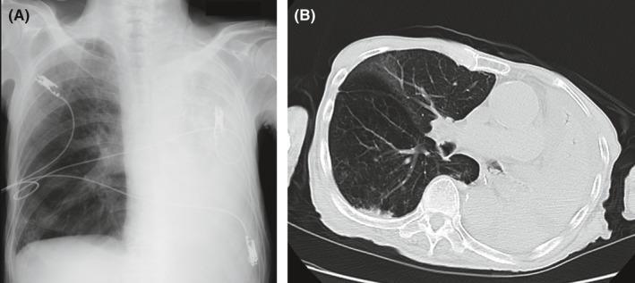
This composite medical image presents a clinical comparison of total left lung collapse using two diagnostic modalities: a frontal chest radiograph (A) and an axial thoracic CT scan (B). In image A, the chest X-ray shows complete opacification of the left hemithorax with a notable absence of air-filled lung markings, indicative of massive atelectasis. The right lung remains well-aerated with visible pulmonary vasculature. Medical hardware, including ECG leads and potentially a nasogastric tube, are visible. In image B, the axial CT scan at the level of the main bronchi confirms complete left-sided atelectasis, appearing as a dense, consolidated mass of soft tissue density occupying the posterior hemithorax. The right lung demonstrates compensatory hyperinflation. A key diagnostic feature shown in the CT is the obstruction of the left main bronchus by high-density foreign material (aspirated enteral formula), leading to distal resorption atelectasis. This visual set is essential for teaching the radiographic signs of obstructive lobar/total lung collapse and the clinical complications associated with pulmonary aspiration in patients receiving enteral nutrition.
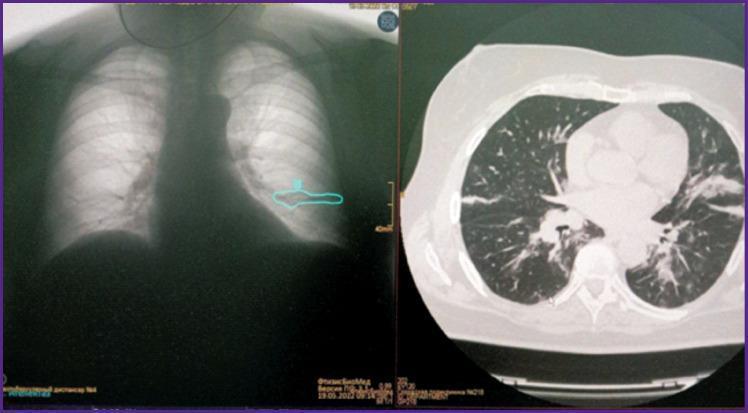
A comparison image displaying a posterior-anterior chest X-ray (left) and a corresponding axial computed tomography (CT) scan (right) of the same patient. The chest X-ray illustrates a horizontal, linear opacity in the lower lobe of the right lung, highlighted by a cyan contour from an AI diagnostic tool, suggesting localized lung collapse. The right panel shows a high-resolution axial CT scan in a lung window, which definitively characterizes the pathology as discoid atelectasis. Visible features on the CT include a band-like, transverse opacity located in the right lower lobe, parallel to the diaphragm, accompanied by focal volume loss and mild adjacent pleural thickening. This image set demonstrates the diagnostic transition from a projectional screening modality (X-ray) to a cross-sectional confirmatory modality (CT) for identifying plate-like atelectasis and differentiating it from other pulmonary infiltrates or pleural pathologies.
Pulmonary Atelectasis (Collapse)
- Robbins & Kumar Pathologic Basis of Disease, p. 630
- Goldman-Cecil Medicine, p. 918
Types of Acquired Atelectasis
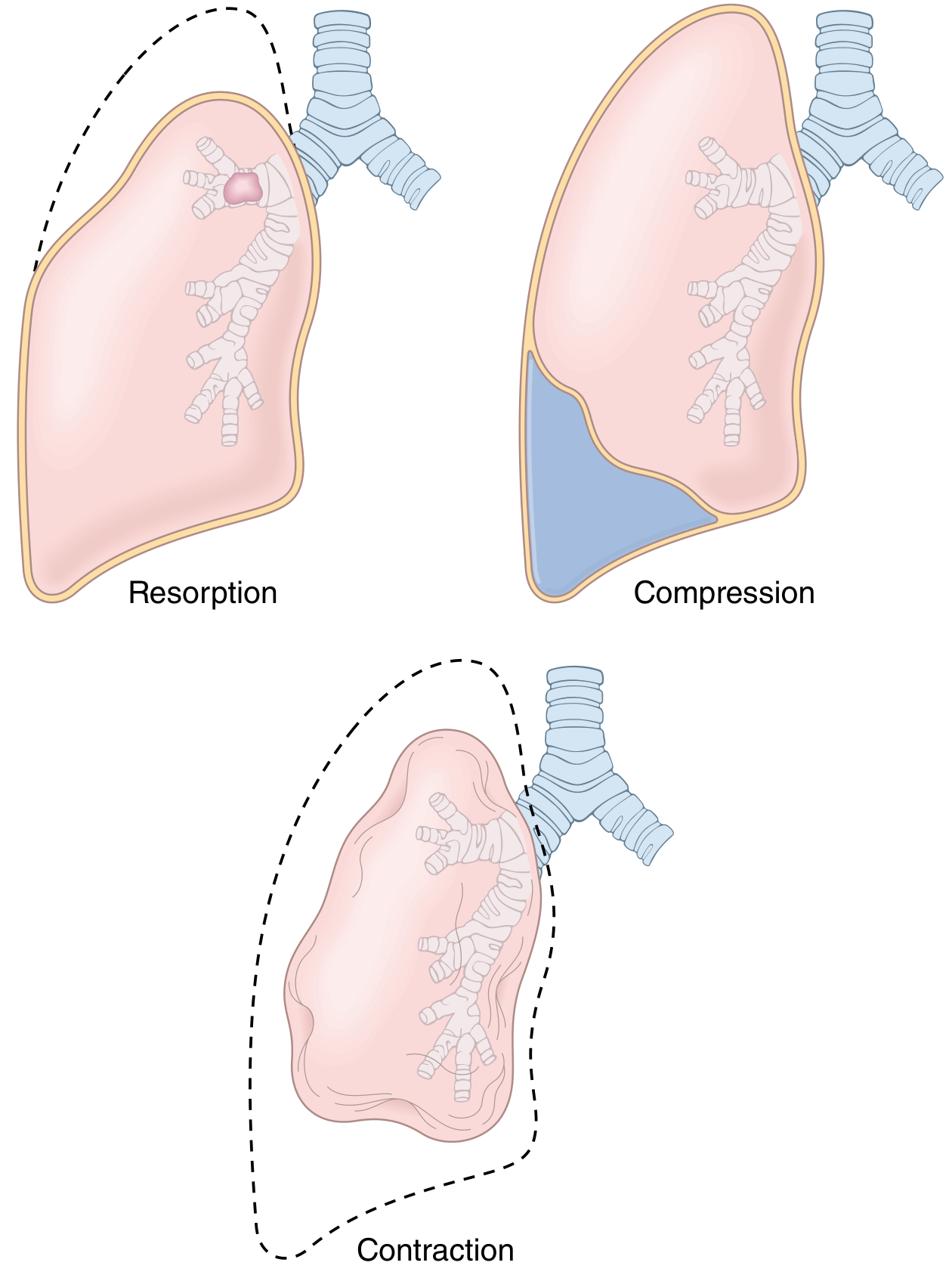
1. Resorption (Obstructive) Atelectasis
- Caused by obstruction of an airway that prevents air from reaching distal lung units
- Trapped air is gradually absorbed, causing alveolar collapse
- Since lung volume is diminished, the mediastinum shifts toward the atelectatic lung
- Common causes:
- Mucus plugs or mucopurulent secretions (most common, especially postoperative)
- Bronchial asthma, chronic bronchitis, bronchiectasis
- Foreign body aspiration (especially in children)
- Intrabronchial tumor (atelectasis may be the first sign of malignancy)
2. Compression Atelectasis
- Caused by accumulation of fluid (pleural effusion), blood, tumor, or air (pneumothorax) in the pleural cavity, which mechanically collapses adjacent lung
- The mediastinum shifts away from the affected lung (opposite to resorption type)
- Common in congestive heart failure (pleural effusions) and pneumothorax
- "Basal atelectasis" from failure to breathe deeply occurs in bedridden patients, those with ascites, and post-surgical patients
3. Contraction (Cicatrization) Atelectasis
- Caused by focal or diffuse pulmonary or pleural fibrosis that prevents full lung expansion
- This form is irreversible
4. Rounded Atelectasis (special form)
- Caused by pleural thickening that invaginates and traps adjacent lung
- Strongly associated with asbestos-related pleural disease
- Appears as an ovoid mass-like density abutting the pleura on imaging
- Does not require treatment; CT helps distinguish it from tumor
Pathophysiology
- Atelectatic lung is still perfused by blood but cannot exchange gases
- This creates a true intrapulmonary shunt (blood passes through non-ventilated lung without oxygenation)
- The result is hypoxemia that does not fully correct with supplemental oxygen alone (in large shunts)
- Sustained atelectasis also predisposes to pulmonary infection because stagnant secretions become a growth medium for bacteria
Clinical Features and Diagnosis
- Often asymptomatic and found incidentally on imaging
- May cause dyspnea, tachypnea, and measurable oxygen desaturation
- In postoperative patients, it is a common cause of low-grade fever in the first 24-48 hours
- SpO2 ≤96% after 30 minutes on Venturi-mask oxygen is a moderately sensitive test for postoperative atelectasis
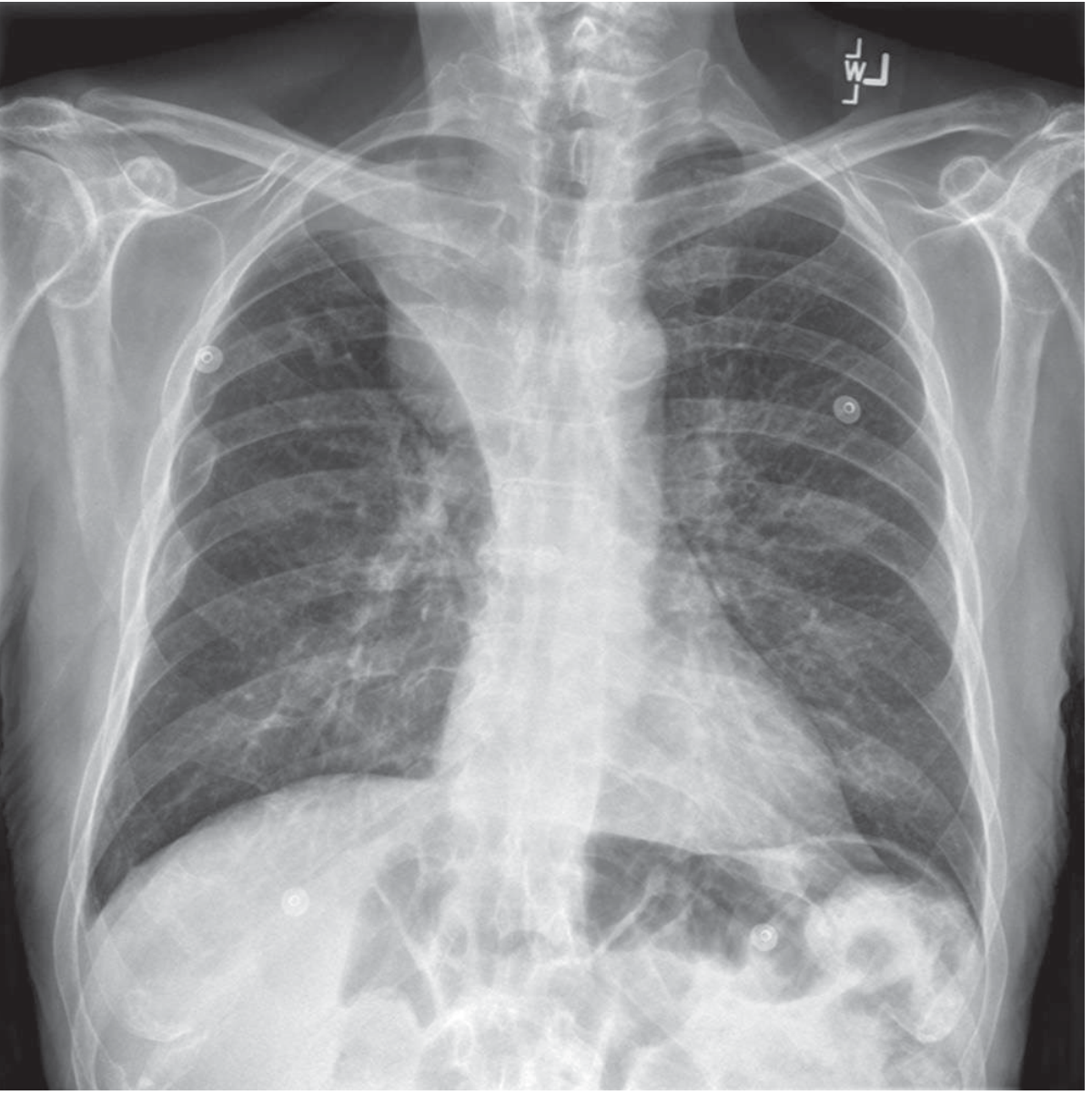
- Plain chest X-ray: loss of lung volume, displacement of lobar fissures, mediastinum, or diaphragm toward the involved area; "plate-like" (discoid) atelectasis appears as horizontal curvilinear lines
- CT scan: more precise delineation of extent and cause; distinguishes rounded atelectasis from tumor
- Bronchoscopy: required to confirm and treat intrinsic vs. extrinsic airway obstruction
Prevention and Treatment
| Approach | Details |
|---|---|
| Incentive spirometry | Commonly prescribed post-surgery; encourages deep breathing, though RCT evidence is limited |
| Preoperative inspiratory muscle training | Shown to reduce atelectasis in upper abdominal and cardiac surgery |
| Noninvasive ventilation (NIV) | Prophylactic use may reduce post-lung-resection pulmonary dysfunction |
| Recruitment maneuvers | Used intraoperatively/postoperatively in mechanically ventilated patients |
| Bronchoscopy | Required for diagnosis and treatment of obstructive/resorptive atelectasis from mucus plugs or tumors; secretions often reaccumulate until the underlying condition improves |
| Treat the underlying cause | Drain pleural effusion for compression atelectasis; manage fibrosis for contraction atelectasis |
| Rounded atelectasis | No treatment needed; monitored with CT |
- Goldman-Cecil Medicine, p. 918
- Robbins Basic Pathology
- Robbins & Kumar Pathologic Basis of Disease