
Hypertension from basics to clinical with medicine
Reading File
Now I'll run parallel searches across the library for all major aspects of hypertension.
Finding Sources
Searching PubMed
"hypertension"[MeSH Terms] AND "management"[MeSH Terms]
Finding Sources
Searching PubMed
"hypertension" AND "guidelines"
Now let me pull the key textbook sections in parallel - pathophysiology, classification, pharmacology, and clinical management:
Reading File
Reading File
Reading File
Reading File
Reading File
Finding Sources
Reading File
Reading File
Reading File
Reading File
Finding Sources
Reading File
Finding Sources
Reading File
Reading File
Reading File
Reading File
I now have comprehensive data from multiple authoritative textbooks. Let me compile the full, detailed response.
Hypertension: Basics to Clinical Medicine
1. Definition
Hypertension is a sustained elevation of systemic arterial blood pressure (BP) above the threshold associated with increased cardiovascular and end-organ risk. Blood pressure is a continuously distributed variable - there is no sharp cutoff that is "safe" below and "dangerous" above, but thresholds are used clinically for practical decision-making.
"Like height and weight, blood pressure is a continuously distributed variable; moreover, detrimental consequences increase progressively as the pressure rises, with no rigidly defined threshold dependably predicting total safety." - Robbins & Kumar Basic Pathology, p. 547
2. Classification (ACC/AHA 2017 Guidelines)
| Category | Systolic BP (mmHg) | Diastolic BP (mmHg) | |
|---|---|---|---|
| Normal | < 120 | and | < 80 |
| Elevated BP | 120-129 | and | < 80 |
| Stage 1 Hypertension | 130-139 | or | 80-89 |
| Stage 2 Hypertension | ≥ 140 | or | ≥ 90 |
Source: ACC/AHA 2017; NKF Primer on Kidney Diseases, 8e; Washington Manual of Medical Therapeutics
Key note: When systolic and diastolic fall in different categories, use the higher category. This classification applies to individuals not taking antihypertensive drugs and not acutely ill.
Special Forms
- Isolated systolic hypertension: SBP ≥ 140 with DBP < 90. Common in the elderly due to arterial stiffening.
- White coat hypertension: Elevated only in clinical settings; normal on ambulatory monitoring.
- Masked hypertension: Normal in office; elevated on ambulatory monitoring.
- Resistant hypertension: BP ≥ 130/80 on ≥ 3 antihypertensive agents (including a diuretic) at maximal doses, or controlled BP on ≥ 4 agents.
- Hypertensive urgency: Severely elevated BP (often > 180/120) without acute end-organ damage.
- Hypertensive emergency: Severely elevated BP WITH evidence of acute end-organ damage (brain, heart, kidneys, retina, large vessels).
- Malignant hypertension: BP > 200/120 with bilateral advanced retinopathy (flame hemorrhages, papilledema).
3. Epidemiology
- Over 40% of U.S. adults are hypertensive by ACC/AHA 2017 criteria (approx. 116 million adults).
- 73.9% of hypertensives in the U.S. have uncontrolled BP.
- For non-hypertensive individuals aged 55-65 years, lifetime risk of developing hypertension is 90%.
- African Americans have the highest prevalence in the U.S. due to combined genetic and environmental factors.
- Without treatment, ~50% of hypertensives die from ischemic heart disease or heart failure; ~33% from stroke.
- Nearly one-third of the world's adults have hypertension.
4. Etiology and Types
Primary (Essential) Hypertension
- Accounts for 90-95% of all cases.
- No single identifiable cause; multifactorial (genetic + environmental).
- Key contributing factors: obesity/visceral adiposity, sedentary lifestyle, high sodium intake, insulin resistance, excessive alcohol, stress.
Secondary Hypertension (5-10%)
| Category | Causes |
|---|---|
| Renal | Acute/chronic glomerulonephritis, polycystic kidney disease, renal artery stenosis, renin-producing tumors, renal vasculitis |
| Endocrine | Primary hyperaldosteronism, Cushing syndrome, pheochromocytoma, congenital adrenal hyperplasia, acromegaly, hypo/hyperthyroidism, OCP/estrogen use |
| Cardiovascular | Coarctation of the aorta, polyarteritis nodosa, increased cardiac output states |
| Neurologic | Increased intracranial pressure, obstructive sleep apnea (OSA), acute stress |
| Drug-induced | NSAIDs, sympathomimetics, cocaine, steroids, cyclosporine, erythropoietin, venlafaxine, MAOIs, TCAs |
Source: Robbins & Kumar Basic Pathology; Washington Manual of Medical Therapeutics
5. Pathophysiology
The Core Formula
BP = Cardiac Output (CO) × Total Peripheral Resistance (TPR)
This simple formula drives all pharmacological targets. In chronic hypertension, CO is usually normal and TPR is elevated - increased TPR is partly secondary to raised BP (via autoregulation of tissue blood flow) rather than always the primary driver.
Renal-Pressure Natriuresis (The Long-Term Regulator)
The kidney is the dominant long-term regulator of BP. Normally, a rise in mean arterial pressure (MAP) of as little as 1-3 mmHg triggers increased urinary NaCl and water excretion ("pressure natriuresis"), returning BP toward normal. In hypertension, this curve is reset - the kidney requires a higher BP to excrete the same salt load.
Salt sensitivity is particularly common in:
- Black patients
- The elderly
- Patients with CKD
- Those with low plasma renin activity
Renin-Angiotensin-Aldosterone System (RAAS)
- Reduced renal perfusion or sympathetic activation → renin released from juxtaglomerular cells
- Renin cleaves angiotensinogen → Angiotensin I
- ACE (in lungs) converts Ang I → Angiotensin II (Ang II)
- Ang II effects:
- Vasoconstriction (AT1 receptor) → ↑ TPR
- Adrenal cortex → aldosterone → Na+ and water retention → ↑ blood volume → ↑ CO
- Proximal tubule → Na+/H+ exchange → ↑ Na+ reabsorption
- CNS → vasopressin release, sympathetic activation, thirst
Sympathetic Nervous System
- Catecholamines cause vasoconstriction (α1) and increase heart rate/contractility (β1)
- Chronic sympathetic overactivation is a feature of essential hypertension
- Renal sympathetics enhance tubular Na+ reabsorption and stimulate renin release
Endothelial Dysfunction
- Nitric oxide (NO) normally causes vasodilation and inhibits vascular smooth muscle growth
- NO deficiency impairs pressure natriuresis and promotes vasoconstriction
- Endothelin-1 (ET-1) is a potent vasoconstrictor - elevated in salt-sensitive hypertension
- Atrial natriuretic peptide (ANP) enhances pressure natriuresis and lowers BP; its blockade produces salt-sensitive hypertension
Vascular Remodeling
- Chronic elevated BP leads to structural changes: vessel wall thickening, capillary rarefaction
- Long-term mechanisms maintain tissue blood flow via angiogenesis adaptations
- This vascular remodeling becomes self-perpetuating
Immune/Inflammatory Component
- Innate and adaptive immunity contribute to hypertension
- T-cell infiltration in kidneys and vessels promotes sodium retention and oxidative stress
6. Cardiovascular Risk Factors in Hypertension
| Modifiable | Relatively Fixed |
|---|---|
| Cigarette smoking | CKD |
| Diabetes mellitus | Family history |
| Dyslipidemia | Increased age |
| Overweight/Obesity (BMI > 30) | Male sex |
| Physical inactivity | Obstructive sleep apnea |
| Unhealthy diet | Psychosocial stress |
| Albuminuria ≥ 30 mg/g | |
| Left ventricular hypertrophy |
7. Target Organ Damage
Sustained hypertension damages multiple organs:
Heart:
- Left ventricular hypertrophy (LVH) - from pressure overload
- Coronary artery disease and myocardial infarction
- Heart failure (HFpEF and HFrEF)
- Atrial fibrillation
Brain:
- Ischemic stroke (most common)
- Intracerebral hemorrhage (hypertension is the leading cause)
- Lacunar infarcts, white matter changes
- Hypertensive encephalopathy (in emergency: lethargy, seizures, cortical blindness, coma)
- Vascular dementia
Kidneys:
- Hypertensive nephrosclerosis (thickening of afferent arterioles, glomerular ischemia)
- CKD progression - BP control is the #1 intervention to slow CKD progression
- Proteinuria (a marker of glomerular damage and cardiovascular risk)
Eyes:
- Arteriolar narrowing (Grade I)
- AV nicking (Grade II)
- Flame hemorrhages and cotton wool spots (Grade III)
- Papilledema (Grade IV - malignant hypertension)
Large Vessels:
- Aortic aneurysm and dissection
- Peripheral arterial disease
- Accelerated atherosclerosis
8. Diagnosis and Evaluation
Blood Pressure Measurement
- Must be performed on multiple occasions under unstressful conditions (seated, legs uncrossed, empty bladder, after 5 minutes of rest)
- Do not diagnose hypertension on a single reading unless BP > 180/120 or end-organ damage is present
- Two or more abnormal readings over several weeks before initiating therapy
- Ambulatory BP monitoring (ABPM) is the gold standard for ruling out white coat hypertension and masked hypertension
- Osler sign (Osler maneuver): Palpable artery after cuff inflation suggests pseudohypertension (common in elderly with stiff vessels)
Diagnostic Workup
Basic labs:
- Urinalysis (proteinuria, casts)
- Hematocrit
- Fasting glucose, HbA1c
- Serum potassium (hypokalemia → consider primary aldosteronism)
- Serum creatinine/eGFR
- Calcium, uric acid
- Fasting lipid panel
Cardiovascular assessment:
- ECG (LVH, ischemia, arrhythmias)
- Chest X-ray
- Echocardiography (for LVH, structural abnormalities, valvular disease)
Secondary cause workup (when suspected):
- Plasma aldosterone:renin ratio (primary aldosteronism)
- 24-hour urine metanephrines/catecholamines (pheochromocytoma)
- Renal Doppler/CTA (renal artery stenosis)
- TSH (thyroid disease)
- Overnight dexamethasone suppression test (Cushing's)
- Sleep study (OSA)
9. Treatment
Treatment Goals
- Target BP: < 130/80 mmHg for most patients (ACC/AHA 2017)
- Based on SPRINT trial data, lower targets reduce CVD events
- Older adults (≥ 65): SBP < 130 mmHg as tolerated
Lifestyle Modifications (First Line for All)
| Modification | Approximate SBP Reduction |
|---|---|
| Weight reduction (per 10 kg loss) | 5-20 mmHg |
| DASH diet | 8-14 mmHg |
| Dietary sodium reduction (< 2 g/day) | 2-8 mmHg |
| Physical activity (150 min/week) | 4-9 mmHg |
| Alcohol moderation (< 2 drinks/day) | 2-4 mmHg |
Source: Washington Manual of Medical Therapeutics, Table 3-3
When to Start Pharmacotherapy
- Elevated BP (120-129/< 80): Lifestyle only; reassess in 3-6 months
- Stage 1 (130-139/80-89):
- Low risk (no ASCVD, 10-year CVD risk < 10%): Lifestyle first; reassess 3-6 months
- High risk (ASCVD or 10-year risk ≥ 10%): Lifestyle + pharmacotherapy; target < 130/80
- Stage 2 (≥ 140/≥ 90): Always start pharmacotherapy + lifestyle; reassess in 1 month
10. Antihypertensive Drug Classes
Four First-Line Drug Classes
1. Thiazide/Thiazide-Like Diuretics
- Mechanism: Block Na/Cl cotransporter in distal convoluted tubule → ↓ plasma volume
- Agents: Hydrochlorothiazide (HCTZ), Chlorthalidone (preferred - longer acting), Indapamide
- Preferred in: Black patients (often used as initial monotherapy or in combination), elderly, isolated systolic HTN, osteoporosis, calcium nephrolithiasis
- Side effects: Hypokalemia, hypomagnesemia, hyperuricemia, hyperglycemia, hyperlipidemia, hyponatremia, hypercalcemia
- Note: Chlorthalidone has greater risk of hypokalemia and glucose intolerance than HCTZ but is more effective
2. Calcium Channel Blockers (CCBs)
- Mechanism: Block L-type voltage-gated calcium channels → vasodilation (dihydropyridines) or reduce heart rate/contractility (non-dihydropyridines)
- Classes:
- Dihydropyridines (amlodipine, nifedipine, felodipine) - primarily vascular
- Non-dihydropyridines: Verapamil (phenylalkylamine) - more cardiac; Diltiazem (benzothiazepine) - intermediate
- Use long-acting agents only - short-acting dihydropyridines increase ischemic cardiac events
- Preferred in: Black patients, angina, isolated systolic HTN, elderly
- Side effects: Peripheral edema (dihydropyridines), bradycardia/AV block (non-dihydropyridines), constipation (verapamil), flushing, gingival hyperplasia
3. ACE Inhibitors (ACEi)
- Mechanism: Block ACE → ↓ Ang II → vasodilation + ↓ aldosterone + efferent arteriolar dilation → ↓ intraglomerular pressure
- Agents: Enalapril, lisinopril, ramipril, perindopril, benazepril
- Compelling indications: Diabetes with proteinuria, CKD with proteinuria, post-MI LV dysfunction, heart failure with reduced EF (HFrEF), high cardiovascular risk
- Special property: Reduce proteinuria independently of BP effect - key for renoprotection
- Side effects: Dry cough (bradykinin accumulation, 10-15%), angioedema (rare but serious), hyperkalemia, acute kidney injury (bilateral renal artery stenosis - contraindicated)
- Contraindicated in: Pregnancy, bilateral renal artery stenosis, prior angioedema with ACEi
4. Angiotensin Receptor Blockers (ARBs)
- Mechanism: Block AT1 receptor directly → similar effects to ACEi without bradykinin accumulation
- Agents: Losartan, valsartan, irbesartan, olmesartan, telmisartan, candesartan
- Same compelling indications as ACEi; preferred when ACEi causes cough
- Side effects: Hyperkalemia, AKI; no cough, angioedema very rare
- Key rule: Never combine ACEi + ARB - dual RAAS blockade increases hyperkalemia and AKI risk without additional cardiovascular benefit
Other Drug Classes
Beta-Blockers:
- Mechanism: Block β1 (heart) → ↓ HR, ↓ contractility → ↓ CO; also ↓ renin release
- Selective agents: Atenolol, bisoprolol, metoprolol, nebivolol (also vasodilatory via NO)
- Non-selective: Propranolol, nadolol
- Compelling indications: Post-MI, HFrEF, angina, tachyarrhythmias
- Avoid in: Decompensated HF, severe bradycardia, high-degree AV block, asthma (non-selective)
- Do not stop abruptly (rebound hypertension and angina)
Aldosterone Antagonists (Potassium-Sparing Diuretics):
- Spironolactone, eplerenone - competitive aldosterone inhibitors
- Especially useful in resistant hypertension (add-on therapy)
- Finerenone is nonsteroidal MRA approved for HF/CKD but NOT for HTN
- Triamterene/Amiloride - ENaC blockers; often combined with HCTZ
- Side effects: Hyperkalemia; spironolactone causes gynecomastia/breast tenderness (eplerenone does not)
Loop Diuretics:
- Furosemide, bumetanide, torsemide, ethacrynic acid
- Block Na/K/2Cl cotransporter in thick ascending limb of loop of Henle
- Preferred when eGFR < 35 mL/min (thiazides lose efficacy in advanced CKD)
- Side effects: Hypokalemia, hypomagnesemia, hypocalcemia, ototoxicity (dose-related, especially IV)
Alpha-Blockers:
- Prazosin, doxazosin, terazosin - block α1 receptors → peripheral vasodilation
- Useful in resistant hypertension; also treat benign prostatic hypertrophy (BPH)
- Risk of first-dose orthostatic hypotension
Central Alpha-2 Agonists:
- Clonidine, methyldopa
- Reduce central sympathetic outflow → ↓ HR, ↓ TPR
- Methyldopa is drug of choice in pregnancy
- Abrupt discontinuation causes rebound hypertension (especially clonidine)
Direct Vasodilators:
- Hydralazine (arterial), minoxidil (arterial) - used in refractory hypertension
- Hydralazine: Used IV in hypertensive emergencies of pregnancy
- Minoxidil: Very potent; causes reflex tachycardia, hirsutism, fluid retention
11. Drug Selection by Clinical Scenario
| Condition | Preferred Agents |
|---|---|
| Diabetes with microalbuminuria | ACEi or ARB (first line) |
| CKD with proteinuria | ACEi or ARB |
| Post-MI / CAD | Beta-blocker + ACEi |
| HFrEF | ACEi/ARB + beta-blocker + MRA + diuretic |
| Black patients | CCB or thiazide-like diuretic (ACEi less effective as monotherapy) |
| Elderly / Isolated systolic HTN | Thiazide-like diuretic or CCB |
| Pregnancy | Methyldopa, labetalol, nifedipine (CCB); avoid ACEi/ARB |
| BPH | Alpha-blocker |
| Angina | Beta-blocker or CCB |
| Atrial fibrillation (rate control) | Beta-blocker or non-DHP CCB |
| Resistant hypertension | Add spironolactone; consider alpha-blocker or centrally acting agent |
Combination Therapy
- Most stage 2 hypertensives need ≥ 2 drugs
- Black patients and those with SBP ≥ 150 or DBP ≥ 90 often need 2 drugs initially
- Effective combinations:
- Thiazide + ACEi/ARB
- CCB + ACEi/ARB
- Thiazide + CCB
- Avoid: ACEi + ARB together
- Triple therapy: Thiazide + CCB + ACEi or ARB
12. Hypertensive Emergency - Management
When BP is severely elevated with acute target-organ damage:
- Admit to ICU for parenteral therapy
- Reduce MAP by 10-20% in first hour, then a further 5-15% over the next 23 hours
- Avoid overly rapid reduction - causes ischemia (cerebral, coronary, renal)
Exceptions requiring different targets:
- Acute ischemic stroke: Permissive hypertension (do not aggressively lower unless > 220/120, or thrombolysis planned)
- Acute aortic dissection: Target SBP < 120 mmHg within minutes (IV beta-blocker + nitroprusside)
- Intracerebral hemorrhage: Controlled BP lowering (target SBP < 140 in first 24 hours)
IV Agents Used:
| Agent | Best for |
|---|---|
| Sodium nitroprusside | Most hypertensive emergencies; risk of cyanide toxicity (prolonged use) |
| Labetalol | Most emergencies; safe in pregnancy |
| Nicardipine (IV CCB) | Hypertensive encephalopathy, post-op HTN |
| Nitroglycerin | ACS, acute pulmonary edema |
| Esmolol | Aortic dissection, perioperative |
| Hydralazine | Eclampsia/pre-eclampsia |
| Phentolamine | Pheochromocytoma, cocaine-induced HTN (alpha-blockade first before any beta-blocker) |
13. Secondary Hypertension - Key Clues
| Cause | Clinical Clue | Test |
|---|---|---|
| Primary aldosteronism | Hypokalemia, resistant HTN, adrenal adenoma | Aldosterone:renin ratio |
| Renovascular (renal artery stenosis) | Young woman (fibromuscular dysplasia) or older atherosclerotic patient; flash pulmonary edema; AKI on ACEi | Renal Doppler/CTA/MRA |
| Pheochromocytoma | Episodic HTN, headache, sweating, palpitations (the "5 Ps") | 24-hour urine metanephrines |
| Cushing syndrome | Central obesity, striae, buffalo hump, moon face | 24-hour urine cortisol, dexamethasone suppression |
| Coarctation of aorta | Young patient, BP difference between arms and legs, rib notching on CXR | Echo, CTA chest |
| Obstructive sleep apnea | Obese, snoring, daytime somnolence, resistant HTN | Polysomnography |
| Hypothyroidism | Weight gain, fatigue, diastolic HTN | TSH |
14. Hypertension in Special Populations
Pregnancy:
- Gestational hypertension: New-onset HTN ≥ 20 weeks without proteinuria
- Pre-eclampsia: HTN ≥ 20 weeks + proteinuria (> 300 mg/24h) or other organ dysfunction
- Eclampsia: Pre-eclampsia + seizures
- Safe agents: Methyldopa, labetalol, nifedipine
- Contraindicated: ACEi, ARBs (teratogenic - fetal renal agenesis, oligohydramnios)
CKD:
- ACEi or ARB is first-line (reduces proteinuria beyond BP effect)
- Add thiazide-like diuretic as second line; switch to loop diuretic when eGFR < 35
- Target BP < 130/80
Elderly:
- Isolated systolic HTN is common (stiff arteries)
- CCB or thiazide-like diuretic preferred
- Caution with overly aggressive lowering → orthostatic hypotension, falls
Diabetes:
- ACEi or ARB is first line (renoprotection)
- Target BP < 130/80 mmHg
Recent Evidence (PubMed, 2024)
A 2024 AHA Scientific Statement (PMID: 38804130) on elevated BP management in the acute care setting emphasizes individualized targets and gradual pressure reduction to avoid ischemic injury. A 2024 BMJ review (PMID: 39059997) on hypertensive emergency confirms that MAP reduction of 10-20% in the first hour remains the standard approach, with organ-specific exceptions.
Quick Reference Summary
| Topic | Key Point |
|---|---|
| Formula | BP = CO × TPR |
| Most common type | Primary/essential (90-95%) |
| Long-term BP regulator | Kidney (pressure natriuresis) |
| ACC/AHA Stage 1 | 130-139/80-89 mmHg |
| ACC/AHA Stage 2 | ≥ 140/≥ 90 mmHg |
| Treatment target | < 130/80 mmHg (most patients) |
| First-line drugs | Thiazide diuretics, CCBs, ACEi, ARBs |
| Renoprotective drug | ACEi or ARB |
| Drug of choice in pregnancy | Methyldopa; also labetalol, nifedipine |
| Hypertensive emergency | ↓ MAP 10-20% in first hour (IV agents) |
| Resistant HTN add-on | Spironolactone |
| Never combine | ACEi + ARB |
Sources: Fuster & Hurst's The Heart 15e; Washington Manual of Medical Therapeutics; NKF Primer on Kidney Diseases 8e; Robbins & Kumar Basic Pathology; Brenner & Rector's The Kidney 2-Vol Set; Goodman & Gilman's Pharmacological Basis of Therapeutics 14e; ACC/AHA 2017 Hypertension Guidelines (Whelton PK et al.)
Process of heart attack
Reading File
This is a broad, visual topic. Running parallel searches across pathology, pathophysiology, clinical features, and diagnosis/treatment of myocardial infarction.
Finding Sources
Finding Sources
Searching PubMed
"myocardial infarction"[MeSH Terms] AND "pathophysiology"[MeSH Terms]
Reading File
Reading File
Reading File
Reading File
Reading File
Finding Sources
Reading File
Finding Sources
Reading File
Finding Sources
Reading File
Reading File
Loading Image
Reading File
Loading Image
Reading File
Loading Image
I now have comprehensive data from multiple authoritative textbooks, along with key images. Let me compile the full, detailed answer.
The Process of a Heart Attack (Myocardial Infarction)
What Is a Heart Attack?
A myocardial infarction (MI) - commonly called a "heart attack" - is the necrosis (death) of heart muscle resulting from ischemia (inadequate blood supply). The 2018 joint U.S./European Cardiology task force defines it as "acute myocardial injury detected by abnormal cardiac biomarkers in the setting of evidence of acute myocardial ischemia."
The heart muscle requires a continuous, uninterrupted supply of oxygen. It cannot survive on stored energy alone - cardiac muscle requires about 1.3 mL O₂/100 g of tissue/min just to stay alive. When blood flow is cut off for long enough, those muscle cells die permanently and are replaced by scar tissue that cannot contract.
Step-by-Step: How a Heart Attack Happens
Stage 1 - The Silent Build-Up: Atherosclerosis
Long before any symptoms appear, the coronary arteries are silently narrowing over years and decades. This process - atherosclerosis - is the root cause of over 90% of heart attacks.
How a plaque forms:
- Repeated endothelial injury (from hypertension, smoking, high LDL, diabetes) allows lipoproteins to accumulate beneath the arterial intima
- Oxidized LDL triggers macrophage recruitment; macrophages engulf lipid and become foam cells
- Foam cells accumulate → form the fatty streak (earliest visible lesion)
- Smooth muscle cells migrate in, secrete collagen → fibrous cap forms over a lipid-rich necrotic core
- Over time, the plaque grows, narrows the lumen, and may calcify
- A plaque narrowing 70% or more causes symptoms only during exertion (stable angina)
Stage 2 - The Trigger: Plaque Rupture or Erosion
The classic MI is not caused by gradual blockage. It is caused by a sudden, acute event at the plaque:
The sequence of coronary occlusion:
-
Plaque disruption - An atheromatous plaque is eroded or suddenly disrupted by endothelial injury, intraplaque hemorrhage, or mechanical shear forces. This exposes the subendothelial collagen and the highly thrombogenic necrotic plaque contents to circulating blood.
-
Platelet activation and aggregation - Platelets adhere to the exposed collagen, activate, and release:
- Thromboxane A₂ (potent vasoconstrictor + platelet aggregator)
- ADP (recruits more platelets)
- Serotonin (vasoconstriction) This triggers a cascade of further platelet aggregation and vasospasm.
-
Coagulation cascade activation - Exposed tissue factor activates the extrinsic coagulation pathway → thrombin generated → fibrin mesh formed around the platelet plug → the thrombus rapidly grows.
-
Complete occlusion - Within minutes, the enlarging thrombus may completely occlude the coronary artery lumen, cutting off all blood flow to the downstream myocardium.
Angiography performed within 4 hours of MI onset demonstrates coronary thrombosis in nearly 90% of cases. - Robbins & Kumar Basic Pathology
In ~10% of MIs, there is no occlusive thrombus - these are caused by coronary vasospasm, embolization (from mural thrombi in atrial fibrillation, or valve vegetations), or cocaine-induced vasospasm.
Stage 3 - Ischemia: The Clock Starts
Once flow is cut off:
| Time | What Happens |
|---|---|
| Seconds | Aerobic metabolism ceases → ATP drops → lactic acid accumulates |
| < 2 minutes | Contractile function stops - the muscle stops beating in the affected zone |
| < 20 minutes | Changes are still reversible - if flow is restored now, the muscle can recover |
| 20-40 minutes | Irreversible cell death begins - coagulative necrosis of myocytes |
| Hours | Wavefront of necrosis spreads from subendocardium → outward toward epicardium |
| 3-7 days | Necrotic tissue is soft and friable - highest risk of cardiac rupture |
| Days-weeks | Inflammation + granulation tissue formation |
| 6-8 weeks | Dense fibrous scar replaces dead muscle (no regeneration occurs) |
Why does subendocardial muscle die first?
The subendocardium is the most vulnerable zone because:
- It is the last area to receive blood delivered by epicardial vessels (greatest distance from supply)
- It is exposed to the highest intramural pressures during systole, which further impedes blood inflow
- It has the highest oxygen consumption in the heart wall
Stage 4 - The Wavefront of Necrosis
The pattern of infarction depends on which artery is occluded:
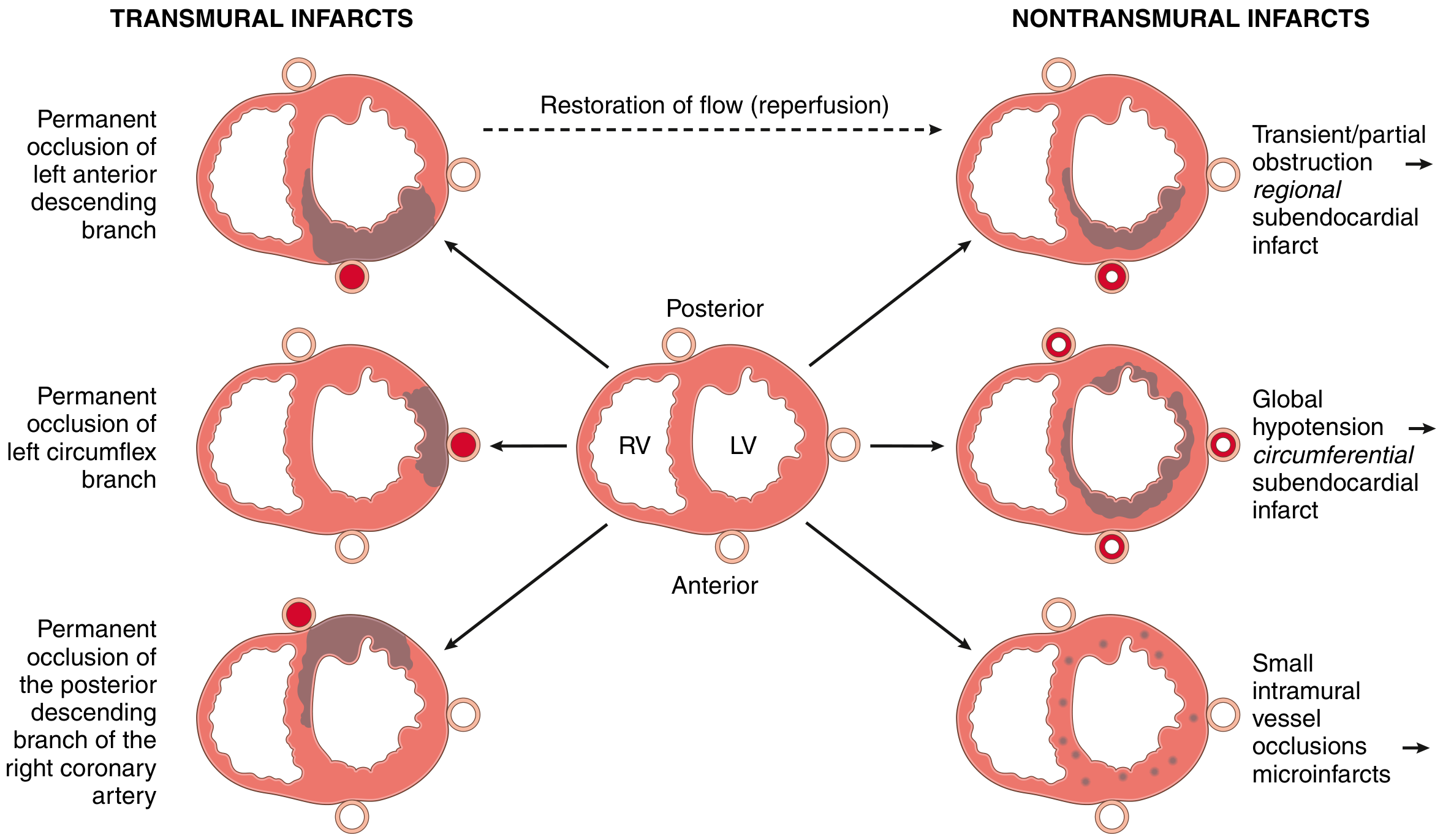
| Occluded Artery | Territory Affected |
|---|---|
| Left Anterior Descending (LAD) | Anterior wall, anterior septum, apex of LV ("widow maker") |
| Left Circumflex (LCx) | Lateral wall of LV |
| Right Coronary Artery (RCA) | Inferior/posterior LV wall, RV; SA/AV nodes |
Cellular and Biochemical Consequences
Ischemia disrupts the cell in multiple ways:
- ATP depletion → Na+/K+ ATPase fails → intracellular Na+ and Ca²+ rise
- Massive Ca²+ influx activates proteases, phospholipases → membrane damage
- Lactic acidosis → enzyme denaturation
- Free radical formation (especially on reperfusion) → oxidative membrane damage
- Sarcolemmal rupture → intracellular proteins (troponin, CK-MB, myoglobin) leak into blood → the basis of cardiac biomarkers
Reactive oxygen species and inflammatory mediators (released from dying cells) fuel the progressive wavefront of necrosis outward from the subendocardium.
ECG Changes: The Electrical Signature
Ischemic and infarcted myocardium loses its normal electrical behavior. Three membrane changes produce the classic ECG findings:
| Defect in Infarcted Cells | Current Flow | ECG Change |
|---|---|---|
| Rapid repolarization (accelerated K+ efflux) | Out of infarct | ST elevation |
| Decreased resting membrane potential (K+ loss) | Into infarct | TQ depression (manifests as ST elevation) |
| Delayed depolarization | Out of infarct | ST elevation |
Timeline of ECG changes:
- Minutes to hours: Hyperacute tall T waves → ST elevation (in STEMI)
- Hours to days: ST elevation persists; T wave inversion develops
- Days to weeks: ST normalizes; Q waves appear (electrically silent scar)
- Weeks: Q waves may persist permanently (evidence of old MI)
Leads showing ST elevation indicate which territory is infarcting:
- V1-V4: Anterior (LAD territory)
- I, aVL, V5-V6: Lateral (LCx territory)
- II, III, aVF: Inferior (RCA territory)
- Posterior MI: Tall R in V1-V2 + ST depression (reciprocal)
STEMI vs. NSTEMI
| Feature | STEMI | NSTEMI |
|---|---|---|
| Occlusion | Complete occlusion | Partial or transient occlusion |
| Infarct depth | Transmural (full thickness) | Subendocardial (partial thickness) |
| ECG | ST elevation → Q waves | ST depression / T wave changes / normal |
| Troponin | Elevated | Elevated |
| Urgency | Immediate reperfusion | Urgent but more flexible timing |
| Mortality | ~10% in-hospital | ~6% in-hospital |
Cardiac Biomarkers: Timing and Significance
When myocytes die, their internal proteins leak into the bloodstream. This is how we diagnose MI with blood tests.
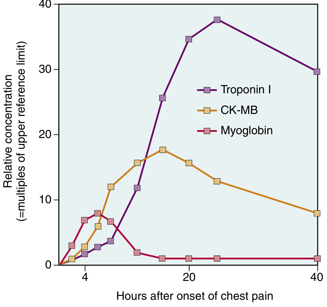
| Biomarker | Rises | Peaks | Returns to Normal | Notes |
|---|---|---|---|---|
| Myoglobin | 1-3 hrs | 6-9 hrs | 24 hrs | First to rise; NOT cardiac-specific |
| CK-MB | 2-4 hrs | 24-48 hrs | ~72 hrs | Useful for detecting reinfarction |
| Troponin I / T | 2-4 hrs | 48 hrs | 7-10 days | Most sensitive and specific; preferred marker |
| LDH | 24-48 hrs | 3-6 days | 8-14 days | Historically used; rarely tested now |
Key point: High-sensitivity troponin assays can detect MI within 1-2 hours and have largely replaced older markers. With successful reperfusion, both troponin and CK-MB peak earlier due to washout from restored flow.
Clinical Presentation
Classic symptoms:
- Severe, crushing substernal chest pain - often described as "pressure," "tightening," or "elephant on my chest"
- Pain radiates to the left arm, jaw, neck, epigastrium, or back
- Pain lasts minutes to hours (unlike angina, it does NOT resolve with rest or nitrates)
- Diaphoresis (profuse sweating)
- Nausea and vomiting (especially inferior MI - vagal activation)
- Dyspnea (from impaired LV function → pulmonary congestion)
- Rapid, weak pulse
Silent MIs (25% of cases):
Particularly common in diabetics (autonomic neuropathy blocks pain perception) and the elderly. Discovered incidentally on ECG or echo.
Women often present atypically: Fatigue, jaw pain, nausea, shortness of breath without classic chest pain - leading to delayed diagnosis and worse outcomes.
Complications of MI
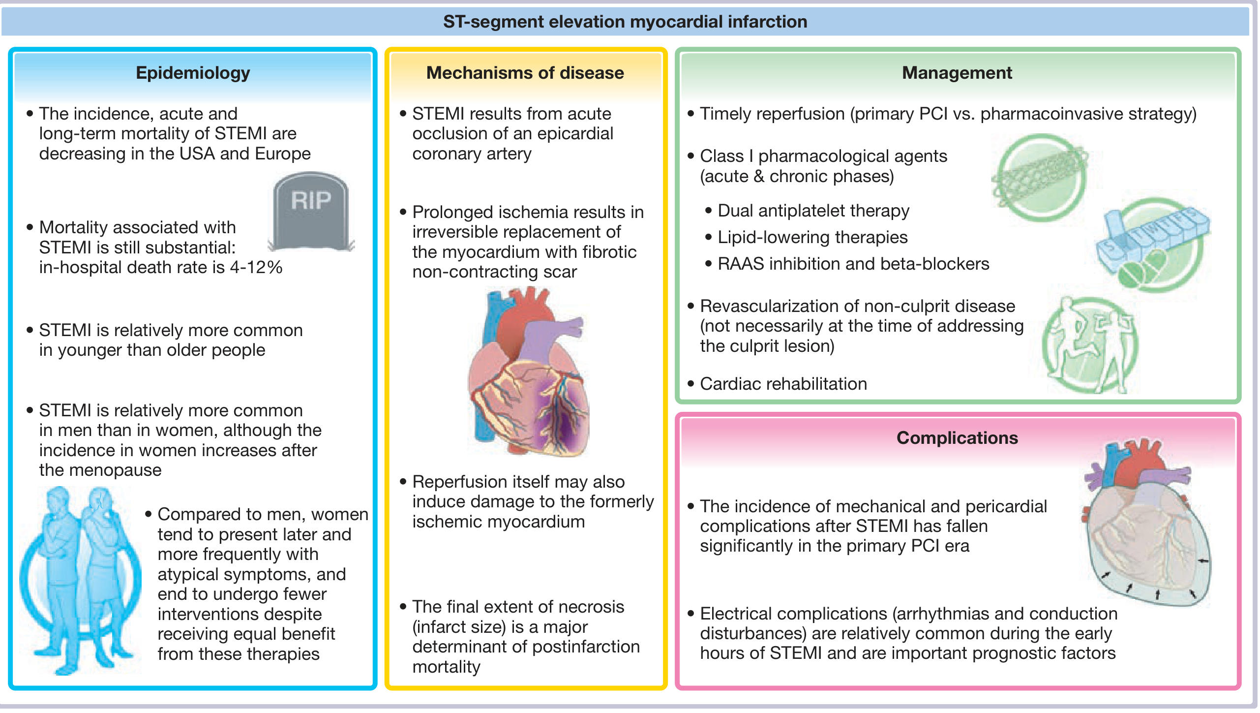
Electrical (Most Dangerous in First Hour)
- Ventricular fibrillation (VF) - leading cause of pre-hospital MI death; risk is highest in first 10 minutes, then again at 1-2 hours
- Ventricular tachycardia (VT)
- Heart block (especially with inferior MI affecting AV node via RCA)
- Bradycardia, asystole
Why does VF occur?
- Potassium leaks from ischemic cells → elevated extracellular K+ → increased myocardial irritability
- Injury current flows between ischemic and normal zones → triggers ectopic beats
- Sympathetic reflex activation (from reduced BP) → further irritability
- Dilated ventricle → longer conduction pathways → re-entry circuits
Mechanical
| Complication | Timing | Features |
|---|---|---|
| Cardiogenic shock | Early | >40% LV infarcted; mortality 40-50% despite treatment |
| Acute mitral regurgitation | Day 2-7 | Papillary muscle necrosis/rupture; sudden pulmonary edema |
| Ventricular septal rupture | Day 3-7 | New harsh pansystolic murmur; VSD; most common rupture |
| Free wall rupture | Day 3-7 | Hemopericardium, tamponade, instant death; rare |
| Right ventricular failure | Inferior MI | Hypotension, raised JVP, clear lungs |
| Acute pulmonary edema | Days | Impaired LV function → pulmonary venous congestion |
Rupture occurs most often 3-7 days after MI - when lysis of necrotic myocardium is maximal and much of the infarct has been converted to soft, friable granulation tissue. - Robbins & Kumar Basic Pathology
Late Complications
- Pericarditis - Day 2-3; friction rub, pleuritic chest pain; from transmural infarct triggering surface inflammation
- Dressler syndrome - Weeks later; autoimmune pericarditis (antibodies against injured myocardium)
- Mural thrombus - Stasis in akinetic LV + endocardial damage → thrombus formation → risk of systemic embolism/stroke
- Ventricular aneurysm - Late; thinned scar bulges during systole; risk of thrombus, arrhythmia, HF (does NOT rupture)
- Chronic ischemic cardiomyopathy / HFrEF - Progressive heart failure from cumulative myocardial loss
Reperfusion: Salvage and Its Paradox
The cornerstone of treatment is restoring blood flow as fast as possible. The formula is:
"Time is muscle" - every minute of ischemia = more irreversible muscle death
If blood flow is restored before ~20-40 minutes of ischemia, muscle can be fully saved. After that, a wavefront of necrosis is already underway, but earlier reperfusion still saves the outer layers.
Reperfusion injury - Paradoxically, restoring flow to ischemic-but-still-viable cells can itself cause damage:
- Calcium overload (intracellular Ca²+ floods in when ATP-dependent pumps restart unevenly)
- Oxygen free radical burst on reoxygenation
- Neutrophil infiltration
- Contraction band necrosis - irreversibly damaged cells exposed to calcium influx develop hypercontracted sarcomeres fixed in a permanent agonal state - this is the microscopic hallmark of reperfused infarction
Despite reperfusion injury, early restoration of flow is overwhelmingly beneficial. Stunned myocardium - viable muscle that survived ischemia but remains temporarily non-contractile for days after reperfusion - will gradually recover function.
Treatment: The Clinical Response
Immediate Goals
- Relieve ischemia and pain
- Restore blood flow (reperfusion)
- Prevent thrombus extension
- Treat life-threatening arrhythmias
STEMI - Time-Sensitive Reperfusion
Primary PCI (Percutaneous Coronary Intervention) - preferred:
- Balloon angioplasty + stent placement
- Door-to-balloon (first medical contact to device) time: ≤ 90 minutes at PCI-capable center; ≤ 120 minutes if transfer required
- Success rate > 90% for restoring flow
Fibrinolytic therapy (if PCI not available within time targets):
- Within 30 minutes of ED arrival
- Agents: Alteplase (tPA), tenecteplase, streptokinase
- Contraindicated in prior hemorrhagic stroke, recent surgery, uncontrolled hypertension
Drug Treatment (STEMI and NSTEMI)
| Drug | Purpose |
|---|---|
| Aspirin 162-325 mg (immediate) | Irreversible COX-1 inhibition → ↓ thromboxane A₂ → prevents platelet aggregation |
| P2Y12 inhibitor (clopidogrel, ticagrelor, prasugrel) | Block ADP receptor → additional antiplatelet effect (DAPT = dual antiplatelet therapy) |
| Anticoagulant (heparin, enoxaparin, fondaparinux) | Prevents thrombus propagation |
| Beta-blocker | ↓ HR and myocardial oxygen demand; reduces infarct size; prevents VF |
| Nitroglycerin | Vasodilates coronary arteries; relieves chest pain (NOT if inferior MI with RV infarct + hypotension) |
| Statin (atorvastatin 80 mg) | Plaque stabilization + lipid lowering; start immediately |
| ACE inhibitor/ARB | Start within 24 hrs; prevents LV remodeling, reduces mortality (especially with reduced EF) |
| Morphine | Pain relief; use selectively (may interfere with antiplatelet absorption) |
| Oxygen | Only if SpO₂ < 90%; routine supplemental O₂ not beneficial and may be harmful |
Post-MI (Long-term secondary prevention)
- DAPT for 12 months post-stent (aspirin + P2Y12 inhibitor)
- Lifelong aspirin
- High-intensity statin (e.g., atorvastatin 40-80 mg)
- Beta-blocker (especially if EF reduced)
- ACE inhibitor/ARB (especially if EF reduced, diabetic, or hypertensive)
- Aldosterone antagonist (eplerenone/spironolactone) if EF ≤ 40% with HF or diabetes
- Cardiac rehabilitation - exercise-based program; reduces mortality and rehospitalization
- Lifestyle: Smoking cessation, DASH diet, weight management, physical activity
Mortality and Prognosis
- 1/3 of STEMI patients die before reaching hospital - usually from VF within 1 hour of onset
- In-hospital mortality: ~10% (STEMI), ~6% (NSTEMI)
- Cardiogenic shock (>40% LV infarcted): 40-50% short-term mortality
- Long-term prognosis depends primarily on residual LV function (ejection fraction) and extent of remaining coronary disease
Summary: The Heart Attack Process in a Nutshell
Atherosclerosis (years)
        ↓
Plaque rupture / erosion (minutes)
        ↓
Platelet activation + thrombus formation (minutes)
        ↓
Complete coronary occlusion
        ↓
Ischemia begins → ATP depletion, loss of contractility (seconds)
        ↓
Irreversible cell death starts at subendocardium (20-40 min)
        ↓
Wavefront of necrosis spreads outward (hours)
        ↓
Inflammatory response → granulation tissue (days)
        ↓
Dense fibrous scar replaces dead muscle (6-8 weeks)
        ↓
Permanent loss of contractile tissue → risk of HF, arrhythmia, death
The entire reason cardiology races against the clock - "time is muscle" - is because this sequence is irreversible. Every minute of occlusion = ~2 million myocytes lost. Early reperfusion (PCI within 90 minutes) is the single most important intervention.
Sources: Robbins & Kumar Basic Pathology; Guyton & Hall Textbook of Medical Physiology; Ganong's Review of Medical Physiology 26e; Fuster & Hurst's The Heart 15e; Tintinalli's Emergency Medicine; Washington Manual of Medical Therapeutics
This is a shared conversation. Sign in to Orris to start your own chat.