Recurrence of odontogenic keratocyst and complication of dentigerous cyst

Reading File
Finding Sources
Finding Sources
Finding Sources
Reading File
Finding Sources
Reading File
Reading File
Reading File
Reading File
Finding Sources
Finding Sources
Reading File
Reading File
Searching PubMed

"odontogenic keratocyst" recurrence

Searching the Web

odontogenic keratocyst recurrence rate risk factors dentigerous cyst complications ameloblastoma transformation

Searching Images

odontogenic keratocyst histology parakeratinized stratified squamous epithelium

Searching Images

dentigerous cyst radiograph unerupted tooth
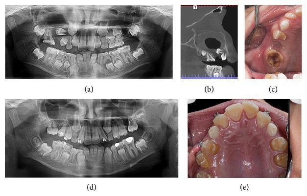
This composite of clinical and diagnostic images documents a case of a dentigerous cyst in the right maxilla and its resolution over 27 months. (a) Initial panoramic radiograph reveals a large, well-defined radiolucency associated with the crown of an unerupted maxillary tooth, typical of a dentigerous cyst. (b) Coronal CT image shows the cystic lesion's expansion within the maxillary sinus region and its relationship to the surrounding cortical bone and developing teeth. (c) Initial intraoral photograph displays a localized defect and swelling in the right maxillary alveolar ridge with visible tooth decay. (d) Follow-up panoramic radiograph at 27 months demonstrates complete bony remodeling and opacification of the previously cystic area, with permanent teeth continuing their eruption into a more normalized alignment. (e) Post-treatment intraoral view shows a fully healed, intact maxillary arch with healthy soft tissue contours and the successful preservation and eruption of teeth in the affected quadrant. The case illustrates the principles of pediatric oral surgery and the regenerative capacity of alveolar bone following cyst management.
This composite of clinical and diagnostic images documents a case of a dentigerous cyst in the right maxilla and its resolution over 27 months. (a) Initial panoramic radiograph reveals a large, well-defined radiolucency associated with the crown of an unerupted maxillary tooth, typical of a dentigerous cyst. (b) Coronal CT image shows the cystic lesion's expansion within the maxillary sinus region and its relationship to the surrounding cortical bone and developing teeth. (c) Initial intraoral photograph displays a localized defect and swelling in the right maxillary alveolar ridge with visible tooth decay. (d) Follow-up panoramic radiograph at 27 months demonstrates complete bony remodeling and opacification of the previously cystic area, with permanent teeth continuing their eruption into a more normalized alignment. (e) Post-treatment intraoral view shows a fully healed, intact maxillary arch with healthy soft tissue contours and the successful preservation and eruption of teeth in the affected quadrant. The case illustrates the principles of pediatric oral surgery and the regenerative capacity of alveolar bone following cyst management.

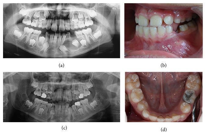
A multi-panel clinical figure showing diagnostic imaging and intraoral photographs of a pediatric patient with a dentigerous cyst (DC). Panel (a) is a baseline panoramic radiograph (orthopantomogram) of a mixed dentition, revealing a well-defined, unilocular radiolucency in the left mandibular region associated with the crown of an unerupted permanent tooth, causing tooth displacement. Panel (b) shows a corresponding clinical photograph of the left mandible with visible alveolar swelling and erythematous mucosa. Panels (c) and (d) provide 12-month follow-up views. The follow-up radiograph (c) demonstrates significant bone regeneration, resolution of the cystic radiolucency, and progression of tooth eruption. The occlusal intraoral photograph (d) shows the successful eruption of the previously affected permanent teeth into the oral cavity with healthy gingival tissue. The series illustrates the clinical management and healing of an odontogenic cyst in a pediatric patient within the specialty of pediatric dentistry and oral maxillofacial surgery.
A multi-panel clinical figure showing diagnostic imaging and intraoral photographs of a pediatric patient with a dentigerous cyst (DC). Panel (a) is a baseline panoramic radiograph (orthopantomogram) of a mixed dentition, revealing a well-defined, unilocular radiolucency in the left mandibular region associated with the crown of an unerupted permanent tooth, causing tooth displacement. Panel (b) shows a corresponding clinical photograph of the left mandible with visible alveolar swelling and erythematous mucosa. Panels (c) and (d) provide 12-month follow-up views. The follow-up radiograph (c) demonstrates significant bone regeneration, resolution of the cystic radiolucency, and progression of tooth eruption. The occlusal intraoral photograph (d) shows the successful eruption of the previously affected permanent teeth into the oral cavity with healthy gingival tissue. The series illustrates the clinical management and healing of an odontogenic cyst in a pediatric patient within the specialty of pediatric dentistry and oral maxillofacial surgery.

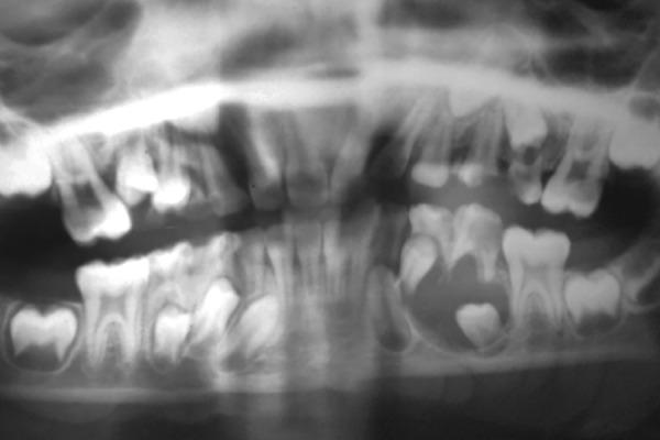
A panoramic dental radiograph (Orthopantomogram) of a pediatric or adolescent patient in the mixed dentition stage. The primary finding is a large, well-circumscribed, unilocular radiolucency located in the left mandibular body. The lesion completely envelopes the crown of the unerupted mandibular left second premolar, with its attachment appearing at the cervical margin (cementoenamel junction), characteristic of a dentigerous cyst. This pathological entity has caused significant inferior apical displacement of the involved tooth bud. Additionally, the adjacent mandibular left second primary molar shows extensive coronal radiolucency indicating deep dental caries with pulp involvement, accompanied by external root resorption and furcation radiolucency. The radiograph illustrates key diagnostic features of odontogenic cysts and the impact of associated primary tooth pathology on developing permanent dentition.

A panoramic dental radiograph (Orthopantomogram) of a pediatric or adolescent patient in the mixed dentition stage. The primary finding is a large, well-circumscribed, unilocular radiolucency located in the left mandibular body. The lesion completely envelopes the crown of the unerupted mandibular left second premolar, with its attachment appearing at the cervical margin (cementoenamel junction), characteristic of a dentigerous cyst. This pathological entity has caused significant inferior apical displacement of the involved tooth bud. Additionally, the adjacent mandibular left second primary molar shows extensive coronal radiolucency indicating deep dental caries with pulp involvement, accompanied by external root resorption and furcation radiolucency. The radiograph illustrates key diagnostic features of odontogenic cysts and the impact of associated primary tooth pathology on developing permanent dentition.

Now I have enough to compile a complete answer.

Recurrence of Odontogenic Keratocyst (OKC)

Recurrence rates and why they occur
OKC (still classified by WHO's ICD-11 as an odontogenic tumor despite the 2017 reversion to "odontogenic keratocyst" terminology) is notorious for its high recurrence rate compared with other jaw cysts.
  • With enucleation alone, historical recurrence rates as high as 60-62.5% have been reported (- Robbins, Cotran & Kumar Pathologic Basis of Disease; - Cummings Otolaryngology Head and Neck Surgery, p. 1622).
  • With modern enucleation plus careful curettage, most contemporary series report recurrence under 10%.
  • Comparative data: enucleation alone 27.8-30.8%, versus bone resection 0-8.4% (per recent case-report literature synthesis).
  • The parakeratinized variant recurs far more often (~47.8%) than the orthokeratinized variant (~2.2%), based on the Crowley et al. series of 449 cases - orthokeratinized OKC is now often considered a separate, less aggressive entity.
Mechanisms proposed for recurrence:
  1. Daughter/satellite cyst formation left behind in adjacent bone or overlying mucosa
  2. Incomplete removal of the thin, friable epithelial lining (which tears easily during enucleation)
  3. Dental lamina rests remaining in the cyst wall or overlying mucosa
  4. Collagenase activity within the cyst
  5. Prostaglandin-induced bone resorption
  6. Increased mitotic activity/intrinsic epithelial proliferative potential (supports the lesion's neoplastic behavior)
Risk factors for recurrence: younger patient age (14-17 years), larger lesion size, posterior mandible location, multilocularity, presence of satellite/daughter cysts, and association with nevoid basal cell carcinoma syndrome (Gorlin syndrome) - caused by germline loss-of-function mutations in the PTCH1 tumor suppressor gene. Patients with multiple or syndromic OKCs are more prone to aggressive/recurrent behavior and should be evaluated for Gorlin syndrome (- Robbins Pathologic Basis of Disease, ch. 16; - Cummings Otolaryngology, p. 1622).
Timing: Most recurrences appear within the first 5-7 years, but recurrence has been documented decades later - Cummings describes a case recurring 40 years after initial treatment, so long-term radiographic follow-up is required indefinitely.
Treatment approaches aimed at reducing recurrence: enucleation with peripheral ostectomy, excision of overlying mucosa, chemical curettage with Carnoy's solution (controversial due to neurotoxicity near the inferior alveolar nerve), 5-fluorouracil, marsupialization/decompression followed by enucleation for large lesions, or en-bloc resection for aggressive/recurrent/syndromic cases. A 2025 systematic review/meta-analysis examined alternative agents to Carnoy's solution for recurrence prevention (PMID: 40269316), and a 2024 systematic review covered management techniques and recurrence risk factors in detail (PMID: 38750607) - both consistent with the textbook data above, with no major contradictions.
Rare but serious complications reported in case literature include intramuscular/soft-tissue extension, extension into adjacent bone, and even death from intracranial extension of a mandibular OKC - though these remain exceptional and shouldn't drive over-aggressive treatment of every lesion.

Complications of Dentigerous Cyst

The dentigerous (follicular) cyst forms from fluid accumulation between the reduced enamel epithelium and the crown of an unerupted tooth (most often third molars and maxillary canines). Unlike OKC, complete surgical excision (enucleation) is curative and simple recurrence is not the typical concern. Instead, its clinically important complications are:
  1. Neoplastic transformation - dentigerous cysts retain the capacity to transform into true neoplasms:
    • Up to 17% of ameloblastomas have been reported arising in association with a pre-existing dentigerous cyst (figure varies across studies)
    • Squamous cell carcinoma and mucoepidermoid carcinoma have both been reported arising within dentigerous cyst linings (- Cummings Otolaryngology, "Neoplastic Potential," p. 1610)
  2. Residual cyst formation - if a dentigerous cyst is incompletely removed during tooth extraction, the remnant can persist and grow as a "residual cyst," histologically and radiographically identical to a residual periapical cyst.
  3. Mechanical/structural effects from expansion - as they enlarge (especially when large or long-standing), dentigerous cysts can cause:
    • Displacement of the involved tooth or adjacent teeth
    • Root resorption of adjacent teeth
    • Cortical bone expansion/thinning, and pathologic fracture risk in large lesions
    • Impingement on adjacent structures (e.g., maxillary sinus, inferior alveolar canal)
  4. Diagnostic overlap/pitfall - inflamed dentigerous cysts can be histologically indistinguishable from pericoronitis or from an odontogenic keratocyst presenting around an unerupted tooth, so radiographic-clinical correlation is required to avoid missing a more aggressive lesion.
Management: usually straightforward enucleation at the time of tooth extraction; large lesions may be managed first with decompression/marsupialization followed by enucleation to preserve adjacent structures and reduce surgical morbidity (- Cummings Otolaryngology Head and Neck Surgery, "Dentigerous Cyst," pp. 1609-1610).
Example radiograph of a dentigerous cyst enveloping an unerupted tooth, with displacement of the tooth bud:
Dentigerous cyst panoramic radiograph
Bottom line comparison: OKC's clinical problem is recurrence driven by its intrinsically aggressive, neoplasm-like epithelium and satellite cyst formation, even after apparently complete removal. Dentigerous cyst's clinical problem is less about recurrence (excision is usually curative) and more about what it can transform into (ameloblastoma, SCC, mucoepidermoid carcinoma) or leave behind if incompletely excised (residual cyst), plus mechanical effects of expansion.
This is a shared conversation. Sign in to Orris to start your own chat.