
Nodule in pathology
pulmonary nodule CT scan pathology
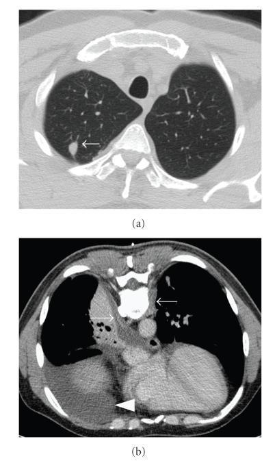
This diagnostic figure consists of two axial CT scan sections of the chest demonstrating pulmonary and pleural pathology. (a) A non-contrast axial CT in lung window settings reveals a discrete, approximately 1.5 cm nonspecific pulmonary nodule in the right upper lobe, indicated by a white arrow. The nodule is well-circumscribed and located within the peripheral lung parenchyma. (b) A contrast-enhanced axial CT scan performed in the prone position at the level of the 9th thoracic vertebra. It displays bilateral latero-spinal subpleural thickening (indicated by arrows) and a localized right-sided pleural effusion (indicated by a white arrowhead). The image demonstrates typical radiological findings associated with systemic inflammatory or granulomatous diseases such as Granulomatosis with Polyangiitis (GPA). These diagnostic images serve as an educational example of how multisystemic conditions present with varied intrathoracic manifestations, including parenchymal nodules and pleural involvement, requiring contrast-enhanced imaging for better soft tissue characterization.
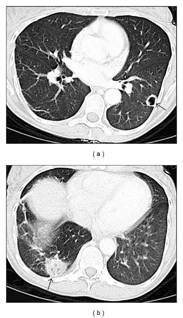
Two axial CT scan slices of the chest in lung window demonstrate multifocal pulmonary pathology. Panel (a) shows a 1.7 cm cavitary nodule (indicated by an arrow) located peripherally in the left lower lobe. The nodule features a relatively thick wall and a central area of lucency, suggesting necrosis or cavitation. Panel (b) reveals a larger 3 cm mass-like infiltrate or consolidation (indicated by an arrow) in the right lower lobe, also positioned peripherally near the pleural surface. The infiltrate displays irregular borders and increased density compared to normal lung parenchyma. These findings are characteristic of infectious processes such as disseminated mycobacterial infection (e.g., Mycobacterium fortuitum) or nocardiosis, which often manifest with peripheral nodules, cavitation, and multifocal pneumonia in immunocompromised or critically ill patients. The image is suitable for medical education in pulmonology, radiology, and infectious diseases.
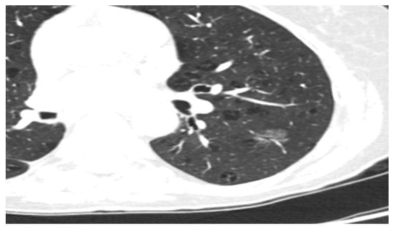
Diagnostic axial CT scan of the chest presented in lung window settings, demonstrating a localized area of pathology within the pulmonary parenchyma. The primary finding is a solitary, pure ground-glass nodule (GGN) located in the peripheral region of the left lung. This nodule is characterized by a hazy increase in lung density that does not obscure the underlying bronchial and vascular structures, consistent with adenocarcinoma in situ (AIS). The nodule exhibits a rounded morphology with ill-defined borders that blend into the surrounding normal lung tissue. There are no associated signs of solid components, pleural retraction, or spiculated margins. The background lung parenchyma appears otherwise clear with normal vascular markings. This image serves as a clinical example of early-stage lung adenocarcinoma screening and the radiographic features used to differentiate non-solid nodules from solid pulmonary masses.
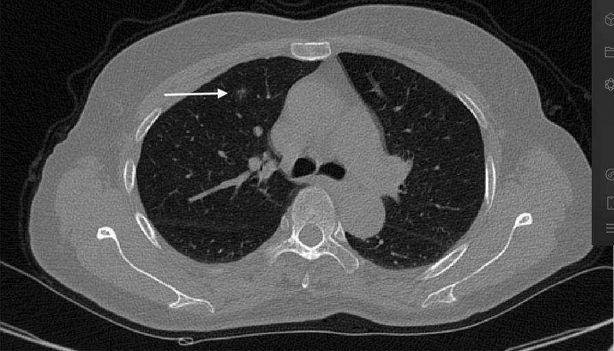
An axial-view non-contrast chest computerized tomography (CT) scan illustrating thoracic anatomy and pulmonary pathology. The image displays a cross-section of the thoracic cavity including the lungs, mediastinum, heart, and musculoskeletal framework (vertebral body, ribs, and scapulae). A white arrow points to a solitary, small pulmonary nodule with ground-glass opacity characteristics located in the anterior segment of the right upper lobe. The nodule is situated within the peripheral lung parenchyma, in relative proximity to the anterior chest wall and the fourth rib. The imaging demonstrates varying radiodensities: low-density (black) air-filled lung tissue, intermediate-density (grey) soft tissues of the heart and thoracic wall musculature, and high-density (white) cortical bone. This diagnostic image is used in medical education to teach anatomical localization of lung lesions and the visual identification of early-stage pulmonary nodules during radiological screening or pre-operative planning.
Nodule in Pathology
Definition
- Micronodule: < 3 mm
- Nodule: 3 mm - 20 mm (some sources use 3-30 mm)
- Mass: > 20-30 mm (sometimes called a "tumor" regardless of biological behavior)
Pulmonary Nodule (Most Clinically Significant)
Classification by Attenuation (CT)
| Type | CT Appearance | Significance |
|---|---|---|
| Solid | Homogeneous soft-tissue attenuation; obscures underlying vessels | Most common; variable malignant risk |
| Non-solid (Ground Glass) | Hazy increased density; does NOT obscure vessels/bronchi | Lower risk but associated with adenocarcinoma spectrum; volume doubling time ~813 days |
| Part-solid | Mixed: non-solid region + solid component | Highest malignant risk per unit size; volume doubling time ~457 days |
Classification by Morphology / Contour
| Margin | Likelihood of Malignancy |
|---|---|
| Smooth/well circumscribed | More likely benign |
| Lobulated | More likely malignant (LR ~11 vs smooth) |
| Spiculated ("corona radiata") | Strongly predictive of malignancy (LR ~5.5-7) |
| Pleural retraction | Associated with malignancy (LR ~1.9) |
| Perifissural / lentiform | Usually an intrapulmonary lymph node - benign |
- Vascular convergence - vessels angling abnormally toward the nodule
- Pseudocavitation ("bubble" / soap-bubble appearance) - seen in adenocarcinoma with lepidic growth
- True cavitation with thick, irregular wall (>15 mm wall thickness: ~95% malignant; <5 mm: ~92% benign)
Classification by Calcification Pattern
| Pattern | Significance |
|---|---|
| Central / diffuse | Benign (post-infectious granuloma) |
| Laminar / concentric | Benign (classic for histoplasmosis) |
| Popcorn + internal fat | Highly specific for hamartoma |
| Punctate or eccentric | Indeterminate - seen in 6-14% of malignant nodules |
CT Images of Pulmonary Nodules
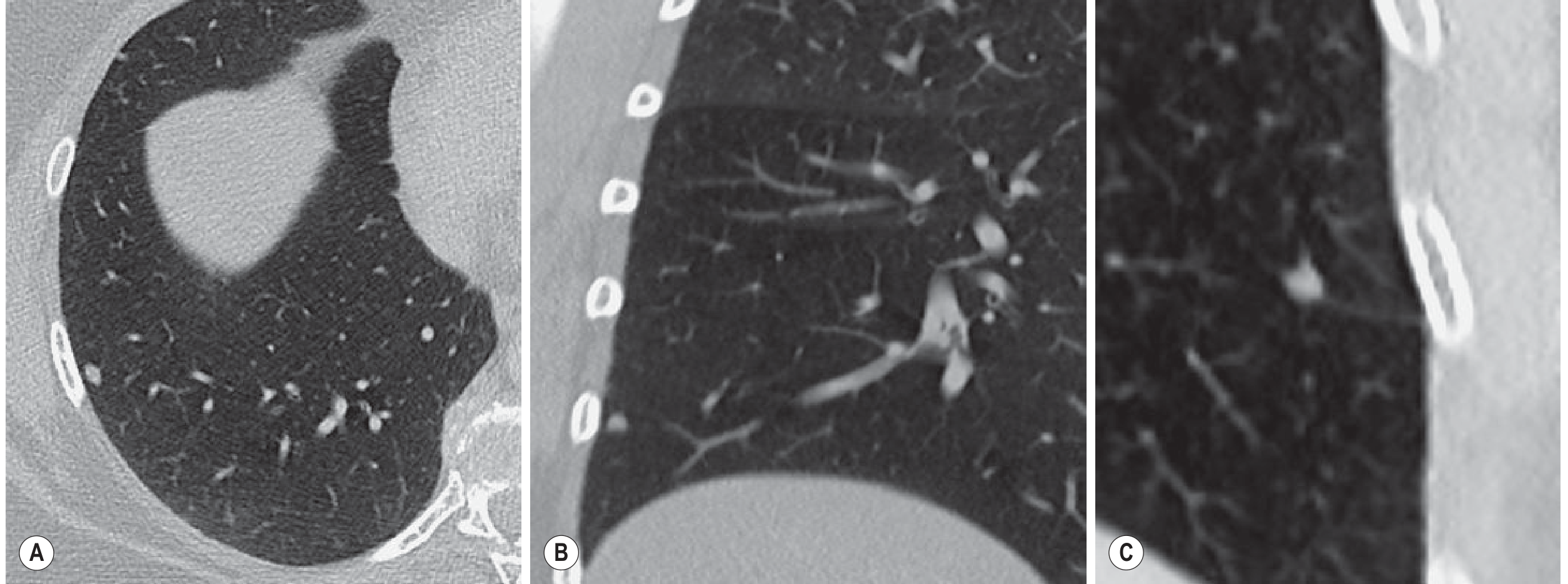
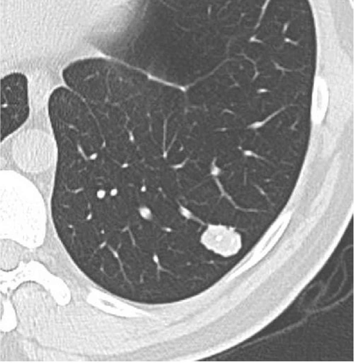
Size and Malignancy Risk
- Solid nodule ≥6 mm bidirectional average: positive screen result
- Non-solid nodule ≥30 mm: positive screen result (increased threshold in Lung-RADS 1.1, reflecting indolent behavior)
- Solid nodule stable for ≥2 years on CT: almost certainly benign - no further workup needed
- Malignancy prevalence for nodules 4-6 mm: <1%
Lung-RADS Scoring System
- Nodule size
- Nodule attenuation (solid vs. subsolid)
- Nodule morphology (margins, calcification)
- Growth on serial studies (baseline vs. interval scan)
Nodules in Other Organs
Thyroid Nodule
- Very common; most are benign
- Cold nodule (non-functioning on radionuclide scan): higher malignancy risk - evaluated with FNAC
- Hot/toxic nodule (hyperfunctioning): almost always benign adenoma; causes mild thyrotoxicosis when >3 cm
- Ultrasound U-classification guides biopsy: U1 (normal) through U5 (highly suspicious) - Scott-Brown's Otorhinolaryngology
- Hard, fixed nodule, rapid growth, or hoarseness (vocal cord paralysis) suggests malignancy - Washington Manual
Skin Nodule
- Firm, palpable, rounded dermal or subcutaneous lesion, typically 0.5-2 cm
- Dermatofibroma (fibrous histiocytoma): single, round/oval papule or nodule ~0.5-1 cm; benign - Andrews' Diseases of the Skin
- Erythematous nodules can be the initial lesion in pyoderma gangrenosum
- Skin-colored or pink nodule on sun-exposed skin with lack of classic melanoma features: may be desmoplastic melanoma
Key Pathological Principles for Any Nodule
- Size matters - larger = higher malignancy risk (though not absolute)
- Margins matter - spiculated > lobulated > smooth in terms of malignancy risk
- Density matters - calcification pattern can confirm benignity or raise concern
- Growth matters - stability over 2 years (CT) is highly reassuring; rapid growth is concerning
- Context matters - patient history (smoking, age, prior malignancy, endemic fungal exposure) shifts pre-test probability dramatically
Stromal
stromal tumor histology GIST gastrointestinal
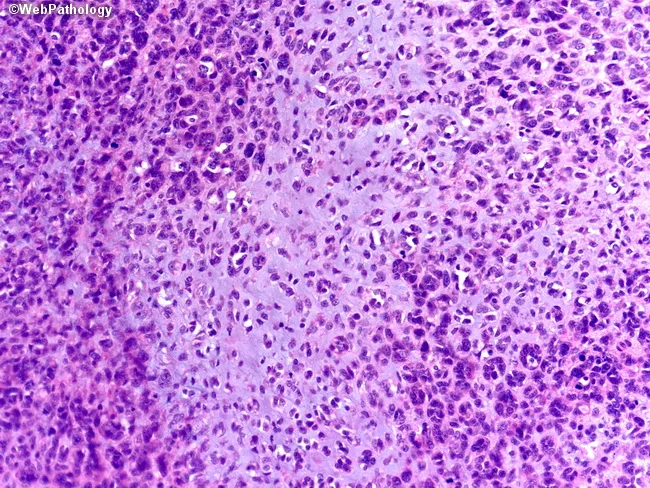
This histology image depicts a gastrointestinal stromal tumor (GIST) in a hematoxylin and eosin stained tissue section. The slide shows background myxoid matrix with focal decreased cellularity, a feature more common in epithelioid GISTs than spindle cell GISTs. Tumor cells are arranged in clusters within a loose, mucoid stroma; epithelioid-appearing cells with round to polygonal nuclei and eosinophilic cytoplasm predominate in the myxoid zones, while denser cellular areas display spindle-shaped cells. The intercellular network includes a delicate capillary network, and mitotic figures are variable, often scarce in myxoid regions but may be present in denser tumor nests. Such myxoid change can complicate histologic interpretation and broaden the differential to other myxoid mesenchymal neoplasms (myxoid leiomyosarcoma, schwannoma, liposarcoma); confirmation with KIT (CD117) and DOG1 immunostains and KIT/PDGFRA mutational analysis is recommended. Clinically, recognizing this histologic variant supports tumor localization within the GI tract, influences risk stratification, and informs targeted therapy decisions (imatinib). The image emphasizes the spectrum of GIST architecture, including myxoid stroma, variable cellularity, epithelioid morphology, and the necessity for ancillary testing to establish a definitive diagnosis and guide management. These features have implications for prognosis and response to therapy. Ancillary testing, including immunohistochemistry and mutational profiling, is essential.
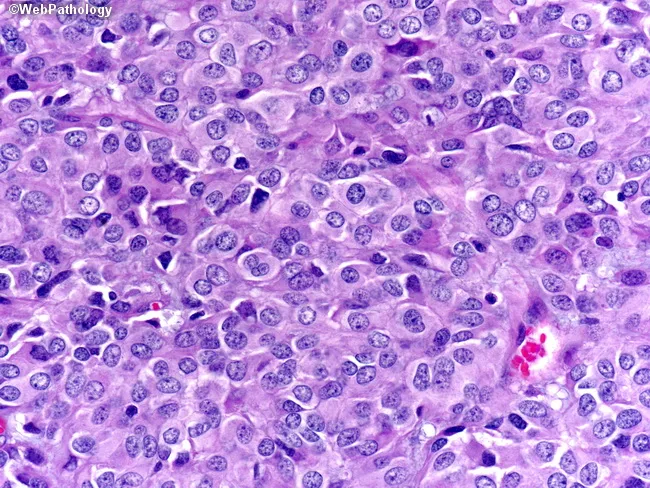
Histology image acquired by bright-field microscopy of a gastrointestinal stromal tumor (GIST), epithelioid variant, in a high-power field after hematoxylin and eosin staining. The tumor shows high cellularity with sheets and nests of round to polygonal epithelioid cells, abundant eosinophilic to clear cytoplasm, and conspicuous nucleoli. Nuclear crowding and marked pleomorphism are evident, with occasional mitotic figures noted at this magnification. The cells are separated by a delicate to moderate amount of hyalinized stroma and exhibit a relatively uniform, cohesive architecture despite cellular pleomorphism; there is minimal necrosis and no evident keratinization. This epithelioid morphology can closely resemble epithelioid carcinomas or melanoma, contributing to a broad differential diagnosis. In practice, immunohistochemical profiling (e.g., CD117/c-KIT, DOG1 positivity; cytokeratins and melanocytic markers typically negative) is essential to confirm GIST and exclude carcinoma or melanoma. Clinically, epithelioid GISTs may present in the stomach or small intestine and can be associated with activating KIT or PDGFRA mutations; prognosis depends on tumor size, mitotic rate, and location, impacting management with surgical resection and targeted therapy (imatinib). This image illustrates the histopathologic challenge of distinguishing epithelioid GIST from metastatic melanoma or epithelial malignancies on routine H&E, underscoring the need for ancillary testing in diagnostic workflows today.
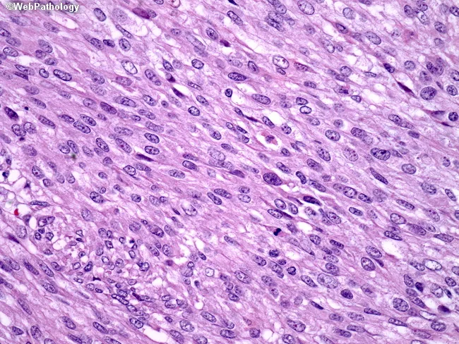
This is a high-magnification histopathology image of a gastrointestinal stromal tumor (GIST) showing a spindle cell-dominant morphology. Tissue is examined by light microscopy after Hematoxylin and Eosin staining, at ×400 magnification, highlighting elongated, fusiform cells arranged in intersecting fascicles with ovoid to elongated nuclei and eosinophilic cytoplasm. The stroma appears fibrous to lightly myxoid, with preserved cellular detail and minimal necrosis in this field. The spindle cell population is characteristic of many GISTs and supports a mesenchymal neoplasm of the gastrointestinal tract. In clinical practice, GISTs commonly harbor activating mutations in the c-KIT (CD117) gene in about 80% of cases or in PDGFRA (~8%), leading to constitutive tyrosine kinase signaling. Immunohistochemical CD117 positivity is a key diagnostic surrogate; DOG1 is frequently positive and may aid in challenging cases. Accurate molecular testing for c-KIT and PDGFRA mutations informs prognosis and eligibility for targeted therapy with tyrosine kinase inhibitors such as imatinib. This image serves as a reference for recognizing spindle cell GIST histology, differentiating it from smooth muscle or neural tumors, and illustrating how morphology guides ancillary testing, mutation profiling, and therapeutic decision-making in gastroenterology oncology and surgical pathology. This education-oriented caption emphasizes diagnostic workflow and targeted therapy implications for practice.
Stroma in Pathology
Definition
"All tumors, benign and malignant, have two basic components: (1) parenchyma, made up of transformed or neoplastic cells; and (2) stroma, the supporting host-derived, nonneoplastic connective tissue, inflammatory cells, and blood vessels."
"Within most organs, connective tissue proper forms the supportive stroma, which supports the organ's unique functional components or parenchyma."
Components of Stroma
| Component | Role |
|---|---|
| Fibroblasts / fibrocytes | Synthesize collagen, elastin, and ground substance |
| Extracellular matrix (ECM) | Protein fibers (collagen, elastin) + ground substance (GAGs, proteoglycans) |
| Blood vessels | Deliver oxygen and nutrients to parenchymal cells |
| Inflammatory / immune cells | Macrophages, lymphocytes, mast cells - homeostasis and defense |
| Smooth muscle / myofibroblasts | Structural support, wound contraction |
Stroma in Tumors (Tumor Microenvironment)
- Desmoplasia - dense fibrous stroma, characteristic of some carcinomas (e.g., scirrhous breast cancer, pancreatic ductal adenocarcinoma)
- Angiogenesis - new vessel formation driven by tumor-secreted VEGF
- Cancer-associated fibroblasts (CAFs) - activated fibroblasts that support tumor invasion and suppress immune responses
Stromal Tumors by Organ System
1. Breast - Stromal Neoplasms
| Stroma Type | Tumors Arising |
|---|---|
| Intralobular stroma | Fibroadenoma (benign), Phyllodes tumor (variable) |
| Interlobular stroma | Lipoma, angiosarcoma, myofibroblastoma |
- Driver mutations in MED12 (~2/3 of cases) - encodes a subunit of the Mediator transcription complex (same gene mutated in uterine leiomyoma)
- Additional mutations in RARA (retinoic acid receptor alpha) in ~1/3
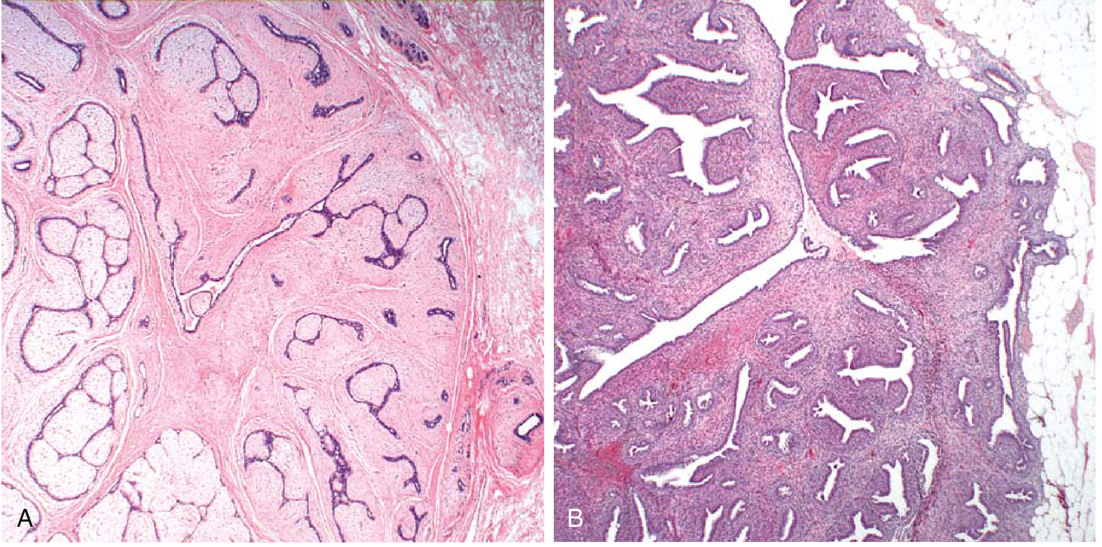
- Morphology: well-circumscribed, rubbery, gray-white nodule; delicate myxoid stroma surrounds slit-like epithelial spaces
- Low-cellularity stromal proliferation with reactive (non-neoplastic) epithelium
- Stromal cells outgrow epithelial cells, forming bulbous "leaf-like" (phyllodes) nodules
- Range from benign to borderline to malignant (rare); may recur after excision
- High-grade: sarcomatous appearance with scant/absent epithelium
2. Uterus / Endometrium - Endometrial Stromal Tumors
| Type | Features |
|---|---|
| Benign stromal nodule | Well-circumscribed; does not invade |
| Low-grade endometrial stromal sarcoma | Infiltrating; JAZF1-SUZ12 fusion gene (chromosomal translocation disrupts polycomb complex); 5-yr survival: ~90% (stage I/II), ~50% (stage III/IV) |
| High-grade endometrial stromal sarcoma | YWHAE-NUTM2A/B or ZC3H7B-BCOR fusions; more aggressive; distant metastases may appear decades later |
3. Gastrointestinal Stromal Tumor (GIST)
- ~80%: activating gain-of-function mutation in KIT (CD117) - a transmembrane tyrosine kinase receptor
- ~8%: PDGFRA mutations - another tyrosine kinase receptor; often epithelioid morphology
- Small subset: neither KIT nor PDGFRA (other kinase mutations)
- Spindle cell (most common): elongated fusiform cells in intersecting fascicles
- Epithelioid: round to polygonal cells with prominent nucleoli; harder to distinguish from carcinoma/melanoma on H&E alone
- Mixed
| Risk Level | Size | Mitoses/50 HPF |
|---|---|---|
| Very low | < 2 cm | < 5 |
| Low | 2-5 cm | < 5 |
| Intermediate | < 5 cm / 5-10 cm | 6-10 / < 5 |
| High | > 5 cm OR > 10 cm | > 5 / Any |
4. Ovarian Stroma
- Virilization (elevated testosterone, DHT, androstenedione)
- Hyperinsulinemia and glucose intolerance in up to 90% of patients
- HAIR-AN syndrome (hyperandrogenism, insulin resistance, acanthosis nigricans)
- Normal (not elevated) gonadotropins - distinguishes from PCOS
Summary Table: Stromal vs. Parenchymal Tumors
| Feature | Stromal (Mesenchymal) Tumors | Epithelial / Parenchymal Tumors |
|---|---|---|
| Cell of origin | Fibroblasts, smooth muscle, endothelium, ICC | Epithelial cells |
| Naming (benign) | -oma (fibroma, lipoma, leiomyoma) | Adenoma, papilloma |
| Naming (malignant) | Sarcoma | Carcinoma |
| Spread | Hematogenous (blood-borne) | Lymphatic (then blood) |
| Examples | GIST, fibroadenoma, phyllodes, leiomyoma, angiosarcoma | Breast carcinoma, colon adenocarcinoma |