
Tonsilitis
tonsillitis inflamed tonsils throat exudate
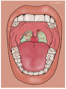
Summary : This illustration shows the inside of a human mouth with a focus on the throat, depicting inflamed tonsils with white-greenish patches, suggestive of tonsillitis or a similar infection. photo: Scene Overview : • Main subject is the open mouth and throat, viewed from the front. • The tonsils are visibly enlarged and covered with white-greenish exudate. • The uvula is centered and appears slightly swollen. • The tongue is prominent in the lower half of the image. • Teeth are visible along the top and bottom edges of the mouth. • The background is a solid, muted color, emphasizing the oral cavity. Technical Details : • No scale bar or magnification indicated. • No staining or medical imaging modality used; this is a digital illustration. • No on-image text except a faint watermark in the upper right corner. Spatial Relationships : • Tonsils are located on either side of the uvula, at the back of the throat. • White-greenish patches are localized to the tonsils, not extending to other oral structures. • The tongue occupies the foreground, with the throat and tonsils in the background. Analysis : • The key visual message is the presence of inflamed, exudate-covered tonsils, which is characteristic of acute tonsillitis or pharyngitis. • The illustration highlights the typical appearance of infected tonsils, useful for educational or diagnostic reference.
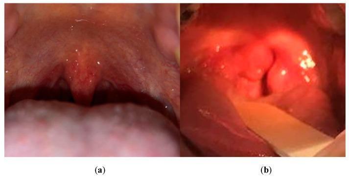
This comparison clinical photograph depicts a healthy throat (a) and a throat with pharyngitis, specifically strep throat (b). Image (a) shows normal oropharyngeal anatomy with a uniform pink mucosal lining, a neutral-sized uvula, and smooth tonsillar pillars without exudate or swelling. In contrast, image (b) demonstrates classic clinical signs of streptococcal pharyngitis, including significant erythema (redness) of the oropharynx, a swollen (edematous) uvula, and enlarged tonsils. Notably, whitish-yellow spots or patches, consistent with tonsillar exudates and petechiae, are visible on the inflamed tissue. The comparison highlights the transition from healthy smooth texture and consistent color to the irregular, highly vascularized, and exudative appearance characteristic of bacterial inflammation. This visual is used in medical education to assist in the clinical diagnosis of upper respiratory infections and to differentiate between normal anatomical variations and pathological inflammatory states.
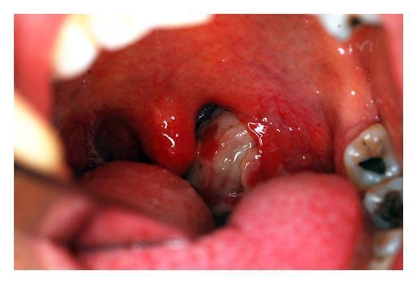
This clinical photograph shows an intraoral view of the oropharynx, demonstrating acute tonsillopharyngitis. There is generalized, intense hyperemia (redness) and congestion of the pharyngeal mucosa and palatine tonsils. Significant edema is visible in the uvula and the anterior tonsillar pillars. The left tonsil is prominently enlarged and almost entirely covered with a thick, diffuse, whitish-yellow purulent exudate. The right tonsil also appears hyperemic and swollen, though the exudate is more localized on the left. The image serves as an educational example of severe pharyngeal infection, illustrating physical findings such as exudative tonsillitis, which can be seen in various bacterial (e.g., Chlamydia trachomatis, Group A Streptococcus) or viral (e.g., Infectious Mononucleosis) etiologies. The educational focus is on the visual identification of tonsillar hypertrophy, purulent coating, and surrounding soft tissue inflammation.
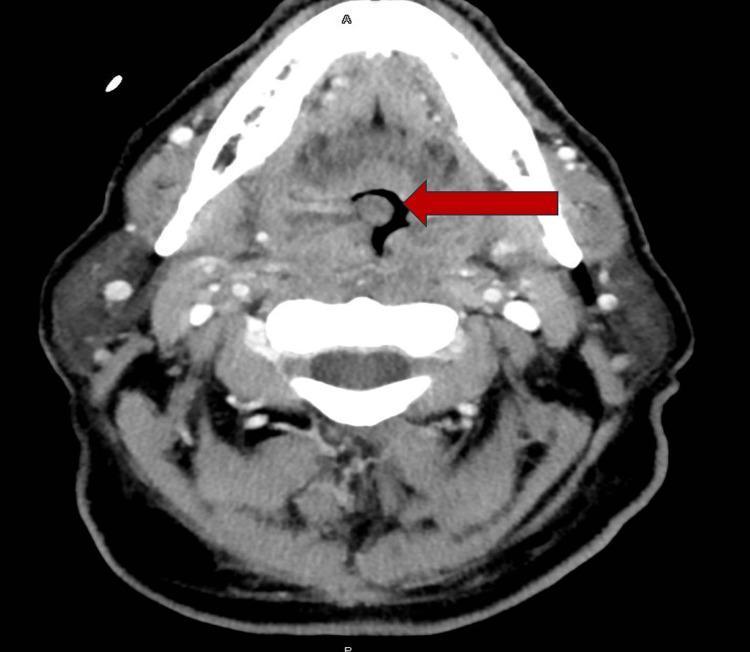
This diagnostic image is an axial computed tomography (CT) scan of the neck with intravenous contrast. The primary finding is significant bilateral palatine tonsillar enlargement (tonsillar hypertrophy). The enlarged tonsillar tissue is characterized by heterogeneous attenuation, with distinct internal hypoattenuating areas (measuring approximately 0.5 cm) suggestive of small intratonsillar abscesses or microabscesses. A large red arrow points to the resulting severe narrowing of the oropharyngeal airway, which is significantly compromised by the mass effect of the inflamed tonsils. Surrounding soft tissue structures demonstrate inflammatory changes consistent with infectious tonsillitis or pharyngitis. This imaging modality is used in Otolaryngology to differentiate between uncomplicated cellulitis and abscess formation requiring surgical drainage, and to assess for airway obstruction in severe upper respiratory infections.
tonsillitis streptococcal management
Tonsillitis
Definition & Anatomy
Etiology
| Pathogen | Syndrome | ~% of Cases |
|---|---|---|
| Streptococcus pyogenes (GABHS) | Pharyngitis, tonsillitis, scarlet fever | 10-23% |
| Fusobacterium necrophorum | Tonsillitis, Lemierre's syndrome | 5-10% |
| Group C Streptococcus | Pharyngitis, tonsillitis | 3-6% |
| Neisseria gonorrhoeae | Pharyngitis | <1% |
| Corynebacterium diphtheriae | Diphtheria | <1% |
| Pathogen | Syndrome | ~% of Cases |
|---|---|---|
| Rhinovirus | Common cold | 15-20% |
| Coronavirus | Common cold | >5% |
| Adenovirus | Pharyngoconjunctival fever | 6% |
| Herpes simplex virus | Gingivitis, stomatitis, pharyngitis | 4% |
| Parainfluenza virus | Common cold, croup | 2% |
| Influenza A/B | Influenza | 2% |
| Epstein-Barr virus (EBV) | Infectious mononucleosis | <1% |
| HIV type 1 | Acute retroviral syndrome | <1% |
Clinical Features
- Sore throat - usually sudden onset
- Fever and general malaise
- Dysphagia / odynophagia - painful swallowing
- Referred otalgia - ear pain (via glossopharyngeal nerve)
- Voice change - "hot potato" voice in severe cases
- On exam: tonsils are swollen and erythematous; yellow/white exudates or pustules may be visible ("follicular tonsillitis")
- Jugulodigastric (tonsillar) lymph node enlargement - tender, palpable
Diagnosis
- Primarily clinical
- Throat swab for culture and sensitivity (bacterial vs. viral)
- Blood for EBV testing (Monospot test) - to rule out infectious mononucleosis - sensitivity <50% in children, 70-90% in adults; confirm with specific antibody titres
- Full blood count: leukocytosis with bacterial infection; atypical lymphocytes in EBV
- Rapid antigen detection tests (RADT) for Group A Strep
Centor / McIsaac Scoring (GABHS probability)
- Tonsillar exudate
- Tender anterior cervical lymphadenopathy
- Fever (>38°C)
- Absence of cough (Score 3-4 = likely bacterial; score 0-1 = likely viral)
Types
1. Acute Tonsillitis
2. Recurrent Tonsillitis
3. Chronic Tonsillitis
- Whitish/yellow semisolid debris from tonsil crypts (tonsilloliths)
- Halitosis / foul taste
- Chronic sore throat
- Presence of Actinomyces indicates chronic infection requiring tonsillectomy (Goldman-Cecil Medicine)
Management
Supportive (All Cases)
- Paracetamol / NSAIDs for analgesia and fever
- Saline gargles (soothing)
- Adequate hydration
Antibiotics
- First-line: Penicillin V (phenoxymethylpenicillin) or benzylpenicillin - given until sensitivities are established
- AVOID ampicillin/amoxicillin if infectious mononucleosis is suspected - causes a florid rash in ~90% of EBV patients (amoxicillin causes rash in ~30%)
- If no improvement within 48-72 hours, antibiotics should be started
- If clinically severe at presentation, start antibiotics early
Corticosteroids
- Oral or intramuscular corticosteroids, in addition to antibiotics, expedite resolution of pain - especially in severe cases
- Particularly useful in EBV tonsillitis with significant tonsillar swelling compromising the airway (Scott-Brown's ORL)
Chronic Tonsillitis Specific
- Frequent gargling with hydrogen peroxide mouthwash
- Manual expression of debris
- Long-term amoxicillin (500 mg TID x 21 days) or clindamycin (300 mg TID x 21 days)
- Tonsillectomy if Actinomyces present or if medical therapy fails
Complications
Local / Suppurative
- Peritonsillar abscess (Quinsy) - most common complication; collection of pus lateral to the tonsil; presents with severe unilateral sore throat, odynophagia, and trismus (pterygoid muscle spasm); displaced uvula; treat with IV penicillin/cephalosporins, needle aspiration or incision and drainage
- Parapharyngeal abscess
- Retropharyngeal abscess
- Lemierre's syndrome - septic thrombophlebitis of the internal jugular vein (especially with F. necrophorum)
- Septicaemia
Non-suppurative (Post-streptococcal, GABHS only)
- Acute Rheumatic Fever - antibody cross-reactivity with cardiac tissue; may lead to rheumatic heart disease
- Post-streptococcal glomerulonephritis - immune complex deposition in glomeruli
- Scarlet fever - erythrogenic toxin-mediated
Tonsillectomy Indications (Paradise Criteria)
- ≥7 episodes in 1 year, OR
- ≥5 episodes per year for 2 consecutive years, OR
- ≥3 episodes per year for 3 consecutive years
- Plus: each episode must include fever, tonsillar exudate, positive throat culture, or cervical lymphadenopathy
- Peritonsillar abscess (interval tonsillectomy)
- Obstructive sleep apnoea (most common indication in children)
- Tonsillar asymmetry with concern for lymphoma
- Presence of Actinomyces in chronic tonsillitis
- Adenotonsillectomy decreases sore throat episodes and days in children in the first year; benefit in adults is less certain (Goldman-Cecil Medicine)
Special Consideration: Infectious Mononucleosis (EBV)
- Significant systemic upset (fatigue, malaise)
- Splenomegaly - risk of splenic rupture
- Haematological derangement - atypical lymphocytes
- Liver function derangement - transaminitis
- Monospot test (heterophile antibodies)
- Management: high-dose IV penicillin or cephalosporins (NOT ampicillin/amoxicillin); corticosteroids if airway compromise; antivirals (acyclovir) only in severe/immunocompromised cases