
Urticaria
urticaria hives skin wheals
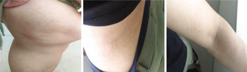
A series of three clinical photographs demonstrating the dermatological resolution of urticaria (hives) across three anatomical regions: the knee joint (A), the lateral waist (B), and the elbow (C). The images show characteristic wheals—edematous, circumscribed, and slightly elevated skin lesions. Panel A displays the knee area with subtle, flesh-colored to slightly erythematous elevations and an uneven skin texture consistent with resolving wheals. Panel B shows the lateral waist with faint, linear, and confluent dermal edema. Panel C depicts the elbow where the lesions have largely flattened, leaving only minimal, faint markings on the skin surface. The progression illustrates the transient nature of urticarial lesions as they gradually diminish in size and elevation. This visual serves as an educational reference for identifying skin morphology in the recovery phase of an acute urticarial reaction, emphasizing the characteristic lack of secondary skin changes (such as scaling or scarring) during the resolution of wheals.
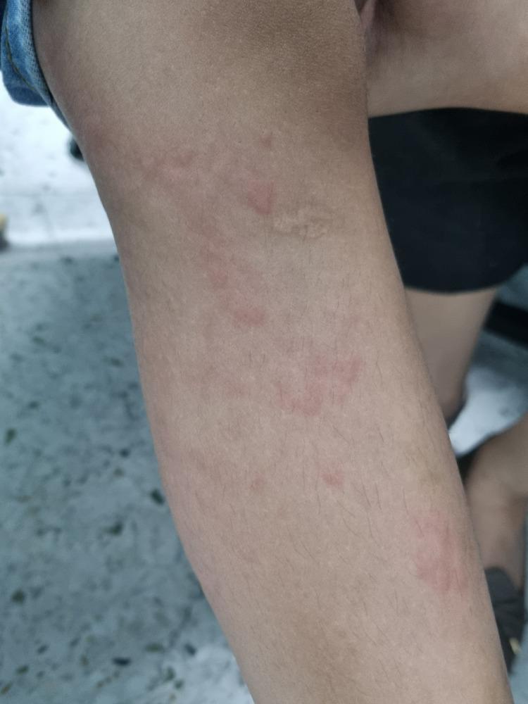
A clinical photograph showing a close-up of a child's lower extremity demonstrating urticaria. The image displays multiple raised, edematous skin lesions known as wheals or hives. These lesions are characterized by pale-to-skin-colored central swellings of varying sizes and irregular shapes, surrounded by a distinct zone of erythema (redness). The distribution is scattered across the leg, illustrating the typical appearance of a hypersensitivity reaction or allergic response. This image serves as an educational reference for identifying cutaneous manifestations of urticaria, focusing on the morphology of superficial dermal edema and associated inflammatory flare.
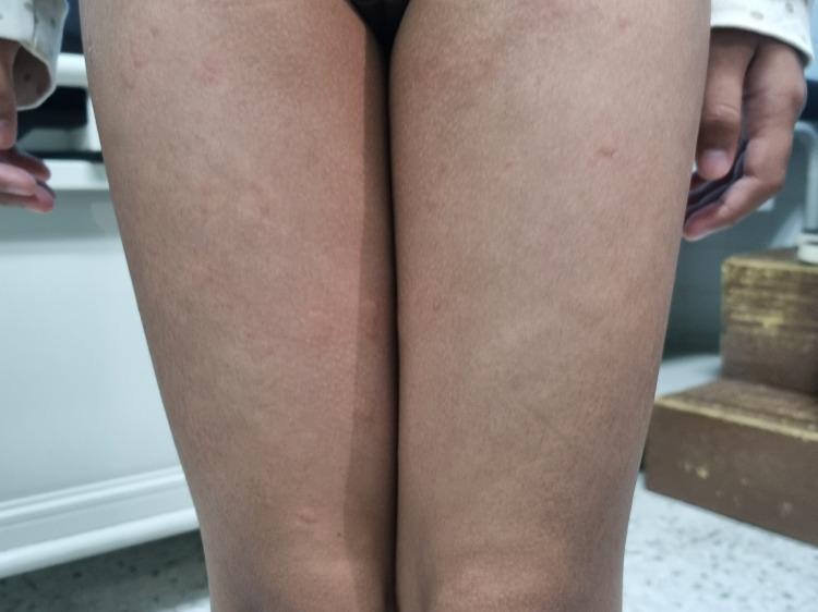
Clinical photograph of a pediatric patient's bilateral lower limbs demonstrating classic urticaria (hives). The image shows multiple discrete and confluent wheals distributed across the thighs. Morphologically, the lesions are characterized by raised, edematous, superficial swellings of varying sizes. The central portions of the wheals appear pale or skin-colored, while the peripheries exhibit varying degrees of erythema. These findings represent a type I hypersensitivity reaction or mast cell degranulation, leading to dermal edema. This visual evidence is a key diagnostic clinical sign for acute urticaria, commonly used in dermatology and pediatric curricula to differentiate transient wheals from other types of papular eruptions or inflammatory skin conditions.
urticaria management guidelines
Urticaria (Hives)
Definition
Epidemiology
- Affects ~15-20% of the population during their lifetime - Rosen's Emergency Medicine, p. 2412
- Acute urticaria occurs at any age; chronic urticaria peaks in the 3rd-4th decade (or 3rd-5th decade per Harrison's)
- Chronic urticaria is more common in women (40s-50s); half of chronic urticaria patients have disease for 5+ years
- Acute urticaria is frequently seen in children with atopic dermatitis - Harrison's, p. 2851
Classification
By Duration
| Type | Duration |
|---|---|
| Acute | < 6 weeks |
| Chronic | ≥ 6 weeks (recurrent) |
By Cause
- Drug reactions (antimicrobials, NSAIDs, radiocontrast media, opioids, ACE inhibitors)
- Food reactions (shellfish, tree nuts, eggs, peanuts, strawberries, lobster)
- Environmental/inhalant allergens
- Transfusion reactions
- Stinging/biting insects
- Infections - viral (rhinovirus, rotavirus, hepatitis, mononucleosis, coxsackievirus), bacterial, parasitic
- Vaccine reactions
-
Spontaneous/idiopathic - no identifiable trigger (most common in chronic urticaria)
-
Autoimmune - functional IgE autoantibodies; associated with thyroid autoimmunity, rheumatoid arthritis, Sjögren's, celiac disease, SLE
-
Inducible urticaria (physical stimuli):
- Dermatographism (2-5% of population; linear wheal with stroking)
- Cholinergic urticaria (small 1-2 mm wheals; triggered by heat, exercise, hot bath)
- Cold urticaria (can progress to vascular collapse with cold water immersion)
- Solar urticaria (6 subtypes by light spectrum)
- Pressure urticaria (sustained pressure - shoulder straps, belts)
- Vibratory, aquagenic
- Exercise-induced anaphylaxis
-
Vascular disease - urticarial vasculitis
-
Mastocytosis (cutaneous or systemic)
-
Hereditary angioedema (HAE), Schnitzler's syndrome, Muckle-Wells syndrome
-
Fitzpatrick's Dermatology, p. 717; Harrison's, p. 2851
Pathophysiology
- Histamine (primary mediator)
- Slow-reacting substance of anaphylaxis (leukotrienes)
- Bradykinin, kallikrein, acetylcholine
| Mechanism | Examples |
|---|---|
| IgE-mediated (Type I hypersensitivity) | Food, drug, insect venom allergens |
| Autoimmune | Functional IgG anti-FcεRIα or anti-IgE autoantibodies → mast cell activation |
| Immune complex / complement | Serum sickness, transfusion reactions |
| Direct mast cell degranulation (nonimmunologic) | Opioids, NSAIDs (aspirin), radiocontrast agents, strawberries, lobster |
| Bradykinin-mediated | ACE inhibitors, hereditary angioedema (C1-INH deficiency) |
Clinical Features
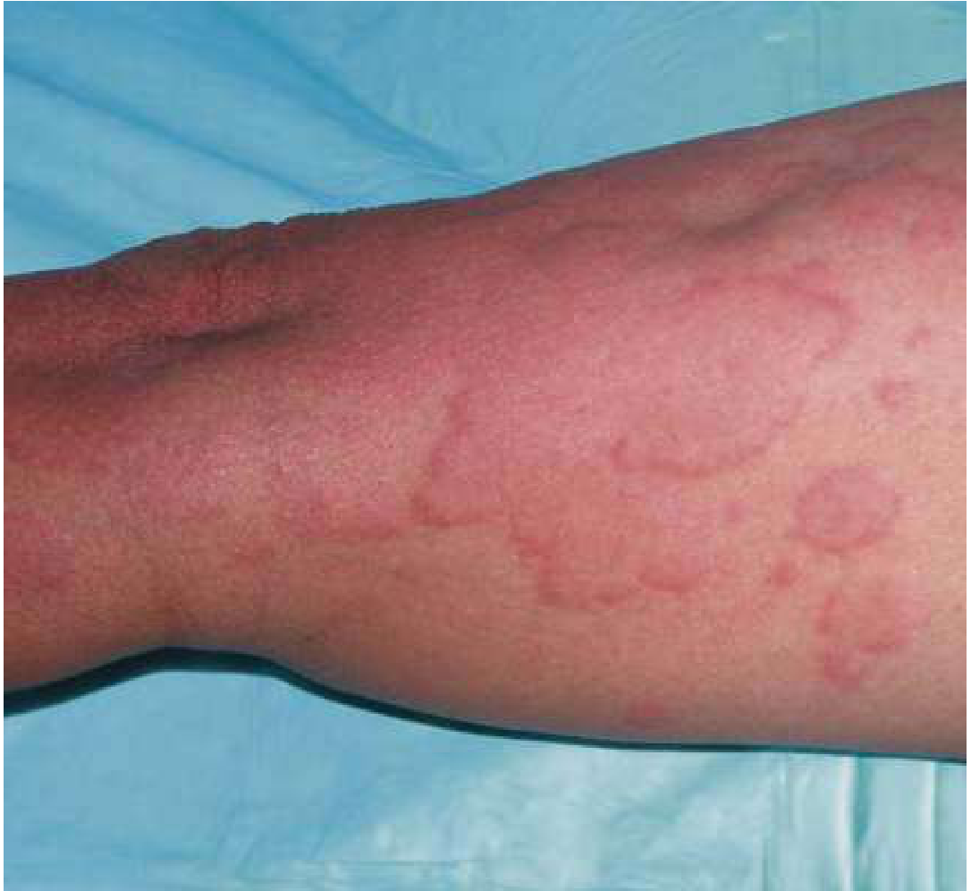
- Pruritic, raised, blanching wheals - pink to light red
- Individual lesions typically resolve within 24 hours (a hallmark); if >36 hours, suspect urticarial vasculitis
- May be accompanied by angioedema (deeper dermal/subcutaneous edema) - lips, eyelids, tongue, larynx, bowel
- Angioedema in HAE: notably lacks pruritus and urticaria, has GI involvement, and does NOT respond to antihistamines
- Cholinergic urticaria: distinctive tiny (1-2 mm) wheals with large surrounding erythema
Diagnosis
- Minimum: Differential blood count, ESR/CRP
- Extended (based on history):
- Thyroid hormones and antibodies
- H. pylori testing
- Autologous serum skin test (functional autoantibodies)
- Allergy skin tests / avoidance diet
- Tryptase (rule out mastocytosis)
- Lesional skin biopsy if vasculitis is suspected
- Fitzpatrick's Dermatology, Table 41-1; Harrison's, p. 2852
Treatment
Step-wise Management for Chronic Urticaria (EAACI & AAAAI/ACAAI Guidelines)
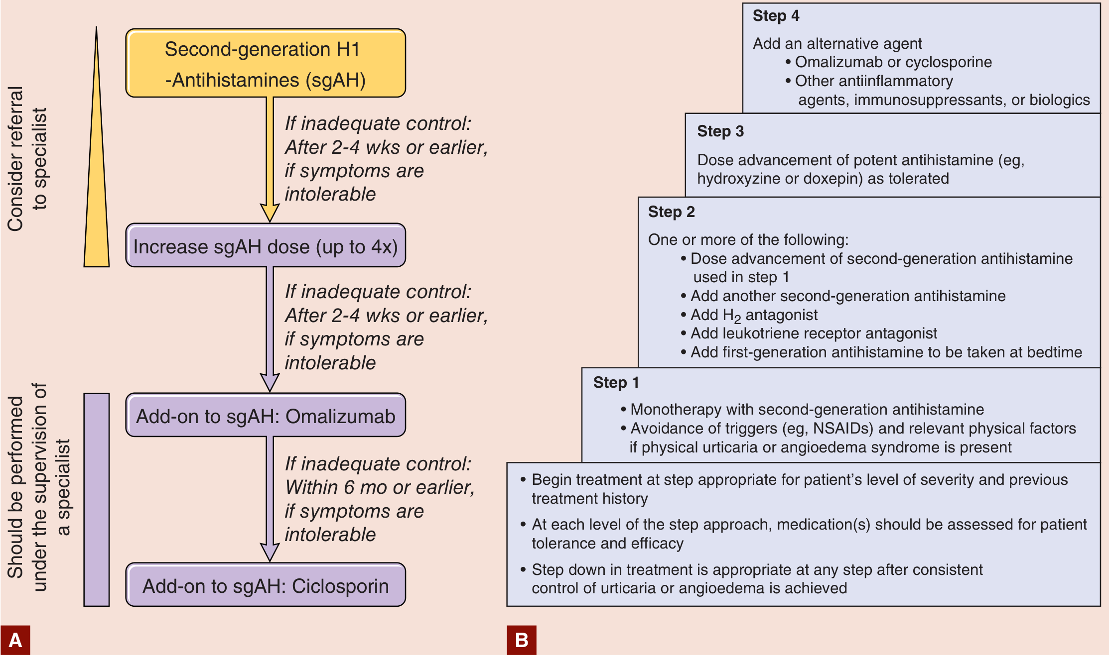
Step 1 - First-line
- Second-generation (non-sedating) H1 antihistamines as monotherapy: loratadine, fexofenadine, cetirizine, desloratadine
- Trigger avoidance (NSAIDs, physical factors)
Step 2 - If inadequate control after 2-4 weeks
- Increase second-generation H1 antihistamine dose up to 4x the standard dose
- Add H2 antagonist (ranitidine or famotidine)
- Add leukotriene receptor antagonist (montelukast 10 mg/day)
- Add first-generation antihistamine at bedtime (hydroxyzine, diphenhydramine)
Step 3 - Dose advancement
- Potent antihistamines: hydroxyzine or doxepin (also has antidepressant properties)
Step 4 - Refractory disease (specialist supervision)
- Omalizumab (anti-IgE monoclonal antibody) - first-line biologic, highly effective for chronic spontaneous urticaria
- Cyclosporine - for severe, poorly responsive chronic spontaneous urticaria
A recent 2025 systematic review and meta-analysis confirmed omalizumab's efficacy and safety in pediatric chronic spontaneous urticaria as well (PMID 40545961).
Other agents (refractory/special cases)
- Urticarial vasculitis / delayed pressure urticaria: Systemic corticosteroids; hydroxychloroquine, dapsone, colchicine
- Dapsone, methotrexate, tacrolimus, IVIG, plasmapheresis, narrow-band UVB (anecdotal evidence)
- Note: Systemic corticosteroids are generally avoided in idiopathic/allergen-induced urticaria due to long-term toxicity; reserved for pressure urticaria, vasculitic urticaria, debilitating disease
First-generation antihistamines - cautions
Emergency / Acute Setting
- For mild-moderate acute urticaria: H1 antihistamines ± short course of oral steroids
- For severe urticaria with angioedema or anaphylaxis: epinephrine, IV/IM antihistamines, systemic corticosteroids
- Chronic urticaria: H1 + H2 antihistamine combination - Rosen's Emergency Medicine, p. 2414
Hereditary Angioedema (HAE) - Special Case
- Caused by C1-inhibitor (C1-INH) deficiency (Type 1) or dysfunction (Type 2)
- Bradykinin-mediated, NOT histamine-mediated - does not respond to antihistamines or steroids
- Lab: Chronically low C4 and C2; low C1-INH antigen (Type 1) or functional assay (Type 2)
- Acute treatment: C1-INH concentrate (plasma-derived or recombinant), icatibant (bradykinin B2 receptor antagonist), ecallantide (kallikrein inhibitor)
- Prophylaxis: Lanadelumab (anti-plasma kallikrein), berotralstat, C1-INH infusions, attenuated androgens
- Harrison's, p. 2852; Fitzpatrick's Dermatology
Differential Diagnosis
| Condition | Distinguishing Feature |
|---|---|
| Urticarial vasculitis | Lesions persist >36h, painful not pruritic, may scar; biopsy shows fibrinoid necrosis |
| Urticarial bullous pemphigoid | Urticarial phase precedes bullae; DIF positive |
| Erythema multiforme | Targetoid lesions, epidermal necrosis |
| Mastocytosis | Hyperpigmented papules that urticate with stroking (Darier's sign), elevated tryptase |
| HAE | No urticaria, no pruritus, GI attacks, laryngeal edema, family history |
| Fixed drug eruption | Recurs at same site, leaves pigmentation |
- Fitzpatrick's Dermatology, p. 46; Rosen's Emergency Medicine
Summary Pearls
- The defining lesion: Transient pruritic wheal resolving within 24 hours, no scarring.
- Chronic = ≥6 weeks; >50% of chronic cases are idiopathic/autoimmune.
- Mast cell degranulation with histamine release is the final common pathway.
- First-line treatment for all urticaria: Non-sedating second-generation H1 antihistamines (can be quadrupled in dose).
- Omalizumab is the established biologic for antihistamine-refractory chronic spontaneous urticaria.
- Painful, persistent (>36h) urticaria → biopsy to exclude vasculitis.
- Angioedema without urticaria or pruritus + no antihistamine response → think HAE, check C4 levels.
- Recent evidence (2024 systematic review [PMID 38420865]): Antihistamines remain the evidence-based first-line for acute urticaria; epinephrine is reserved for anaphylaxis.