
1. Describe the female sexual cycle. What is LH surge? [6+1][2013][2011][2015] [2017] page 463,464 2. Describe spermatogenesis. What is Blood-Testis-Barrier?[5+2][2014] [2012] page 456-458 3. What is spermatogenesis? Describe the hormonal control of it. [2+5] [2010] 4. What are the hormones of placenta? Though genetically different, why is not fetus rejected immunologically? [3+4] [2018] page 472-474 5. Define ovulation. How does interplay of different hormones lead to ovulation? How is it detected?[2+3+2] [2019] [2014 supple.] page 463,464,466 6. What is the menstrual cycle? Explain the ovarian changes taking place during menstrual cycle. [3+7] [NEW PATTERN 2021] PAGE 464-466 7. What are the functions of placenta? What are the hormones secreted from it? What do you mean by the feto-placental unit? Though genetically different, why is not fetus rejected immunologically? [2+1+2+2] [2013 supple.] 8. What is the fetoplacental unit? [7] [2018 supple.] 9. Name the key hormones secreted by Leydig cells and Sertoli cells. Steps involved in spermatogenesis. [2019 supple.] 10. Define spermatogenesis. Explain the phases of spermatogenesis. Describe the factors affecting spermatogenesis. [1+5+4][2023] PAGE 456-458 11. Discuss the various hormonal changes that take place in ovarian cycle. [2023] 12. Define menstrual cycle. Describe ovarian cycle with the help of diagram. Discuss about indicators of ovulation. Explain the basis of pregnancy test.(2+4+2+2)[2024]
spermatogenesis stages seminiferous tubule diagram
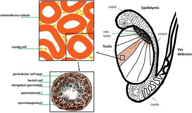
This medical illustration depicts the gross anatomy and histology of the human male reproductive system, specifically the testis and its associated ducts. The main diagram shows a sagittal-like section of the testis, highlighting internal lobules containing seminiferous tubules that converge at the rete testis. The epididymis is shown as a coiled ductal system attached to the posterior testis, divided into the caput (head), corpus (body), and cauda (tail), which then continues as the vas deferens. A magnified histological inset focuses on the interstitial space, showing Leydig cells clustered between cross-sections of seminiferous tubules. A second, high-magnification cross-section of a single seminiferous tubule illustrates the stages of spermatogenesis. Labeled structures include the peritubular cell layer (basal lamina), Sertoli cells for structural support, and germ cells in various stages of maturation: spermatogonia at the periphery, followed by spermatocytes I, and elongated spermatids positioned near the central lumen. This diagram serves as an educational tool for understanding the relationship between macroscopic anatomy and microscopic gametogenesis.

A multi-panel medical infographic illustrating the sequential stages of male reproductive function and the pharmacological impact of small molecule medications. The diagram is divided into four annotated sections linked by arrows: (A) Testicular weight, featuring a schematic of the male reproductive tract including the testes and vas deferens; (B) Testicular microenvironment, showing a cross-sectional view of a seminiferous tubule with various germ cell layers and a central lumen containing spermatozoa; (C) Spermatogenesis, a branching lineage diagram depicting the differentiation from spermatogonia through meiosis to mature spermatozoa; and (D) Sperm capacitation & Fertilization, illustrating sperm cell activation and interaction with an oocyte (egg). Each section includes an icon of a medication bottle and pills, symbolizing the points at which clinical drugs (e.g., SSRIs, antihypertensives, analgesics) can exert positive or negative effects. Panel D specifically includes a red upward arrow, signifying enhanced sperm motility or activity. This educational graphic serves as a summary of drug-induced changes in male fertility, from organ morphology to gamete function.
female sexual cycle menstrual cycle hormones FSH LH estrogen progesterone graph