
SVT its causes and treatment 10 mark answer
supraventricular tachycardia SVT ECG
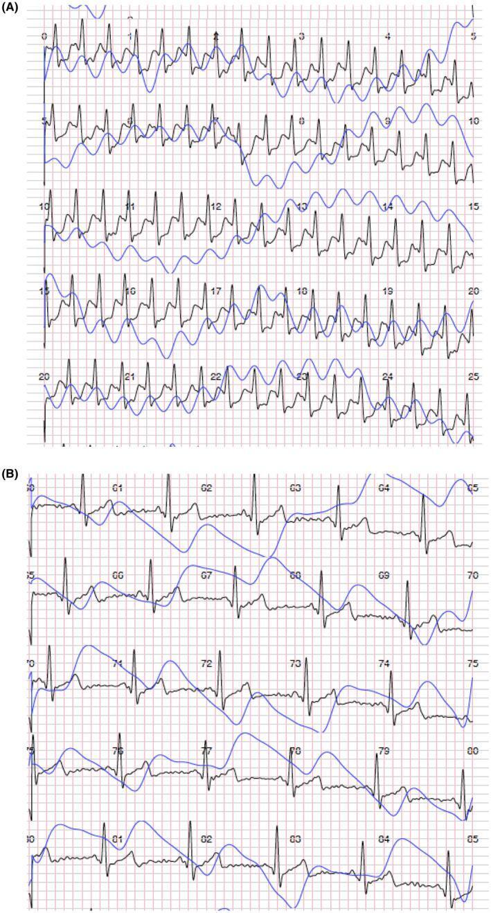
Comparison of diagnostic physiological traces illustrating supraventricular tachycardia (SVT) and its conversion to normal sinus rhythm. Panel A displays a continuous single-track electrocardiogram (ECG, black line) and synchronized photoplethysmography (PPG, blue line). The ECG shows a narrow-complex tachycardia characterized by a rapid, regular ventricular rate and absent P-waves, consistent with SVT. The corresponding PPG trace in Panel A exhibits high-frequency, low-amplitude oscillations, reflecting reduced stroke volume during the tachycardic event. Panel B demonstrates the physiological state following a successful Valsalva maneuver intervention. The ECG shows a restoration of normal sinus rhythm with identifiable P-waves, QRS complexes, and T-waves at a significantly slower heart rate. The PPG trace in Panel B shows a corresponding decrease in frequency and an increase in pulse wave amplitude, indicating improved hemodynamics and stroke volume consistency. This comparison highlights the efficacy of vagal maneuvers in managing acute supraventricular arrhythmias and demonstrates the utility of integrated ECG-PPG monitoring in clinical cardiology for real-time cardiac rhythm analysis.
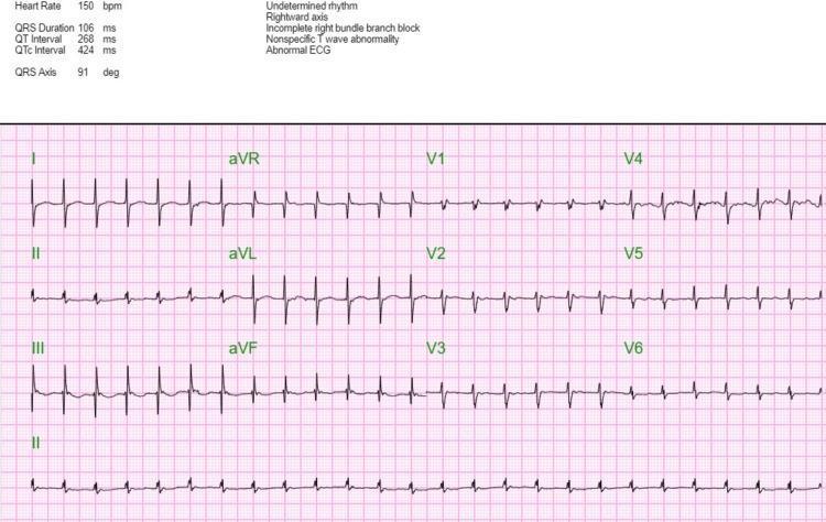
A 12-lead diagnostic electrocardiogram (ECG) demonstrating supraventricular tachycardia (SVT) with a heart rate of 150-151 beats per minute. The rhythm exhibits a regular tachycardia with narrow QRS complexes, although a right bundle branch block (RBBB) morphology is present, particularly visible as an RSR' pattern in the precordial leads V1-V3. Numerical data on the tracing indicates a QRS duration of 106 ms, a QT interval of 268 ms, and a QTc of 424 ms. A rightward axis of approximately 91 degrees is noted. Secondary findings include non-specific T-wave abnormalities across multiple leads, characterized by flattening or slight inversion. The image includes both standard limb leads (I, II, III), augmented limb leads (aVR, aVL, aVF), and precordial leads (V1-V6), plus a rhythm strip from lead II. This ECG provides a classic clinical representation of SVT combined with conduction system delays, serving as an educational tool for identifying tachyarrhythmias and morphological variations in cardiac electrical activity.
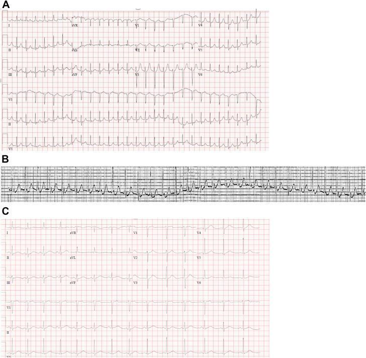
This composite diagnostic image presents a 12-lead electrocardiogram (ECG) comparison and rhythm strip illustrating supraventricular tachycardia (SVT). Panel A displays a standard 12-lead ECG showing a narrow-complex, regular tachycardia at approximately 170 beats per minute. Key diagnostic features include the absence of visible anterograde P waves and the presence of retrograde P waves, manifested as 'pseudo-r' waves' in the inferior leads (II, III, aVF), highly suggestive of Atrioventricular Nodal Reentrant Tachycardia (AVNRT). Panel B provides a continuous cardiac monitor rhythm strip, highlighting the detailed morphology of these retrograde P waves, which appear as sharp, negative deflections immediately following the QRS complexes. Panel C serves as the baseline clinical reference, showing the patient in normal sinus rhythm with a regular rate, clear anterograde P waves preceding each QRS complex, and normal waveform morphology. This comparison is an educational tool for distinguishing between normal sinus conduction and reentry-based arrhythmias, emphasizing the visual identification of retrograde conduction patterns during clinical SVT events.
Supraventricular Tachycardia (SVT) - Causes and Treatment
Definition
Classification and Causes
1. AV Nodal Reentrant Tachycardia (AVNRT) - Most Common
- Accounts for 80% or more of all PSVT cases.
- Caused by a reentrant circuit within the AV node, which has two functionally distinct pathways: a slow pathway (alpha) and a fast pathway (beta).
- A premature atrial beat triggers sustained reentry between these two pathways.
- ECG: P waves are "buried" within the QRS in ~70% of cases. In the remaining 30%, retrograde P waves appear immediately before or after the QRS (pseudo-R' in V1, pseudo-S in inferior leads).
2. AV Reentrant Tachycardia (AVRT) - Accessory Pathway Mediated
- Accounts for the remaining ~15-20% of PSVT.
- Requires an accessory bypass tract (bundle of Kent) connecting atria and ventricles outside the AV node.
- Wolff-Parkinson-White (WPW) syndrome is the prototype - characterized by a short PR interval (<0.12 s) and delta wave.
- Orthodromic AVRT: anterograde conduction through AV node, retrograde through accessory pathway - produces narrow QRS.
- Antidromic AVRT: anterograde through accessory pathway, retrograde through AV node - produces wide QRS.
- Associated with congenital heart disease: Ebstein's anomaly, HCM, ASD, VSD, transposition of great arteries.
3. Atrial Tachycardia (AT)
- Originates from an ectopic atrial focus outside the sinus node.
- Mechanism: abnormal automaticity, triggered activity, or intra-atrial reentry.
- Usually not dependent on AV nodal conduction, so AV block (by adenosine) only transiently slows rate while P waves remain visible.
4. Multifocal Atrial Tachycardia (MAT)
- Characterized by at least 3 distinct P-wave morphologies in a single ECG lead.
- No consistent P-P, PR, or R-R intervals; rate usually 100-180 bpm.
- Common in COPD, pulmonary hypertension, and elderly patients.
5. Junctional Tachycardia
- Arises from the AV junction; uncommon in adults.
- Seen in digitalis toxicity and after cardiac surgery.
Precipitating / Underlying Causes of SVT
- Structural heart disease (congenital, cardiomyopathy)
- Ischemic heart disease
- Hyperthyroidism
- Caffeine, alcohol, stimulants (cocaine, amphetamines)
- Electrolyte imbalances (hypokalemia, hypomagnesemia)
- Fever, sepsis, dehydration (especially in children)
- Medications (digoxin, sympathomimetics)
- Idiopathic (most young patients have no structural disease)
ECG Features (Table)
| Feature | Finding |
|---|---|
| P waves | Absent (buried in QRS) or retrograde |
| QRS width | Narrow (<100 ms), unless aberrant conduction |
| Rate | 130-300 bpm; typically 170-180 bpm |
| Rhythm | Regular |
| Onset | Abrupt |
Clinical Features
- More frequent in females; peak in late teenage and young adult years.
- Most patients have no active cardiovascular disease.
- Symptoms: palpitations, lightheadedness, dyspnea, chest discomfort.
- Can cause syncope if rate is very high (>220 bpm in infants, >180 bpm in children).
- Rarely causes hemodynamic instability in otherwise healthy individuals.
Treatment
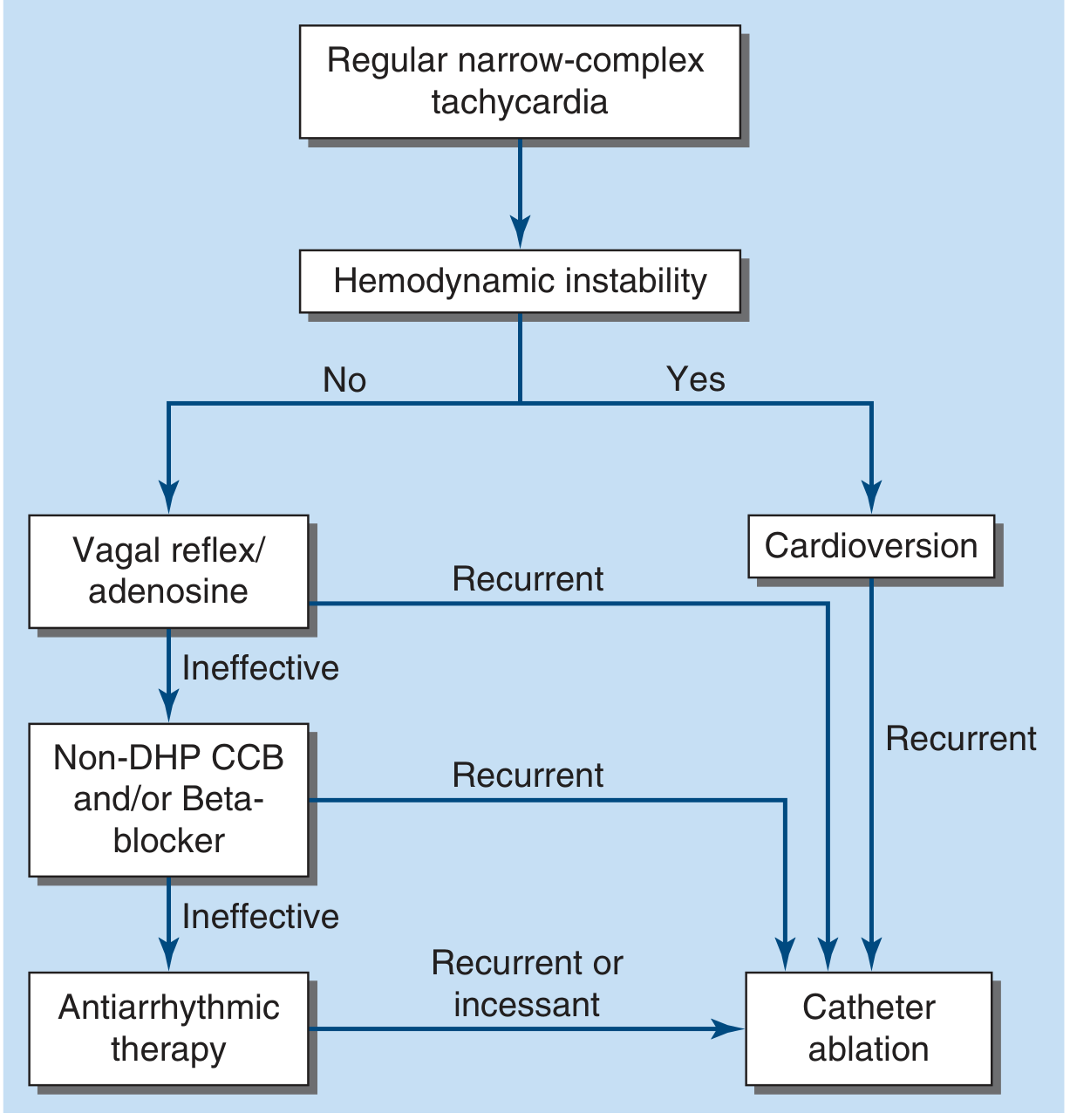
A. Acute Management (Hemodynamically Stable)
- Carotid sinus massage (only if no carotid bruit or prior stroke history).
- Valsalva maneuver - highly effective if applied early; patient can be taught to self-administer.
- Mechanism: increases vagal tone, slows AV nodal conduction, breaks the reentrant circuit.
- Dose: 6 mg IV rapid bolus (followed by NS flush); if ineffective, 12 mg.
- Mechanism: transiently blocks AV nodal conduction, terminating AVNRT and AVRT.
- Terminates the vast majority of PSVT episodes.
- Side effects: transient chest pain, dyspnea, flushing, anxiety.
- Contraindications: prior cardiac transplant (hypersensitivity risk), severe bronchospasm (relative).
- Caution in WPW: can precipitate AF, which may cause hemodynamic instability in WPW.
- Verapamil or diltiazem IV if adenosine fails.
- Also effective but may cause hypotension; have longer duration of action.
- Can also be given orally on an as-needed basis (pill-in-the-pocket approach).
B. Acute Management (Hemodynamically Unstable)
- Synchronized DC cardioversion - QRS-synchronous direct current cardioversion is indicated if the patient has hypotension, unconsciousness, or respiratory distress.
- This is rarely needed because adenosine works promptly in most cases.
C. Wide-Complex SVT
- If wide-complex tachycardia is suspected SVT with aberrancy, treat as ventricular tachycardia until proven otherwise.
- A trial of IV adenosine is reasonable if the rhythm is regular and patient is stable.
- Irregular wide-complex tachycardia (likely preexcited AF/flutter in WPW): treat with cardioversion, IV procainamide, or ibutilide.
- AV nodal blockers (adenosine, verapamil, beta-blockers) are contraindicated in confirmed WPW with AF, as they accelerate anterograde conduction through the accessory pathway.
D. Long-Term / Definitive Management
- Catheter ablation - recommended for most patients with recurrent PSVT; achieves cure in >95% of AVNRT and >90% of AVRT.
- Preferred over chronic antiarrhythmic drug therapy in symptomatic patients.
- Antiarrhythmic drugs (flecainide, propafenone, sotalol) for patients who refuse ablation or where ablation is not suitable.
- Oral beta-blockers or CCBs for rate control and symptom reduction as maintenance.
SVT in Special Populations
- Pregnancy: Adenosine is safe for acute SVT. Beta-blockers (metoprolol) preferred for maintenance. Electrical cardioversion is safe in pregnancy.
- Children: Adenosine is first-line. Vagal maneuvers include ice-to-face (diving reflex in infants).
- WPW + AF: Avoid AV nodal blockers - use procainamide, ibutilide, or cardioversion.
ECG Images for Reference

Summary Table
| Aspect | Key Points |
|---|---|
| Most common type | AVNRT (>80%) |
| 2nd most common | AVRT / WPW (15-20%) |
| ECG hallmark | Narrow QRS, rapid regular rate, absent/retrograde P waves |
| 1st line vagal Rx | Valsalva maneuver, carotid sinus massage |
| 1st line drug (acute) | IV Adenosine 6 mg rapid bolus |
| 2nd line drug | IV Verapamil / Diltiazem or beta-blocker |
| Unstable patient | Synchronized DC cardioversion |
| Definitive cure | Catheter ablation (>90-95% success) |
| WPW + AF - avoid | Adenosine, verapamil, digoxin |
Cardioversion 10 marks answer
cardioversion defibrillation synchronized DC shock paddle placement
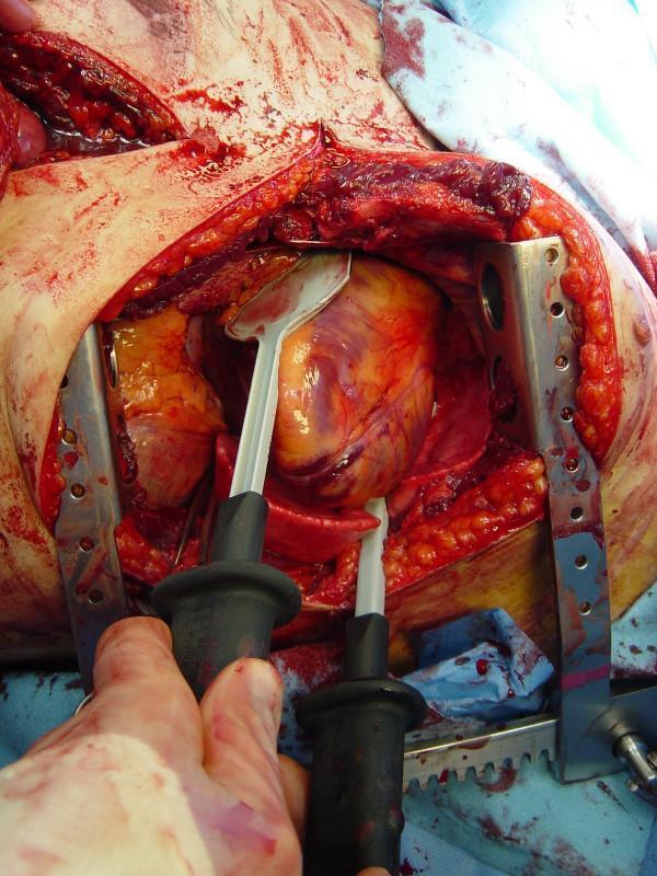
This clinical photograph captures a critical surgical moment during emergency resuscitative thoracotomy. The image shows an open chest cavity with a metal rib retractor in place, providing wide exposure of the mediastinum and the heart. The heart appears mottled with reddish-purple and yellow tones, suggestive of physiological stress or trauma. Two internal defibrillation paddles are actively being used; they feature spoon-shaped, white metallic surfaces with black insulated handles. One paddle is positioned on the anterior surface of the heart while the other is placed posteriorly, 'sandwiching' the myocardium to deliver a direct electrical shock for cardioversion or defibrillation. Significant hemorrhage is visible within the surgical field and on the surrounding drapes, reflecting the acute trauma setting. This visual serves as a primary educational tool for demonstrating the technique of internal cardiac massage and the correct placement of internal paddles in advanced trauma life support and thoracic surgery.

This EKG tracing illustrates the transition from a life-threatening arrhythmia to a paced rhythm following synchronized DC cardioversion. The left portion of the strip displays a monomorphic ventricular tachycardia (VT), characterized by a rapid, regular rhythm (approximately 200 beats/min) with wide, uniform QRS complexes. In the middle of the strip, a distinct high-amplitude deflection signifies the delivery of a 120J biphasic DC shock. Post-cardioversion, the tracing on the right reveals a transition into a slower rhythm (60 beats/min). The wide, regular complexes in the post-shock phase, preceded by sharp vertical pacing spikes, indicate that the patient's intrinsic heart rhythm has been superseded by a ventricular pacemaker. This visual sequence is a classic clinical demonstration of successful electrical conversion of sustained VT in a patient with an implanted cardiac device. It highlights key concepts in advanced cardiac life support (ACLS), arrhythmia management, and medical device interaction in the post-arrest or peri-procedural setting.
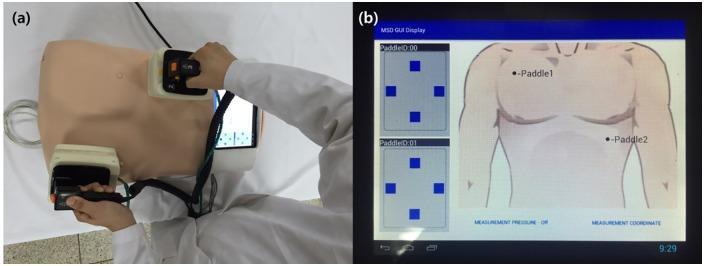
This figure presents a medical training simulator designed for defibrillation education, featuring a position-measuring system for paddle placement. Image (a) shows a physical clinical photograph of a chest manikin with two defibrillator paddles held by an operator in white clinical attire. One paddle is positioned at the upper right sternal border and the other at the cardiac apex. The paddles are wired to an integrated electronic interface. Image (b) shows the Graphical User Interface (GUI) of the position-measuring system. The digital display provides a schematic anatomical diagram of a human torso with markers indicating 'Paddle 1' and 'Paddle 2' to verify correct anatomical placement. To the left, diagnostic signal panels labeled 'PaddleID:00' and 'PaddleID:01' show spatial coordinate grids. The system is designed to provide real-time feedback to medical trainees on the accuracy of electrode positioning, a critical factor in successful cardiac resuscitation. Educational concepts include Advanced Cardiovascular Life Support (ACLS) training and the application of biomedical engineering in clinical simulation.
Cardioversion - 10 Mark Answer
Definition
Physiology / Rationale
- Reduced preload and falling cardiac output (CO = HR × SV)
- Ventricular ischemia and possible infarction
- Rise in pulmonary capillary wedge pressure, leading to pulmonary edema
- Lactic acidosis from hypoperfusion, further worsening arrhythmia
- Takes effect almost immediately
- Fewer side effects than antiarrhythmic drugs
- Often more effective (antiarrhythmics have narrow therapeutic windows and can be proarrhythmic)
- Life-saving in hemodynamic collapse
Types of Cardioversion
| Type | Mechanism | Indication |
|---|---|---|
| Synchronized DC cardioversion | Shock delivered on R-wave peak | Tachyarrhythmias with a pulse |
| Unsynchronized cardioversion (Defibrillation) | Shock delivered at any point in cycle | Pulseless VT, VF (cardiac arrest) |
| Chemical/Pharmacological cardioversion | Antiarrhythmic drugs (IV flecainide, amiodarone, ibutilide) | Elective conversion of AF/flutter |
Indications
- Atrial fibrillation (AF) - with rapid ventricular response not responding to drugs, or hemodynamically unstable
- Atrial flutter - usually requires less than 50 J for conversion
- Supraventricular tachycardia (SVT/PSVT) - when vagal maneuvers and adenosine fail, or if patient is unstable
- Ventricular tachycardia (VT) with a pulse - if hemodynamically unstable or amiodarone fails
- WPW with AF - urgent conversion needed (AV nodal blocking drugs are contraindicated)
- Junctional tachycardia
- Multifocal atrial tachycardia (MAT)
- Sinus tachycardia
- Digoxin toxicity-induced arrhythmias (relative contraindication - high risk of post-shock VF)
Contraindications
- Sinus tachycardia - treat the underlying cause
- Digitalis toxicity - increased risk of VF with cardioversion; correct toxicity first
- Severe electrolyte imbalance (hypokalemia, hypomagnesemia) - correct before elective cardioversion
- Junctional / automatic tachycardias - electrical shock may accelerate the rate
- AF with known atrial thrombus (unless anticoagulation is given for ≥3 weeks or TEE excludes thrombus)
Pre-Procedure Preparation
- IV access, oxygen, continuous ECG, pulse oximetry, and end-tidal CO2 monitoring
- Correct metabolic abnormalities: electrolytes, hypoxia, acidosis should be addressed before elective cardioversion
- Anticoagulation for AF/atrial flutter lasting >48 hours:
- Therapeutic anticoagulation (DOAC or warfarin) for at least 3 weeks before elective cardioversion, OR
- Transesophageal echocardiogram (TEE) to exclude atrial thrombus
- All patients anticoagulated for at least 1 month after cardioversion if AF/flutter >48 hours duration
- Prepare antiarrhythmic drugs (amiodarone, lidocaine) and agents for bradycardia (atropine)
- Sedate all conscious patients (except those rapidly deteriorating)
Sedation Agents (IV)
| Drug | Dose | Notes |
|---|---|---|
| Midazolam | 0.15 mg/kg | Most commonly used; onset ~2 min; flumazenil available as reversal |
| Propofol | 1.5 mg/kg | Small BP drop; rapid onset |
| Etomidate | 0.15 mg/kg | No drop in BP; ideal in hemodynamically compromised patients; painful IV infusion |
| Methohexital | 1 mg/kg | Quicker onset than midazolam; risk of laryngospasm |
Technique (Step-by-Step)
- Place adhesive electrode pads or paddles on the patient:
- Anterolateral: one pad at right infraclavicular area, other at left lateral chest (V5-V6 level)
- Anteroposterior: one pad anteriorly over the heart, one posteriorly between scapulae (better for obese patients)
- Press SYNC button to engage synchronized mode - verify that markers appear on each R wave
- Adjust gain if needed so sync markers consistently align with R waves (not T waves)
- Select appropriate energy level (see below)
- Press CHARGE button; announce "I'm clear, you're clear, everybody's clear!" - ensure all personnel step back
- Press SHOCK button to deliver cardioversion
- Re-check monitor - if rhythm persists, double the energy and repeat
- Re-engage SYNC mode after each shock (most defibrillators revert to unsynchronized mode after a shock)
- If cardioversion produces VF, immediately switch to unsynchronized mode and defibrillate
Energy Levels
| Arrhythmia | Starting Energy | Notes |
|---|---|---|
| Atrial flutter | <50 J | Lowest threshold; usually converts with 25-50 J |
| SVT (reentrant) | 50 J | Double if unsuccessful |
| Atrial fibrillation | ~100 J (biphasic); 200 J (monophasic) | Most energy-demanding among supraventricular rhythms |
| Ventricular tachycardia (pulse) | 50 J | 10-20 J effective in >80%; 50 J in >90% |
| Pulseless VT | 200 J initially, then 300 J | Treat as defibrillation |
| Pediatric | 0.5-1 J/kg; repeat at 2 J/kg | Synchronized mode; amiodarone or procainamide as adjuncts |
- Obese patients may require higher energy; expiratory phase reduces energy requirement
- Biphasic waveforms are more effective and less damaging than monophasic
Post-Procedure
- Reassess rhythm and hemodynamics immediately after shock
- Administer antiarrhythmic drugs (e.g., amiodarone) after VT conversion to prevent recurrence
- Continue ECG monitoring
- Troponin may be mildly elevated post-cardioversion (transient, usually without clinical significance)
- Continue anticoagulation for ≥1 month after AF/flutter cardioversion
Complications
In the Patient:
- Thromboembolism - most important after AF/flutter cardioversion; prevented by adequate anticoagulation
- Post-shock arrhythmias - VT, VF, bradycardia, AV block, asystole (especially with ischemic heart disease or high-dose shocks ≥200 J)
- Hypotension - from sedative drugs or post-conversion myocardial dysfunction
- Pulmonary edema - may occur hours after cardioversion at higher energy levels
- Chest wall burns - superficial from inadequate gel or loose paddle contact; preventable
- Respiratory depression - from sedation; requires airway monitoring and supplemental O2
- Mild troponin rise - common; rarely associated with significant myocardial dysfunction
- Pacemaker/ICD dysfunction - place electrodes as far from device generator as possible; check pacing thresholds after the procedure
In Healthcare Personnel:
- Accidental shocks and minor burns if proper "all-clear" protocol is not followed
Chemical Cardioversion (Pharmacological)
- Atrial fibrillation: IV ibutilide, IV flecainide, IV propafenone, IV amiodarone
- VT: IV amiodarone 150 mg over 10 min (repeat up to 2.2 g/24 h); lidocaine as alternative
- SVT: IV adenosine, verapamil, diltiazem
Summary
| Aspect | Key Point |
|---|---|
| Definition | Synchronized DC shock delivered on R-wave peak |
| Key distinction | Synchronized (has pulse) vs. Defibrillation (no pulse/VF) |
| Most common indication | AF, atrial flutter, SVT, VT with pulse |
| Avoid in | Digitalis toxicity, automatic tachycardias, sinus tachycardia |
| AF >48 h | Anticoagulate ≥3 weeks before or exclude thrombus with TEE |
| Easiest to convert | Atrial flutter (<50 J) |
| Hardest to convert | AF (~100 J biphasic) |
| Sedation | Mandatory in conscious patients; midazolam or propofol |
| Critical step | SYNC mode must be re-engaged after each shock |
| Pediatric dose | 0.5-1 J/kg initially; increase to 2 J/kg if needed |