
Nipple discharge and it's diagnosis
Reading File
Now let me search the medical library for nipple discharge content in parallel.
Finding Sources
Finding Sources
Searching PubMed
"nipple discharge" diagnosis evaluation
Excellent results. Now let me read the most comprehensive sections in parallel.
Reading File
Reading File
Reading File
Reading File
Let me get the remaining sections and fetch the ductogram image.
Reading File
Reading File
Reading File
Loading Image
Now I also want to get the ductogram image from Mulholland and a web search for current guidelines.
Loading Image
Searching the Web
nipple discharge diagnosis evaluation guidelines 2024 2025
I now have comprehensive information from multiple authoritative surgical textbooks. Let me compose a thorough answer.
Nipple Discharge - Causes, Classification, and Diagnosis
Nipple discharge is the third most common breast complaint prompting surgical or radiologic evaluation. The majority of causes are benign, but a systematic approach is needed to identify the small fraction that represent malignancy.
1. Classification: Physiologic vs. Pathologic
The single most important first step is determining whether the discharge is physiologic or pathologic.
Physiologic Discharge
| Feature | Description |
|---|---|
| Onset | Non-spontaneous; requires nipple manipulation/compression to elicit |
| Laterality | Bilateral |
| Ductal origin | Multiple duct openings |
| Color | White, milky, clear, yellow, green, or gray |
| Occult blood | Negative |
| Clinical significance | No pathologic significance; reassure and stop nipple manipulation |
Pathologic Discharge
| Feature | Description |
|---|---|
| Onset | Spontaneous (any spontaneous discharge is considered pathologic by definition) |
| Laterality | Unilateral |
| Ductal origin | Single duct orifice |
| Color | Bloody, serosanguineous, or serous |
| Occult blood | Often positive |
| Risk of malignancy | Increases with age: 3% in <40 yrs, 10% in 40-60 yrs, 32% in >60 yrs |
"Any nipple discharge that is spontaneous is considered pathologic."
- Mulholland and Greenfield's Surgery, 7e
Red flags strongly suggesting cancer: spontaneous discharge, unilateral, single duct, age ≥40, bloody/serous, associated with a palpable mass.
2. Causes of Nipple Discharge
Physiologic / Hormonal
- Galactorrhea (bilateral milky): elevated prolactin from pituitary adenoma, hypothyroidism, chest wall trauma, medications
- Drug-induced: oral contraceptives, metoclopramide, phenothiazines, SSRIs, antipsychotics
Non-neoplastic (Benign Pathologic)
- Intraductal papilloma - the most common cause of pathologic nipple discharge; benign epithelial lesion with stroma growing within a duct
- Duct ectasia - ductal dilatation with loss of elastin, chronic inflammation; typically in older/perimenopausal women; discharge is often green/brown
- Fibrocystic change - often premenopausal, multiduct, bilateral, greenish/brownish, cyclic with menses
- Periductal mastitis / subareolar abscess
- Papillomatosis
Neoplastic
- Ductal carcinoma in situ (DCIS) - most common malignant cause (5-15% of pathologic nipple discharge)
- Invasive carcinoma
- In males: isolated nipple discharge is very abnormal - up to 9% of male breast malignancy presents this way
3. Diagnostic Workup
History (Key Questions)
- Is the discharge spontaneous or expressed?
- Unilateral or bilateral?
- Single duct or multiple ducts?
- Color and nature (bloody, serous, milky)?
- Associated breast mass?
- Relation to menstrual cycle?
- Pre- or postmenopausal?
- Medications (OCP, antipsychotics, SSRIs, metoclopramide)?
- History of trauma, thoracotomy, or endocrine disorders?
Physical Examination
- Inspect nipple and areola for Paget disease, eczema, retraction
- Palpate each quadrant from periphery to nipple-areolar complex to identify the "trigger point" (the quadrant that, when compressed, produces discharge)
- Identify whether single or multiple ducts involved
- Perform occult blood test on the discharge
- Visual field testing if pituitary/chiasmal syndrome suspected
Laboratory Tests
(Indicated when galactorrhea or endocrine cause suspected)
- Serum prolactin (elevated in pituitary adenoma, hypothyroidism, medications)
- Thyroid function tests (TSH, free T4)
- Quantitative beta-hCG (rule out pregnancy)
- For pituitary adenoma: thin-section CT or MRI sella turcica; visual field testing
4. Imaging Investigation
For Physiologic Discharge
- No further imaging required if truly physiologic (nonprovoked, bilateral, multiductal)
For Pathologic Discharge
Step 1: Mammography + Ultrasound
- Mammogram (age-appropriate; recommended for all >30 years)
- Directed subareolar ultrasonography
- These are the first-line investigations for all pathologic discharge
Step 2 (if mammogram + ultrasound negative or inconclusive):
Ductography (Galactography)
- A thin cannula is placed into the discharging duct and a small amount of water-soluble contrast is instilled
- Abnormal findings: intraductal filling defect, ductal wall irregularity, or abrupt duct truncation/obstruction
- The image below shows an intraductal papilloma on ductogram:
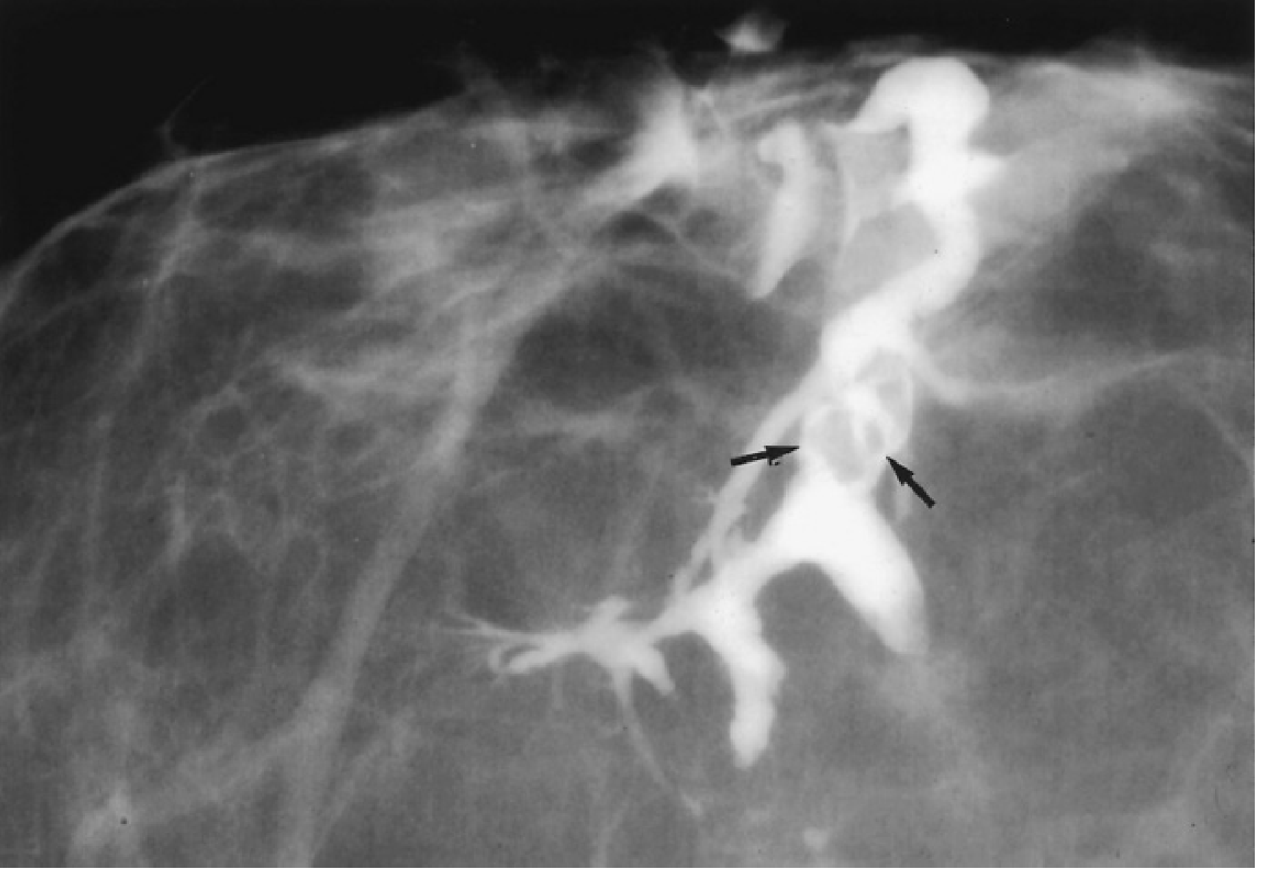
Ductogram: the filling defect (arrows) represents an intraductal papilloma - Mulholland & Greenfield's Surgery, 7e
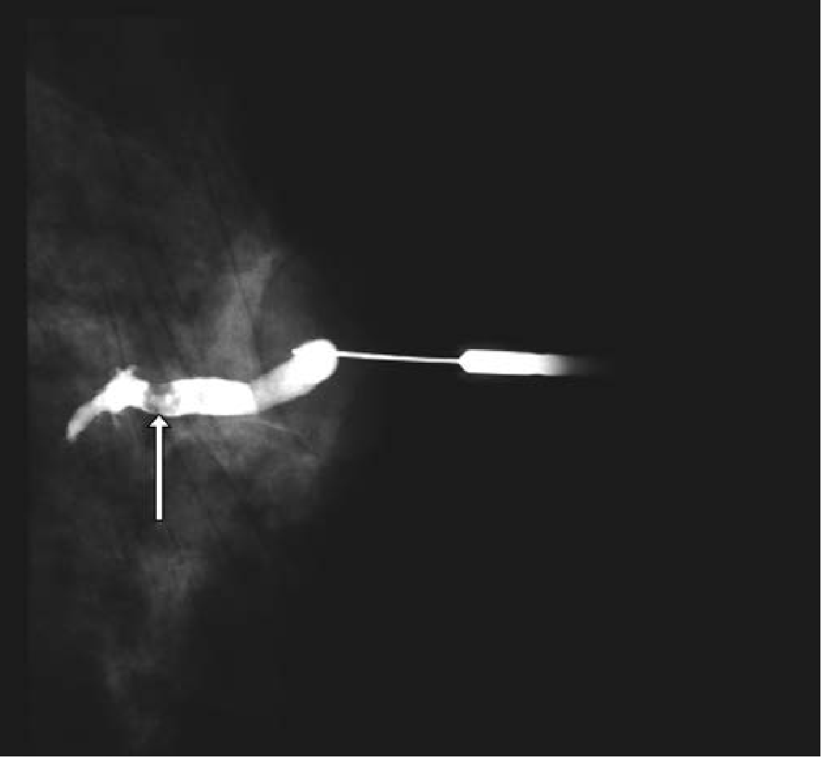
Ductogram with filling defect (arrow) = papilloma on subareolar resection - Current Surgical Therapy 14e
Note: A normal ductogram does not exclude cancer. The procedure is also painful and not commonly performed at many centers.
Breast MRI with contrast
- Has rapidly replaced ductography as the preferred tool for persistent/pathologic discharge when mammogram and ultrasound are negative
- Identifies underlying pathology in 19-96% of cases where other imaging is negative
- Also useful when clinical suspicion remains high despite negative conventional imaging
Ductoscopy
- Fiberoptic ductoscope placed directly into the involved duct (usually in the operating room)
- Allows direct visualization of intraductal lesions
- Enables more precise surgical excision by guiding the outer sheath as a surgical guide
5. Biopsy and Surgical Management
| Situation | Action |
|---|---|
| Imaging abnormality found | Image-guided core needle biopsy |
| Core biopsy = benign, concordant | May observe; further intervention may not be necessary |
| Papilloma found on core biopsy | Surgical excision recommended (may harbor atypia or DCIS) |
| No lesion on imaging / persistent symptoms | Surgical terminal duct excision |
| Malignancy confirmed | Cancer surgery as appropriate |
| Male with isolated nipple discharge | Mammogram + ultrasound; subareolar resection if negative |
Surgical Technique - Terminal Duct Excision
- Periareolar incision
- Intraoperative duct identification: lacrimal duct probe or methylene blue dye injection
- Excise duct from proximal to distal extent (~2-3 cm)
- If multiple ducts involved: central or total terminal duct excision
- Risks: inability to breastfeed, nipple hypersensitivity or hyposensitivity
6. Summary Algorithm
Nipple Discharge
       |
 ┌─────┴──────┐
Physiologic  Pathologic
(bilateral,  (spontaneous,
multiductal, unilateral,
non-spont.)  single duct)
       |           |
  Reassure,   Mammogram + USS
  stop         (age-appropriate)
  manipulation      |
              ┌─────┴──────┐
          Normal       Abnormality
              |             |
          MRI breast    Core needle
          or Ductogram    biopsy
              |
      ┌───────┴────────┐
  Normal/         Focal lesion
  persistent         |
  symptoms      Core biopsy ± excision
      |
  Terminal duct
  excision
Key Points to Remember
- Most nipple discharge is benign - <10% of spontaneous nipple discharge is malignant
- Intraductal papilloma is the #1 cause of pathologic nipple discharge
- Bloody + unilateral + spontaneous + single duct = high suspicion, always investigate
- Galactorrhea in a non-lactating woman requires serum prolactin, TSH, and hCG before breast-focused workup
- Breast MRI has largely replaced ductography in current practice for negative conventional imaging
- Postmenopausal nipple discharge - all cases are significant and warrant specialist referral
- Male nipple discharge - always abnormal; investigate aggressively for malignancy
Sources: Mulholland and Greenfield's Surgery 7e | Current Surgical Therapy 14e | Schwartz's Principles of Surgery 11e | Berek & Novak's Gynecology | ACR Appropriateness Criteria - Evaluation of Nipple Discharge (2022)
Osteo arthritis explain briefly
Reading File
Finding Sources
Reading File
Reading File
Reading File
Reading File
Loading Image
I have everything needed for a clear, concise overview. Here is the answer:
Osteoarthritis (OA) - Brief Overview
Osteoarthritis (also called degenerative joint disease) is the most common joint disease, characterized by progressive degeneration of articular cartilage leading to structural and functional failure of synovial joints. Despite the name, it is primarily a degenerative rather than inflammatory condition - inflammation acts as a secondary contributor.
Definition
Failed repair of synovial joint tissues after intra-articular stress, resulting in cartilage and bone breakdown causing pain, stiffness, and functional disability.
Types
| Type | Description |
|---|---|
| Primary (Idiopathic) | No identifiable cause; age-related; affects a few weight-bearing joints; onset typically >50 years |
| Secondary | Younger patients; predisposing cause identified (joint deformity, prior injury, obesity, diabetes, metabolic disease) |
- Prevalence increases exponentially after age 50 - about 40% of people >70 are affected
- Women are disproportionately affected
Pathogenesis
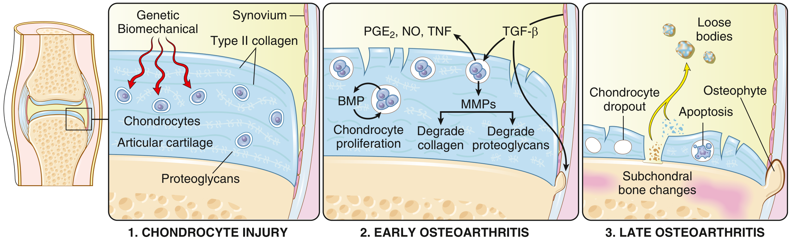
Stages of OA: (1) Chondrocyte injury from genetic/biomechanical stress → (2) Early OA: chondrocytes proliferate but MMPs degrade collagen and proteoglycans, outpacing repair → (3) Late OA: chondrocyte dropout, subchondral bone changes, osteophyte formation, loose bodies - Robbins & Kumar Basic Pathology
Key mechanism: Biomechanical stress on cartilage → chondrocyte injury → release of degradative enzymes (MMPs - matrix metalloproteinases), PGE2, NO, TNF → collagen and proteoglycan degradation exceeds repair capacity → cartilage loss → subchondral bone exposure.
Morphological Changes (Pathology)
| Feature | Description |
|---|---|
| Cartilage fibrillation | Surface cracking and fraying of cartilage (earliest change) |
| Cartilage loss | Full-thickness sloughing in advanced disease |
| Bone eburnation | Exposed subchondral bone is polished to an ivory-like appearance by friction |
| Subchondral cysts | Synovial fluid forced through microfractures (ball-valve mechanism) |
| Osteophytes | Bony outgrowths at joint margins capped by fibrocartilage; gradually ossify |
| Loose bodies (joint mice) | Dislodged cartilage/bone fragments floating in joint space |
| Synovium | Mild congestion and fibrosis; scattered inflammatory cells (minimal) |
Joints Commonly Affected
- Knees and hips (weight-bearing)
- Lower lumbar and cervical vertebrae
- Proximal (PIP) and distal (DIP) interphalangeal joints of fingers
- First carpometacarpal joint (base of thumb)
- First metatarsophalangeal joint (big toe)
Heberden nodes = prominent osteophytes at DIP joints (more common in women)
Bouchard nodes = osteophytes at PIP joints
Clinical Features
- Joint pain that worsens with use and improves with rest (early); later becomes constant
- Morning stiffness (<30 minutes - distinguishes from RA which is >1 hour)
- Crepitus on movement
- Limitation of range of movement
- Joint deformity (no joint fusion - unlike RA)
- Spinal osteophytes → nerve root compression → radicular pain, muscle spasm, neurological deficits
"The level of disease severity detected radiographically does not correlate well with pain and disability."
- Robbins & Kumar Basic Pathology
OA vs Rheumatoid Arthritis (Quick Comparison)
| Feature | OA | RA |
|---|---|---|
| Primary mechanism | Mechanical/degenerative | Autoimmune |
| Inflammation | Secondary | Primary |
| Joints | Weight-bearing (knees, hips) | Small joints (fingers) first |
| Serum antibodies | None | RF, ACPA positive |
| Joint fusion | No | Yes (ankylosis) |
| Systemic involvement | No | Yes (lungs, heart) |
Diagnosis
- Clinical: history + examination (pain pattern, stiffness, crepitus)
- X-ray (gold standard): joint space narrowing, subchondral sclerosis, osteophytes, subchondral cysts
- Labs: no specific markers; used to exclude other arthritis (normal ESR, CRP, RF)
- MRI: detects early cartilage loss and soft tissue changes before X-ray changes appear
Management (Brief)
- Non-pharmacological: exercise, weight loss, physiotherapy, activity modification, assistive devices
- Pharmacological: paracetamol, NSAIDs (oral/topical), intra-articular corticosteroids, duloxetine (for refractory pain)
- Surgical: joint replacement (arthroplasty) for severe, refractory cases
"There are no treatments to prevent or halt the progression of OA."
- Robbins & Kumar Basic Pathology
Sources: Robbins & Kumar Basic Pathology | Goldman-Cecil Medicine | Grainger & Allison's Diagnostic Radiology
This is a shared conversation. Sign in to Orris to start your own chat.