
Uric acid related disorders
Reading File
Searching sources...
Finding Sources
Searching PubMed
"uric acid" AND "hyperuricemia" AND "gout"
Finding Sources
Finding Sources
I have multiple excellent textbook sources. Let me read the most comprehensive ones in parallel.
Reading File
Reading File
Reading File
Reading File
Reading File
Finding Sources
Reading File
Reading File
Loading Image
Finding Sources
I now have comprehensive information from multiple authoritative textbooks. Let me compile the full answer.
Uric Acid-Related Disorders
1. Biochemistry of Uric Acid
Uric acid is the end product of purine catabolism in humans. Unlike most mammals, humans lack the hepatic enzyme uricase, which would otherwise convert uric acid to the more soluble compound allantoin. This evolutionary loss makes humans uniquely susceptible to hyperuricemia and its consequences. - Campbell Walsh Wein Urology, p. 2690
Purines are synthesized via two pathways:
- De novo pathway: purine nucleotides built from non-purine precursors
- Salvage pathway: free purine bases from diet or DNA/RNA degradation are recycled
Uric acid is filtered by the glomerulus, almost completely reabsorbed by the proximal tubule, then a fraction is secreted by the distal nephron and excreted in the urine. - Robbins & Kumar Basic Pathology
Normal serum urate: Hyperuricemia is defined as plasma urate above 6.8 mg/dL (the solubility threshold at physiologic temperature). - Robbins & Kumar
2. Hyperuricemia: Causes and Classification
Primary Hyperuricemia
- In >99% of cases, the cause is uncertain ("essential" hyperuricemia with polygenic basis)
- ~80% of patients: decreased renal tubular secretion of uric acid
- ~25% of patients: metabolic overproduction (increased PRPP amidotransferase activity)
- Very rarely: inherited enzymatic defects in purine metabolism
Secondary Hyperuricemia
| Mechanism | Causes |
|---|---|
| Overproduction | Tumor lysis syndrome (chemotherapy), myeloproliferative disorders, rapid cell turnover |
| Reduced excretion | CKD, diuretics (thiazides, loop), cyclosporine, lead nephropathy, diabetic ketoacidosis (organic acidemia competes with tubular urate secretion) |
| Enzymatic defects | Partial HGPRT deficiency (gout), complete HGPRT deficiency (Lesch-Nyhan syndrome) |
Cyclosporine (CSA) is a notable secondary cause in transplant patients - it both decreases GFR and increases net proximal tubular reabsorption of uric acid. - Comprehensive Clinical Nephrology 7e
3. Disorders Associated with Uric Acid
A. GOUT
Epidemiology and Risk Factors
- Affects men far more than women (95% of cases), typically in the 4th-6th decades; women develop gout mainly postmenopause
- Estrogen promotes renal urate clearance, explaining sex-based differences
- Asymptomatic hyperuricemia is common (~19% of individuals in the USA/UK), but only ~1 in 8 patients with urate between 7-8 mg/dL develop clinical gout over 14 years
- Risk factors for progression to gout: serum urate >9 mg/dL, CKD, hypertension, cardiovascular disease, obesity, diabetes - Grainger & Allison's Diagnostic Radiology
Pathogenesis
- Crystal deposition: Monosodium urate (MSU) crystals precipitate in joints and periarticular tissues
- Inflammasome activation: Resident macrophages phagocytose MSU crystals → activate the NLRP3 inflammasome → activate caspase-1 → produce active IL-1β
- Neutrophil influx: IL-1 recruits neutrophils → release cytokines, free radicals, proteases
- Phagolysosomal damage: Crystals rupture phagolysosomes → lysosomal enzyme leakage → acute inflammation
- Result: Acute arthritis, typically self-limiting in days-weeks; repeated attacks form tophi - Robbins & Kumar Basic Pathology
Clinical Stages
| Stage | Features |
|---|---|
| Asymptomatic hyperuricemia | Elevated urate, no symptoms; majority never progress |
| Acute intermittent gout | Sudden severe pain, erythema, swelling; 50% involve the 1st MTP joint (podagra); also ankle, knee, midfoot; self-resolves |
| Intercritical gout | Asymptomatic periods between attacks |
| Chronic tophaceous gout | No pain-free intervals; tophi in joints and soft tissues; structural joint damage; hands, feet, ankles, knees most affected |
Key note: Serum uric acid is often normal during an acute attack and is NOT part of the diagnostic criteria. Definitive diagnosis requires demonstration of negatively birefringent needle-shaped MSU crystals in joint aspirate (pathognomonic). - Tietz Textbook of Laboratory Medicine 7e
Imaging
- X-ray: Asymmetric soft-tissue swelling, "punched-out" periarticular erosions (overhanging edge sign)
- Ultrasound: Hyperechoic foci in effusion/synovium/cartilage surface ("double contour sign"); tophus as irregular soft-tissue thickening; cortical irregularity at erosion sites
- Dual Energy CT (DECT): Identifies urate crystal deposits; monitors response to treatment
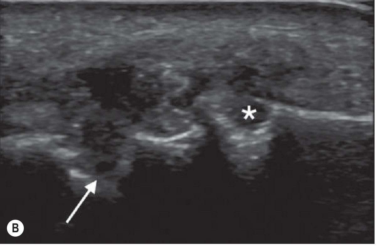
B. URIC ACID NEPHROLITHIASIS (Uric Acid Kidney Stones)
Three main determinants:
- Low urine pH (most important - most patients have normal uric acid excretion but persistently low pH)
- Low urine volume (concentrated urine)
- Hyperuricosuria (excess uric acid excretion)
At pH 5.0, even modest total uric acid exceeds solubility; at pH 6.5, concentrations >1200 mg/L remain soluble. This makes urinary alkalinization the cornerstone of treatment.
Causes:
- Congenital: Defects in renal tubular urate transport or uric acid metabolism
- Acquired: Chronic diarrhea (acidic urine + volume depletion), myeloproliferative disorders, high animal protein intake, uricosuric drugs
- "Gouty diathesis": Decreased fractional excretion of urate; typically absent clinical gout
Uric acid and sodium urate can also serve as nidi for calcium oxalate stones through heterogeneous nucleation - explaining why uric acid lowering can reduce calcium oxalate stone recurrence. - Campbell Walsh Wein Urology
C. URATE NEPHROPATHY
Two forms:
- Acute urate nephropathy: Rapid, massive hyperuricemia (e.g., tumor lysis syndrome) → urate crystals precipitate in renal tubules → acute tubular obstruction → oliguric acute kidney injury. - Harrison's Principles of Internal Medicine 22e
- Chronic urate nephropathy: Long-standing deposition of urate crystals in renal interstitium → chronic kidney disease. Controversial whether it is a distinct entity vs. comorbid CKD + hypertension.
D. LESCH-NYHAN SYNDROME
- X-linked recessive disorder
- Complete absence of HGPRT (hypoxanthine-guanine phosphoribosyltransferase)
- Results in: severe hyperuricemia + neurologic manifestations (intellectual disability, self-mutilation, choreoathetosis, spasticity)
- Neurologic features dominate; classified as secondary gout
- Compare: partial HGPRT deficiency = gout/nephrolithiasis only, no neurologic features - Robbins & Kumar Basic Pathology
4. Treatment
Acute Gout Attack
- NSAIDs (first-line: indomethacin, naproxen) - main focus is pain relief
- Colchicine (most effective when started within 24-36 hrs; inhibits tubulin polymerization → impairs neutrophil migration)
- Corticosteroids (oral, intra-articular, or IV) - when NSAIDs/colchicine contraindicated
- Rest, topical ice
Urate-Lowering Therapy (ULT) - Long-Term
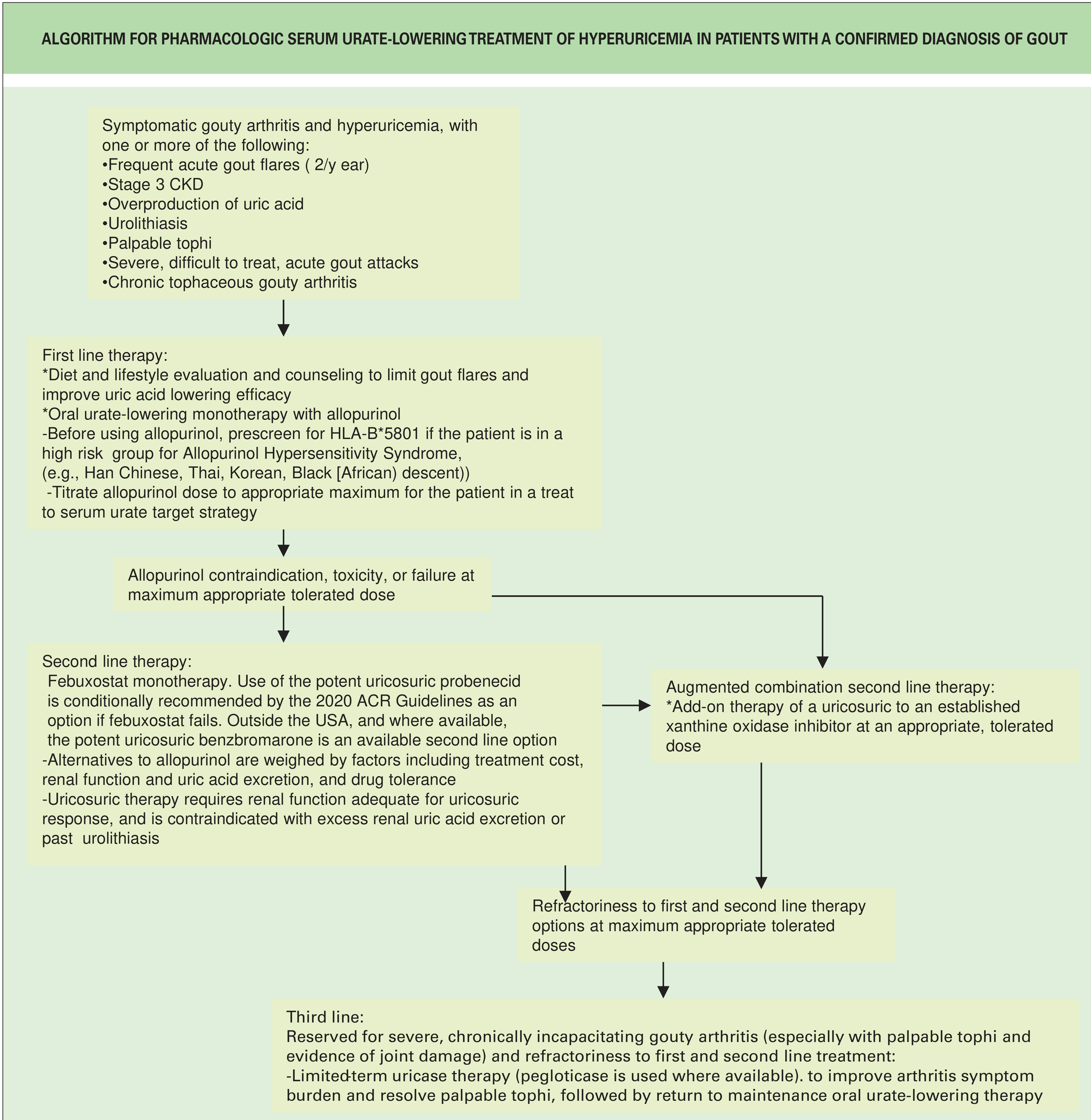
Indications for ULT (per ACR 2020 Guidelines):
- Frequent acute flares (≥2/year)
- Stage 3+ CKD
- Palpable tophi
- Urolithiasis
- Uric acid overproduction
- Severe/difficult-to-treat acute attacks or chronic tophaceous gout
Target: Serum urate < 6 mg/dL (< 0.36 mmol/L) in symptomatic patients; some guidelines recommend < 5 mg/dL for tophaceous gout.
Drug Options
| Drug | Class | Mechanism | Notes |
|---|---|---|---|
| Allopurinol | XOI (1st line) | Inhibits xanthine oxidase → reduces uric acid synthesis | Screen HLA-B*5801 in Han Chinese, Thai, Korean, Black patients (risk of severe hypersensitivity); start low (100 mg/d), titrate; reduce dose in CKD |
| Febuxostat | Non-purine XOI (2nd line) | Selective xanthine oxidase inhibitor | No dose adjustment for renal impairment; higher CV mortality vs. allopurinol in high-risk CV patients; avoid with azathioprine |
| Probenecid / Sulfinpyrazone | Uricosuric | Block proximal tubular urate reabsorption → increase excretion | Requires adequate renal function; contraindicated with urolithiasis or uric acid overexcretion |
| Benzbromarone | Potent uricosuric | Same | Available outside USA; second-line option |
| Pegloticase (Krystexxa) | Recombinant PEGylated uricase | Converts urate → allantoin | FDA-approved 2010 for refractory gout; risk of infusion reactions and loss of efficacy from anti-drug antibodies; third-line |
Important interaction: Both allopurinol and febuxostat inhibit xanthine oxidase, the enzyme that metabolizes azathioprine - concomitant use requires azathioprine dose reduction by ~25% or switching to mycophenolate. - Comprehensive Clinical Nephrology 7e
Lifestyle Modifications
- Avoid high-purine foods: organ meats (liver, kidneys), red meat, sardines, shellfish
- Limit alcohol (especially beer/spirits)
- Avoid drugs that raise urate: thiazide diuretics, salicylates (low-dose aspirin)
- Maintain adequate hydration
- Weight loss in obesity
5. Uric Acid and Cardiovascular/Renal Disease
There is increasing evidence that hyperuricemia is an independent risk factor for:
- Chronic kidney disease progression (elevated urate may accelerate CKD; CKD also raises urate - bidirectional relationship)
- Hypertension
- Cardiovascular disease (metabolic syndrome, coronary artery disease)
- Recent meta-analysis (PMID 39352584) examined CV outcomes of uric acid-lowering medications; urate-lowering therapy appears to have potential cardioprotective effects
- A 2025 network meta-analysis (PMID 40012480) confirms febuxostat and allopurinol are comparably efficacious in CKD stage 3-5 with asymptomatic hyperuricemia, supporting urate management in renal patients
Asymptomatic hyperuricemia: The consensus is that it should not be routinely treated based on urate level alone unless specific indications exist (recurrent gout, urolithiasis, high-risk CKD setting). - Swanson's Family Medicine Review
Summary Table: Key Uric Acid Disorders
| Disorder | Mechanism | Key Feature | Treatment |
|---|---|---|---|
| Acute gout | MSU crystal-triggered inflammasome activation | Podagra; crystals on aspirate | NSAIDs, colchicine, steroids |
| Chronic tophaceous gout | Progressive MSU deposition | Tophi, joint destruction | ULT (allopurinol/febuxostat) |
| Uric acid nephrolithiasis | Low urine pH + hyperuricosuria | Radiolucent stones on X-ray | Urinary alkalinization (K-citrate), hydration |
| Acute urate nephropathy | Tubular obstruction | Tumor lysis syndrome | Rasburicase, allopurinol prophylaxis, hydration |
| Lesch-Nyhan syndrome | Complete HGPRT deficiency | Self-mutilation + hyperuricemia | Allopurinol + neurologic support |
| Gouty diathesis | Low urine pH (no hyperuricosuria) | Uric acid stones without gout | Urinary alkalinization |
Segregate according to hypouricemia and hyperuricemia and also segregate the systems where the disease is involved
Searching sources...
Finding Sources
Finding Sources
Reading File
Reading File
Reading File
Reading File
Finding Sources
Reading File
Reading File
Finding Sources
Reading File
Reading File
Finding Sources
I now have all the data needed for a thorough, well-structured answer. Let me compile it.
Uric Acid-Related Disorders: Segregated by Serum Level and System Involved
REFERENCE RANGES
| Value | |
|---|---|
| Normal serum urate (men) | 3.4 - 7.0 mg/dL |
| Normal serum urate (women) | 2.4 - 6.0 mg/dL |
| Hyperuricemia | > 6.8 mg/dL (above physiologic solubility threshold) |
| Hypouricemia | < 2.0 mg/dL |
PART I: HYPERURICEMIA-RELATED DISORDERS
Caused by overproduction of uric acid, reduced renal excretion, or both.
🦴 MUSCULOSKELETAL SYSTEM
1. Acute Gout (Acute Gouty Arthritis)
- Mechanism: Monosodium urate (MSU) crystals precipitate in supersaturated synovial fluid → phagocytosed by macrophages → NLRP3 inflammasome → caspase-1 → IL-1β → neutrophil recruitment → acute inflammation
- Classic site: 1st metatarsophalangeal joint (podagra) in 50% of cases; also ankle, knee, midfoot
- Features: Sudden onset severe pain, erythema, swelling; typically self-resolves in days-weeks; serum uric acid can be normal during an acute attack
- Diagnosis: Negatively birefringent needle-shaped MSU crystals on joint aspirate (pathognomonic)
- Robbins & Kumar Basic Pathology; Grainger & Allison's Diagnostic Radiology
2. Chronic Tophaceous Gout
- Mechanism: Repeated MSU crystal deposition in joints and periarticular soft tissues when no longer pain-free between attacks
- Features: Tophi (aggregates of urate crystals + inflammatory tissue) in synovial membranes, periarticular tissue, cartilage; progressive joint destruction; affects small joints of hands/feet, ankles, knees
- Imaging: Punched-out periarticular erosions with overhanging edge on X-ray; double contour sign on ultrasound; quantified by dual-energy CT (DECT)
- Grainger & Allison's Diagnostic Radiology; Robbins & Kumar
3. Lesch-Nyhan Syndrome (Secondary Gout - Joint Component)
- Urate deposition in joints contributing to severe gouty arthritis on top of the neurologic syndrome (see Nervous System below)
- Robbins & Kumar Basic Pathology
🫘 RENAL SYSTEM
4. Acute Urate Nephropathy
- Mechanism: Massive, rapid hyperuricemia (classically tumor lysis syndrome) → urate crystals precipitate in renal tubular lumens → acute tubular obstruction → oliguric acute kidney injury
- Cause: Bulky haematologic cancers undergoing chemotherapy; rarely other causes of rapid cell lysis
- Treatment: Rasburicase (recombinant uricase), allopurinol prophylaxis, aggressive IV hydration, urinary alkalinization
- Harrison's Principles of Internal Medicine 22e; Rheumatology 2-Volume Set (Elsevier)
5. Chronic Urate Nephropathy
- Mechanism: Chronic interstitial deposition of urate crystals → tubulointerstitial nephritis → progressive CKD
- Note: This entity is controversial - many cases represent CKD from hypertension/atherosclerosis co-existing with gout; true isolated urate nephropathy is rare
- Tietz Textbook of Laboratory Medicine 7e
6. Uric Acid Nephrolithiasis (Uric Acid Kidney Stones)
- Three determinants: Low urine pH (most important) + low urine volume + hyperuricosuria
- Mechanism of low pH: Insulin resistance → impaired renal ammoniagenesis → unbuffered H⁺ in urine → pH falls → uric acid precipitates (pKa 5.35; at pH 5 even modest uric acid exceeds solubility)
- Strong association: Type 2 diabetes, metabolic syndrome (insulin resistance, hypertriglyceridemia, obesity, hypertension) - uric acid stones represent 36% of stones in T2DM patients vs 11% in non-diabetics
- Complication: Uric acid also acts as a nidus for calcium oxalate stones via heterogeneous nucleation
- Treatment: Potassium citrate (alkalinize urine to pH 6.0-6.5), high fluid intake, dietary protein restriction, allopurinol if hyperuricosuria
- Campbell Walsh Wein Urology
7. Familial Juvenile Hyperuricemic Nephropathy / ADTKD-UMOD
- Now classified as: Autosomal Dominant Tubulointerstitial Kidney Disease (ADTKD)
- Mechanism: UMOD gene mutation → defective uromodulin protein in thick ascending limb → decreased salt reabsorption → compensatory increased proximal tubular reabsorption of NaCl and uric acid → hyperuricemia → progressive CKD
- Features: Early-onset hyperuricemia, gout in young patients, progressive CKD with bland urinary sediment
- Comprehensive Clinical Nephrology 7e
🧠 NERVOUS SYSTEM
8. Lesch-Nyhan Syndrome
- Cause: X-linked recessive; complete absence of HGPRT (hypoxanthine-guanine phosphoribosyltransferase) → no purine salvage → all purines degraded to uric acid → severe hyperuricemia
- Key features:
- Intellectual disability
- Choreoathetosis and spasticity
- Compulsive self-mutilation (pathognomonic - lip and finger biting)
- Gouty arthritis
- Uric acid nephrolithiasis
- Treatment: Allopurinol (reduces uric acid, prevents urate nephropathy) - but has no effect on CNS symptoms; behavioral and symptomatic management for neurologic features
- Adams & Victor's Principles of Neurology 12e; Robbins & Kumar
Compare: Partial HGPRT deficiency (Kelley-Seegmiller syndrome) → hyperuricemia + gout/nephrolithiasis only, NO neurologic features
❤️ CARDIOVASCULAR SYSTEM
9. Hyperuricemia and Cardiovascular Disease
- Elevated urate is associated with hypertension, coronary artery disease, heart failure, and metabolic syndrome
- Proposed mechanisms: oxidative stress via xanthine oxidase, endothelial dysfunction, promotion of vascular inflammation
- Uric acid stone formers share features of metabolic syndrome: hypertriglyceridemia, hyperglycemia, obesity, hypertension
- Urate-lowering therapy has potential CV benefits per recent meta-analysis (PMID 39352584), though causality remains debated
- Brenner & Rector's The Kidney; Campbell Walsh Wein Urology
🩸 HAEMATOLOGY / ONCOLOGY
10. Tumor Lysis Syndrome (Hyperuricemia Component)
- Cause: Rapid destruction of tumor cells (especially leukemia, lymphoma, myeloma) by chemotherapy → massive nucleic acid release → purine catabolism → acute hyperuricemia
- Consequence: Acute urate nephropathy (tubular obstruction), also hyperkalemia, hyperphosphatemia, hypocalcemia
- Tietz Textbook of Laboratory Medicine 7e
11. Hyperuricemia in Myeloproliferative Disorders
- Increased cell turnover → increased nucleic acid catabolism → secondary hyperuricemia
- Secondary gout and uric acid stones can develop
🤰 OBSTETRICS
12. Preeclampsia
- Uric acid is elevated in preeclampsia due to reduced renal urate excretion (decreased GFR + increased tubular reabsorption)
- Historically used as a marker, but now recognized to have limited clinical utility in distinguishing preeclampsia from other hypertensive disorders of pregnancy or predicting adverse outcomes
- Diuretic therapy in pregnancy raises serum uric acid further, invalidating its use as a preeclampsia marker
- Brenner & Rector's The Kidney; Creasy & Resnik's Maternal-Fetal Medicine
🧬 METABOLIC / INHERITED DISORDERS (Causing Hyperuricemia)
13. Glucose-6-Phosphatase Deficiency (Von Gierke Disease, GSD Type I)
- Leads to hyperuricemia via increased purine synthesis and lactic acidosis (which competes with tubular urate secretion)
14. Altered ATP Metabolism / Tissue Hypoxia
- Rapid ATP degradation → AMP → IMP → inosine → hypoxanthine → xanthine → uric acid
- Occurs in strenuous exercise, myocardial infarction, status epilepticus
🩺 SKIN / DERMATOLOGY
15. Psoriasis-Associated Hyperuricemia
- Rapid keratinocyte turnover → increased nucleic acid catabolism → secondary hyperuricemia
- Gout can occur as a complication of psoriasis
- Tietz Textbook of Laboratory Medicine 7e
16. Gouty Tophi (Cutaneous Manifestations)
- Urate crystal deposits in skin and subcutaneous tissue, especially over ears (helix), olecranon bursae, Achilles tendon, fingers
- Appear as firm, chalky white nodules
PART II: HYPOURICEMIA-RELATED DISORDERS
Serum urate < 2.0 mg/dL. Caused by reduced uric acid production or excess renal excretion.
🫘 RENAL SYSTEM
1. Hereditary Renal Hypouricemia (Familial Renal Hypouricemia)
- Inheritance: Autosomal recessive
- Two types:
| Type | Gene | Protein | Prevalence |
|---|---|---|---|
| Type 1 (>90%) | SLC22A12 | URAT1 (apical urate/anion exchanger) | Japanese, Ashkenazi Jewish populations |
| Type 2 | SLC2A9 | GLUT9 (basolateral urate transporter) | More severe |
- Features: Very low serum uric acid (<2.5 mg/dL men, <2.1 mg/dL women); fractional excretion of urate 50-150% (normal ~10%)
- Complications:
- Exercise-induced AKI: Hyperuricosuria + dehydration + low pH → uric acid "gravel" → acute tubular injury → AKI (paradoxically, despite hypouricemia in blood, massive uricosuria causes renal damage)
- Nephrolithiasis: ~25% of patients; only 1/3 of those are uric acid stones (others are calcium stones due to associated hypercalciuria)
- Treatment: High fluid intake; urine alkalinization and allopurinol for persistent stone formers
- Brenner & Rector's The Kidney; Comprehensive Clinical Nephrology 7e
2. Fanconi Syndrome
- Generalized proximal tubular dysfunction → loss of urate reabsorption → hypouricemia + uricosuria (alongside glycosuria, aminoaciduria, phosphaturia, bicarbonate wasting)
- Causes: Cystinosis, Wilson disease, galactosemia, heavy metal toxicity, tenofovir, multiple myeloma (light chains)
- Urolithiasis from uricosuria is rare because high urine flow and alkaline pH prevent precipitation
- Comprehensive Clinical Nephrology 7e
🧬 METABOLIC / INHERITED DISORDERS (Causing Hypouricemia)
3. Hereditary Xanthinuria
- Inheritance: Autosomal recessive
- Type I: Xanthine dehydrogenase (XDH) deficiency
- Type II: Combined XDH + aldehyde oxidase deficiency
- Mechanism: No xanthine oxidase activity → xanthine and hypoxanthine cannot be converted to uric acid → very low serum urate AND very low urine urate (key distinction from renal hypouricemia where urine urate is HIGH)
- Features: Xanthine stones (radiolucent) in ~1/3 of cases; hematuria; occasionally occult renal failure; elevated urinary xanthine + hypoxanthine
- Note: Acquired xanthinuria can occur with allopurinol treatment (xanthine stones may form if allopurinol is overused in Lesch-Nyhan or myeloproliferative disorders)
- Brenner & Rector's The Kidney
4. Molybdenum Cofactor Deficiency
- Combined deficiency of xanthine oxidase + sulfite oxidase (both require molybdenum cofactor)
- Results in hypouricemia + xanthinuria + neurologic damage (from sulfite accumulation)
- Tietz Textbook of Laboratory Medicine 7e
5. Purine Nucleoside Phosphorylase (PNP) Deficiency
- Rare inherited defect → impaired purine catabolism → very low uric acid
- Associated with profound T-cell immunodeficiency (lymphopenia, recurrent infections) - the immunologic features dominate
🫀 HEPATIC SYSTEM
6. Severe Hepatocellular Disease
- Liver is the primary site of xanthine oxidase activity and purine synthesis
- Severe hepatic failure → reduced xanthine oxidase activity → reduced uric acid synthesis → hypouricemia
- Tietz Textbook of Laboratory Medicine 7e
💊 DRUG-INDUCED HYPOURICEMIA
| Drug | Mechanism |
|---|---|
| Allopurinol (overtreatment) | Xanthine oxidase inhibition → reduced synthesis |
| Uricosuric agents (probenecid, sulfinpyrazone) | Block tubular reabsorption → excess excretion |
| 6-Mercaptopurine / Azathioprine | Inhibit de novo purine synthesis |
| Losartan (ARB) | Mild uricosuric property |
| High-dose salicylates | Uricosuric at high doses (paradoxically, low-dose aspirin raises urate) |
| Radiopaque contrast media (acute) | Transient defective tubular reabsorption |
Tietz Textbook of Laboratory Medicine 7e
MASTER SUMMARY TABLE
Hyperuricemia Disorders by System
| System | Disorder | Primary or Secondary |
|---|---|---|
| Musculoskeletal | Acute gout | Both |
| Musculoskeletal | Chronic tophaceous gout | Both |
| Musculoskeletal | Kelley-Seegmiller syndrome (partial HGPRT deficiency) | Primary (genetic) |
| Renal | Acute urate nephropathy | Secondary (tumor lysis) |
| Renal | Chronic urate nephropathy | Both |
| Renal | Uric acid nephrolithiasis | Both |
| Renal | ADTKD-UMOD (familial juvenile hyperuricemic nephropathy) | Primary (genetic) |
| Nervous | Lesch-Nyhan syndrome | Primary (genetic) |
| Cardiovascular | Hypertension, coronary artery disease, metabolic syndrome | Association |
| Haematologic | Tumor lysis syndrome | Secondary |
| Haematologic | Myeloproliferative disorders | Secondary |
| Obstetric | Preeclampsia | Secondary |
| Dermatologic | Psoriasis-related gout | Secondary |
| Metabolic | Von Gierke disease (GSD-I) | Secondary |
Hypouricemia Disorders by System
| System | Disorder | Urine Uric Acid |
|---|---|---|
| Renal | Hereditary renal hypouricemia Type 1 (URAT1 defect) | HIGH (>50% FE) |
| Renal | Hereditary renal hypouricemia Type 2 (GLUT9 defect) | VERY HIGH (up to 150% FE) |
| Renal | Fanconi syndrome | HIGH |
| Metabolic | Hereditary xanthinuria Type I & II | LOW (key distinction) |
| Metabolic | Molybdenum cofactor deficiency | LOW |
| Immunologic | PNP deficiency | Low-normal |
| Hepatic | Severe hepatocellular failure | LOW |
| Iatrogenic | Overtreatment with allopurinol/uricosurics | Varies |
Key Diagnostic Tip: Measuring urine uric acid (or fractional excretion of urate) separates the two main hypouricemia mechanisms:
- High urine uric acid → renal leak (Fanconi, renal hypouricemia)
- Low urine uric acid → reduced production (xanthinuria, hepatic disease, drug inhibition)
Causes of high urea accumulation
Searching sources...
Finding Sources
Finding Sources
Finding Sources
Finding Sources
Reading File
Reading File
Reading File
Finding Sources
Finding Sources
Reading File
Reading File
Reading File
Finding Sources
Finding Sources
Reading File
Reading File
Reading File
Loading Image
Finding Sources
I now have comprehensive data from multiple authoritative textbooks. Here is the complete, well-organized answer:
Causes of High Urea (BUN) Accumulation
Biochemical Basis
BUN (Blood Urea Nitrogen) is the nitrogen component of urea (H₂N-CO-NH₂), the end product of protein catabolism in the liver:
2NH₃ + CO₂ → Urea + H₂O
BUN is directly related to protein catabolism and inversely related to GFR. Normal range: 10-20 mg/dL. Values > 50 mg/dL are generally associated with impaired kidney function. Approximately 40-50% of filtered urea is passively reabsorbed by renal tubules; hypovolemia increases this fraction further, causing a disproportionate BUN rise relative to creatinine.
BUN:Creatinine ratio is the key diagnostic tool:
-
> 20:1 = prerenal state (urea reabsorption + sodium)
-
10-20:1 = intrinsic renal disease (ATN)
-
< 10:1 = low urea production (malnutrition, liver disease) or rhabdomyolysis
-
Morgan & Mikhail's Clinical Anesthesiology 7e; Harrison's Principles of Internal Medicine 22e
Overview Diagram
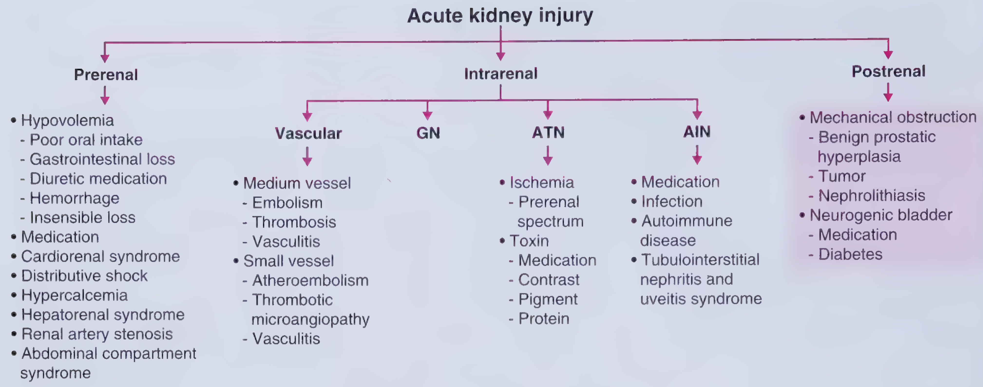
The causes of high urea accumulation fall into two broad categories:
- Increased urea production (non-renal)
- Decreased urea excretion (renal - pre/intra/postrenal)
CATEGORY A: INCREASED UREA PRODUCTION (Non-Renal Causes)
These cause a high BUN with normal or near-normal creatinine - BUN:Cr ratio > 20:1
1. High Dietary Protein Intake
- Excess animal protein → increased hepatic urea synthesis
- BUN rises without kidney disease
- Seen in high-protein diets (bodybuilders, carnivorous diets, hyperalimentation/TPN)
2. Gastrointestinal (GI) Bleeding
- Blood in the GI lumen is digested as protein → massive purine/amino acid load → increased hepatic urea synthesis
- Upper GI bleeding (peptic ulcer, varices, Mallory-Weiss tear) causes more pronounced BUN rise than lower GI bleeding (blood is fully digested in the small intestine)
- BUN:Cr ratio classically > 20:1
- Harrison's Principles of Internal Medicine 22e; Symptom to Diagnosis 4e
3. Increased Tissue Catabolism
| State | Mechanism |
|---|---|
| Fever / infection / sepsis | Accelerated protein breakdown for gluconeogenesis |
| Major trauma / surgery | Stress response → proteolysis |
| Severe burns | Massive protein loss from tissue destruction |
| Starvation (early) | Muscle protein used as fuel |
| Malignancy | Tumor cachexia, increased catabolism |
4. Glucocorticoid / Corticosteroid Use
- Steroids are antianabolic and promote protein catabolism → increased urea production
- Also cause mild volume depletion → increased tubular urea reabsorption
- Frameworks for Internal Medicine; Harrison's Principles
5. Tetracyclines (Antianabolic Effect)
- Tetracyclines block hepatic protein synthesis → shift amino acids toward catabolism → increased urea generation
- Particularly significant in patients with borderline renal function
6. Large Hematoma Reabsorption
- Blood trapped in tissue (post-trauma, post-surgical) is reabsorbed and metabolized as protein → BUN rises
- Similar mechanism to GI bleeding
7. Hypercatabolic States
- Severe inflammation, cancer treatment (tumor lysis), cytokine storms → accelerated nucleic acid and protein breakdown
CATEGORY B: DECREASED UREA EXCRETION (Renal Causes)
Divided into Prerenal, Intrinsic Renal (Intrarenal), and Postrenal - the three categories of AKI.
PRERENAL (Reduced Renal Perfusion)
Mechanism: Reduced blood flow to kidneys → decreased GFR → less urea filtered → urea accumulates. Tubules remain intact and avidly reabsorb urea and sodium. BUN:Cr ratio typically > 20:1.
A. Hypovolemia (True Volume Depletion)
| Cause | Example |
|---|---|
| Poor oral intake | Inability to drink, fasting |
| GI losses | Vomiting, diarrhea, nasogastric drainage |
| Urinary losses | Diuretics (thiazide, loop), diabetes insipidus, osmotic diuresis |
| Hemorrhage | Trauma, surgical blood loss, GI bleed |
| Insensible losses | Burns, fever, excessive sweating |
| Third spacing | Pancreatitis, bowel obstruction, sepsis (capillary leak) |
B. Reduced Cardiac Output (Cardiorenal Syndrome)
- Congestive heart failure, cardiogenic shock, severe valvular disease
- Decreased effective arterial blood volume → renal hypoperfusion despite normal or increased total body fluid
- Smith & Tanagho's General Urology 19e
C. Distributive Shock
- Septic shock, anaphylaxis → vasodilation → effective circulating volume falls → renal underperfusion
D. Hepatorenal Syndrome
- Advanced cirrhosis → splanchnic vasodilation → reduced effective renal perfusion
- Urinary findings mimic prerenal failure; responds to liver transplantation, not fluid resuscitation
- Smith & Tanagho's General Urology 19e
E. Medications Causing Functional Prerenal State
| Drug | Mechanism |
|---|---|
| ACE inhibitors / ARBs | Block angiotensin II → dilate efferent arteriole → drop GFR (especially in bilateral RAS or single kidney) |
| NSAIDs | Block prostaglandin-mediated afferent arteriolar dilation → drop GFR (especially in volume-depleted states) |
| Cyclosporine / Tacrolimus | Intrarenal vasoconstriction |
| Antihypertensive overuse | Systemic hypotension → reduced renal perfusion |
F. Renal Artery Stenosis
- Bilateral (or unilateral in solitary kidney) → chronic prerenal state
- Brenner & Rector's The Kidney
G. Abdominal Compartment Syndrome
- Raised intra-abdominal pressure → compresses renal vasculature → functional prerenal AKI
INTRINSIC RENAL (Parenchymal Causes)
Mechanism: Structural damage to the kidney itself → reduced GFR and impaired tubular function. BUN:Cr ratio typically 10-20:1.
1. Tubular Causes
Acute Tubular Necrosis (ATN) - Most Common Intrinsic Cause
| Subtype | Causes |
|---|---|
| Ischemic ATN | Prolonged prerenal state, cardiac arrest, major surgery, severe hypotension - the "prerenal spectrum" |
| Nephrotoxic ATN | Aminoglycosides, amphotericin B, cisplatin, vancomycin, IV contrast agents |
| Pigment nephropathy | Rhabdomyolysis (myoglobin) → tubular obstruction + direct toxicity; Hemolysis (hemoglobin) |
| Myeloma cast nephropathy | Light chains precipitate in tubules |
2. Glomerular Causes (Glomerulonephritis)
- Rapidly progressive GN (RPGN): Anti-GBM disease, ANCA-associated vasculitis, immune complex GN
- Acute post-infectious GN: Post-streptococcal
- Lupus nephritis
- IgA nephropathy (Berger disease)
3. Vascular Causes
| Vessel Size | Conditions |
|---|---|
| Large vessels | Renal artery thrombosis/embolism, aortic dissection, renal vein thrombosis |
| Medium vessels | Polyarteritis nodosa, Takayasu arteritis |
| Small vessels | Atheroembolic disease, thrombotic microangiopathy (HUS/TTP), malignant hypertension |
4. Interstitial Causes (Acute Interstitial Nephritis - AIN)
- Drug-induced (most common): Penicillins, cephalosporins, NSAIDs, PPIs, sulfonamides, rifampicin
- Infectious: Pyelonephritis, leptospirosis, hantavirus
- Autoimmune: Sarcoidosis, Sjögren syndrome, SLE, TINU syndrome (tubulointerstitial nephritis and uveitis)
5. Chronic Kidney Disease (CKD)
- Any cause leading to progressive nephron loss → sustained reduction in GFR → chronic urea accumulation (uremia)
- Note: 2/3 of renal function must be lost before a significant BUN rise becomes evident
- Common causes: Diabetic nephropathy, hypertensive nephrosclerosis, chronic GN, polycystic kidney disease
- Smith & Tanagho's General Urology 19e
POSTRENAL (Urinary Tract Obstruction)
Mechanism: Obstruction blocks urine outflow → increased intratubular pressure → decreased GFR via afferent vasoconstriction (angiotensin II, thromboxane A2, vasopressin) → urea accumulates. Potentially reversible if relieved early (complete recovery if < 1 week; poor recovery > 12 weeks).
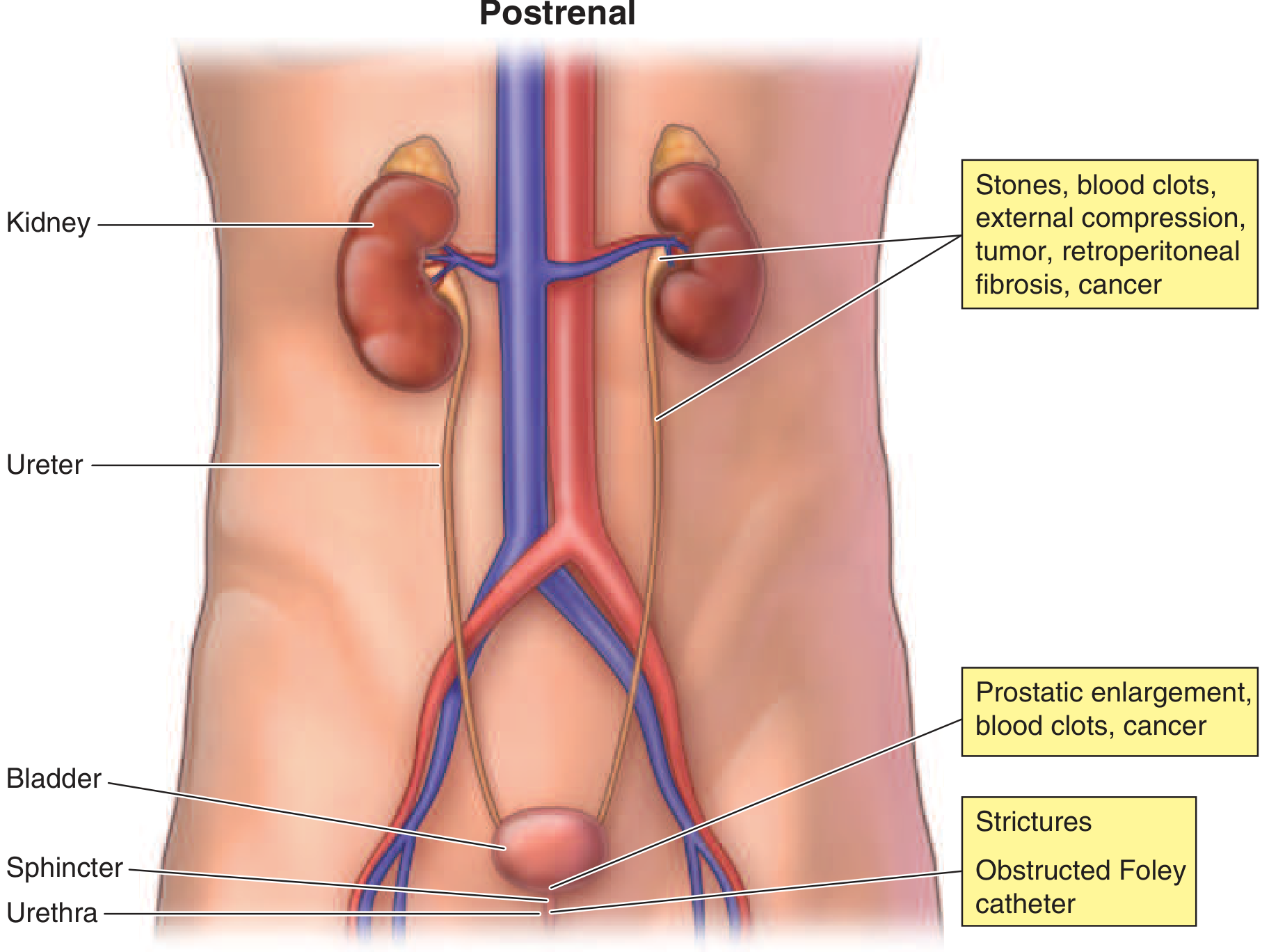
Intraluminal Obstruction
| Location | Cause |
|---|---|
| Ureter | Nephrolithiasis (bilateral or solitary kidney), blood clots, sloughed renal papillae |
| Bladder | Blood clots, calculi, fungal ball |
| Urethra | Strictures, phimosis |
Intramural / Wall Infiltration
- Ureteric/bladder malignancy invading the wall
Extrinsic Compression
| Cause | Notes |
|---|---|
| BPH (Benign Prostatic Hyperplasia) | Most common cause of postrenal AKI in elderly men |
| Prostate cancer | Hard, nodular irregular prostate |
| Retroperitoneal fibrosis | Can compress ureters bilaterally |
| Pelvic/abdominal malignancy | Cervical, colorectal, lymphoma - external ureteric compression |
| Retroperitoneal abscess or hematoma | Post-surgical |
| Inadvertent surgical ureteric ligation | Post-hysterectomy, colorectal surgery |
Functional Obstruction (Neurogenic Bladder)
- Anticholinergic medications (antihistamines, antipsychotics, TCAs)
- Diabetic autonomic neuropathy
- Spinal cord injury
- Harrison's Principles of Internal Medicine 22e; Frameworks for Internal Medicine
MASTER SUMMARY TABLE
| Category | Subcategory | Representative Causes | BUN:Cr Ratio |
|---|---|---|---|
| Increased Production | High protein | Diet, TPN, GI bleed | > 20:1 (with normal Cr) |
| Increased Production | Hypercatabolism | Sepsis, burns, trauma, steroids, tetracyclines | > 20:1 (with normal Cr) |
| Prerenal | True hypovolemia | Vomiting, diarrhea, hemorrhage, diuretics, burns | > 20:1 |
| Prerenal | Reduced CO | Heart failure, cardiogenic shock | > 20:1 |
| Prerenal | Distributive | Septic shock, anaphylaxis | > 20:1 |
| Prerenal | Functional | NSAIDs, ACEi/ARBs, HRS, cyclosporine | > 20:1 |
| Intrinsic Renal | Tubular (ATN) | Ischemia, aminoglycosides, contrast, rhabdomyolysis | 10-20:1 |
| Intrinsic Renal | Glomerular (GN) | RPGN, post-infectious GN, lupus nephritis | 10-20:1 |
| Intrinsic Renal | Vascular | HUS/TTP, atheroembolism, malignant HTN | 10-20:1 |
| Intrinsic Renal | Interstitial (AIN) | Drugs, infection, autoimmune | 10-20:1 |
| Intrinsic Renal | Chronic (CKD) | DM nephropathy, HTN, polycystic kidney | 10-20:1 |
| Postrenal | Mechanical | BPH, stones, tumor, retroperitoneal fibrosis | Variable; initially > 20:1 |
| Postrenal | Neurogenic | Anticholinergics, diabetes, spinal cord injury | Variable |
Clinical Pearl: BUN alone is an unreliable marker of GFR because it is influenced by so many non-renal factors. Serum creatinine (or cystatin C) is more specific for kidney function. However, a rising BUN with a rising BUN:Cr ratio > 20:1 in the absence of GI bleed or catabolic state strongly suggests a prerenal mechanism. - Brenner & Rector's The Kidney; Morgan & Mikhail's Clinical Anesthesiology 7e
This is a shared conversation. Sign in to Orris to start your own chat.