
Sign symptoms of anemia
signs and symptoms of anemia clinical features pallor
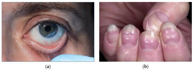
This composite of clinical photographs illustrates physical manifestations of severe anemia. Image (a) shows a close-up of a patient's eye with the lower eyelid retracted to reveal marked conjunctival pallor. The typically pink or reddish palpebral conjunctiva appears significantly pale and porcelain-white, a sign classically associated with a hemoglobin concentration below 90 g/L. Image (b) presents the fingernails of a patient demonstrating pronounced nail bed pallor. The distal nail beds lack the normal healthy pink hue. Additionally, horizontal white bands, known as Muehrcke's lines, are visible across the nail plates; these non-palpable transverse lines are associated with systemic conditions such as hypoalbuminemia and severe anemia. Together, these images demonstrate key external clinical signs used in the bedside assessment of hematological disorders and circulatory efficiency. The content is suitable for medical education regarding the physical examination findings of iron deficiency and other severe anemias.
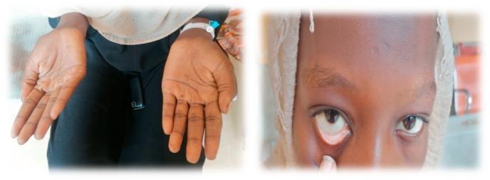
This composite clinical photograph illustrates common physical examination findings associated with severe anemia. The left panel shows the bilateral palms of a patient, demonstrating marked palmar pallor. The skin on the palms and creases appears significantly lighter and lacks the typical pinkish hue of healthy vascularized tissue, suggesting reduced hemoglobin concentration. The right panel is a close-up of the patient's face with the lower eyelid retracted to reveal the palpebral conjunctiva. Instead of the normal vibrant red or pink color, the conjunctiva exhibits conjunctival pallor, appearing whitish or very pale pink. These visual signs are critical diagnostic markers in hematology and general medicine for identifying systemic conditions such as iron-deficiency anemia or other forms of chronic blood loss. The image serves as a teaching tool for medical students and clinicians to recognize mucosal and cutaneous indicators of decreased red blood cell mass or oxygen-carrying capacity.
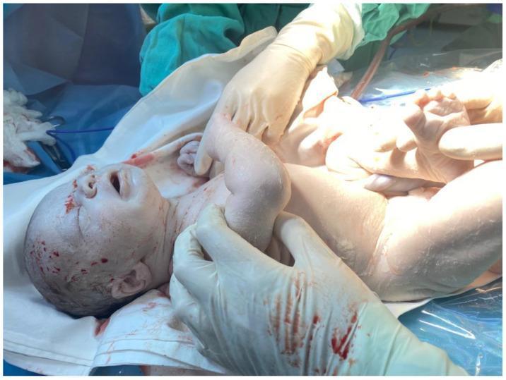
This clinical photograph shows a newborn male infant immediately following delivery, demonstrating severe, generalized pallor of the skin and mucosal surfaces, often described as a 'ghost-like' appearance. The infant is supported by gloved medical personnel in a delivery room setting, surrounded by surgical drapes. Notable features include extremely pale skin across the face, trunk, and extremities, and a marked lack of color in the lips. Superficial dark red-brown spotting and streaks of blood or delivery residue are visible on the scalp, forehead, and chest. The infant's mouth is open, and facial features appear otherwise anatomically normal. Clinically, this extreme pallor is a hallmark sign of severe neonatal anemia or hypovolemic shock, which in this context was caused by massive fetomaternal hemorrhage (FMH). The image serves as a critical educational example of the visual presentation of acute fetal blood loss and the necessity for immediate neonatal resuscitation and volume replacement.
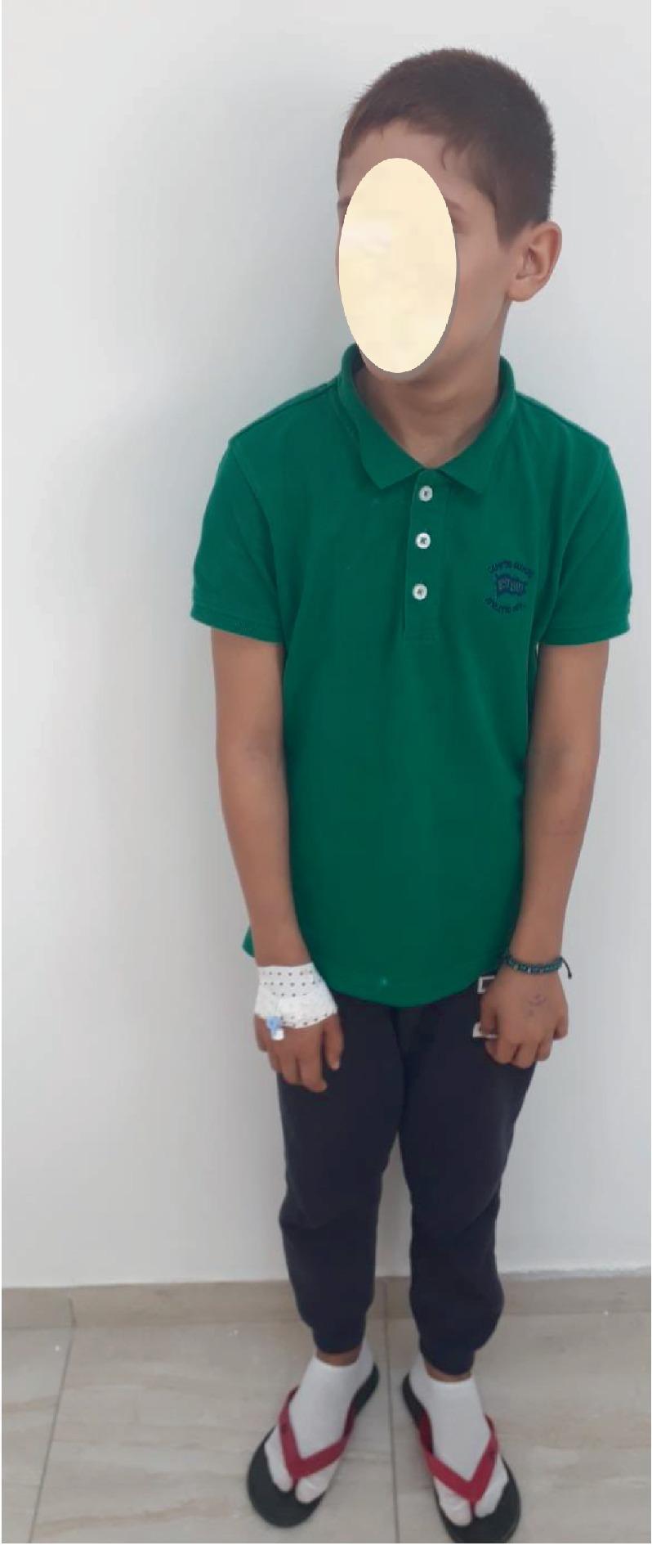
This clinical photograph shows a full-body view of a 10-year-old male patient presenting with physical manifestations suggestive of Fanconi anemia (FA). The most prominent clinical feature is short stature, with the patient appearing significantly smaller than expected for his chronological age. Visible dermatologic findings include generalized skin pallor, likely related to underlying anemia, and several hyperpigmented macules consistent with 'café au lait' spots on the arms and facial region. The patient's right hand is secured with a white compression bandage and an intravenous (IV) catheter, indicating active clinical management or hematologic intervention. Educational focus is placed on the recognition of constitutional features of inherited bone marrow failure syndromes, specifically the triad of short stature, characteristic skin pigmentation, and developmental anomalies in a pediatric hematology context.
Signs and Symptoms of Anemia
Symptoms (What the Patient Feels)
| System | Symptom |
|---|---|
| General | Fatigue, weakness, loss of stamina |
| Cardiovascular | Palpitations, dyspnea on exertion, angina (in atherosclerosis) |
| Neurological | Dizziness, headache, tinnitus, decreased cognitive ability |
| Neurological (B12 deficiency) | Peripheral neuropathy, subacute combined degeneration |
| Gastrointestinal (iron deficiency) | Pica (craving for ice, dirt, etc.) |
| Respiratory | Shortness of breath, dyspnea at rest (severe cases) |
Signs (What the Clinician Finds)
General / Skin
- Pallor - of the skin, mucous membranes, conjunctiva, palmar creases, and nail beds (most classic sign)
Eyes
- Conjunctival pallor - the palpebral conjunctiva appears pale/white instead of its normal pink-red
Cardiovascular
- Resting tachycardia - increased heart rate to compensate
- Wide pulse pressure and bounding/strong peripheral pulses
- Forceful heartbeat
- Systolic flow murmur (high-output state)
- Hypotension (especially in acute blood loss)
- High-output heart failure and peripheral edema (in severe/prolonged anemia)
Nails & Mouth (Severe / Long-standing Anemia)
- Koilonychia (spoon-shaped nails) - classic in iron deficiency
- Brittle nails
- Atrophic glossitis (smooth, red tongue)
- Angular cheilosis (cracks at corners of the mouth)
Clinical Images
Key Clinical Points
- Gradual onset - patients can tolerate surprisingly low hemoglobin levels (sometimes Hgb < 7 g/dL) with few symptoms due to compensatory mechanisms: increased cardiac output, elevated 2,3-DPG (right-shifting oxygen-hemoglobin curve), and expanded plasma volume.
- Acute onset - even moderate anemia can cause shock and hemodynamic instability (hypovolemic shock in acute hemorrhage).
- Signs and symptoms are unreliable at predicting the exact hemoglobin/hematocrit level - laboratory testing is essential.
- Ischemic symptoms (angina, TIA, stroke) may appear if the patient has underlying atherosclerosis.
- Harrison's Principles of Internal Medicine 22E (2025), "Signs and Symptoms of Anemia," p. 488
- Washington Manual of Medical Therapeutics, "Clinical Presentation of Anemia," p. 795
- Frameworks for Internal Medicine, "Anemia - Physical Findings"
Sigb symptoms of jaundice
jaundice clinical signs scleral icterus yellow skin
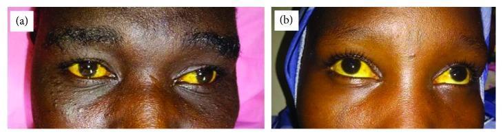
This clinical photograph consists of two panels, (a) and (b), displaying a close-up view of the eyes of two different patients. Both images demonstrate significant conjunctival icterus (scleral jaundice), characterized by a deep, uniform yellow discoloration of the sclera in both eyes. The intensity of the pigmentation is marked, standing in stark contrast to the dark irises. Aside from the jaundice, there are no visible signs of conjunctival injection, chemosis, or discharge. This finding is a clinical hallmark of hyperbilirubinemia, which in these cases is contextually associated with blackwater fever, a severe complication of Plasmodium falciparum malaria characterized by intravascular hemolysis. The visual documentation serves as an educational tool for recognizing advanced jaundice in patients with dark skin tones, where mucosal and scleral examination is critical for detecting hyperbilirubinemia.
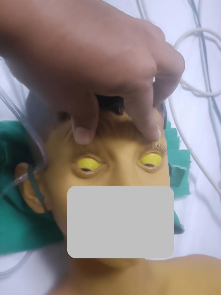
This clinical photograph displays a pediatric patient demonstrating severe systemic jaundice. The primary diagnostic feature is profound scleral icterus, characterized by a bright yellow discoloration of the entire visible sclera in both eyes. The upper eyelids are manually elevated to fully reveal the extent of the icterus. Additionally, the patient's facial skin, forehead, and upper chest exhibit a marked yellowish hue (jaundice), consistent with hyperbilirubinemia. The patient appears to be in a clinical setting, as evidenced by medical tubing and linens visible in the background. This image is an educational example of the physical manifestations of liver dysfunction or hemolytic processes, such as hepatitis, Wilsonian crisis, or acute toxicity, which can lead to significant accumulation of bilirubin in mucosal surfaces and skin.
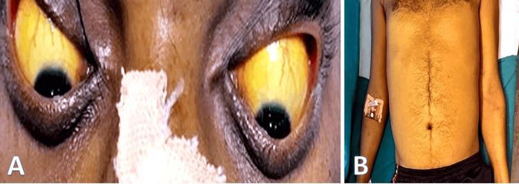
This composite clinical photograph displays physical manifestations of hyperbilirubinemia in a 35-year-old male. Image A provides a close-up view of the patient's eyes, demonstrating severe scleral icterus characterized by a deep yellow discoloration of the bulbar conjunctiva and sclera. The irises, pupils, and surrounding periorbital skin are also visible. Image B is a frontal view of the patient's torso and upper limbs, showing generalized jaundice (icterus) manifested as a diffuse yellowing of the skin across the chest, abdomen, and arms. Visible landmarks include the umbilicus and sparse terminal hair on the chest and linea alba. An intravenous (IV) catheter is noted on the right forearm, secured with medical tape. These findings are classic clinical signs of hepatic or biliary pathology, such as alcoholic hepatitis, cirrhosis, or obstructive jaundice, reflecting a systemic buildup of bilirubin.
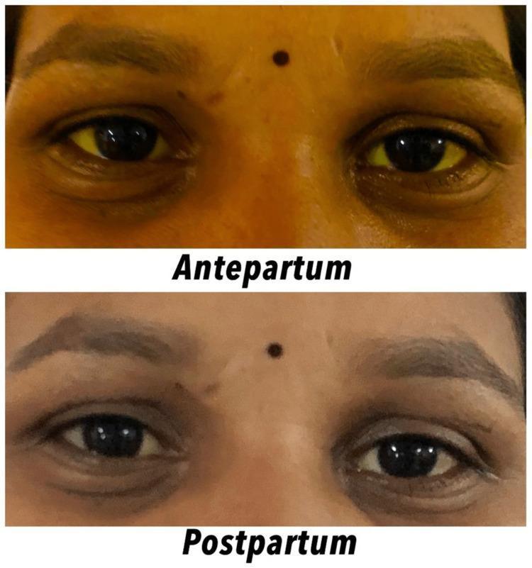
This clinical photograph provides a side-by-side comparison of facial features in a female patient, illustrating the resolution of icterus (jaundice) from the antepartum to the postpartum period. The top panel, labeled 'Antepartum,' displays bilateral scleral icterus, characterized by a distinct yellow discoloration of the ocular sclera. Additionally, the periorbital and facial skin exhibits a generalized yellowish hue. The bottom panel, labeled 'Postpartum,' shows the same patient after delivery, demonstrating a significant clinical improvement. The sclerae have returned to a normal white color, and the skin tone has transitioned back to a typical pigmentation, indicating a reduction in serum bilirubin levels. This comparison is used to visualize the clinical manifestation and subsequent resolution of hepatobiliary or hemolytic conditions during pregnancy, such as intrahepatic cholestasis of pregnancy (IHCP) or other pregnancy-related liver disorders. The image serves as an educational tool for identifying physical signs of hyperbilirubinemia and monitoring treatment response in an obstetric context.
Signs and Symptoms of Jaundice
Threshold for Detection
| Bilirubin Level | Clinical Finding |
|---|---|
| > 2.5 - 3.0 mg/dL | Scleral icterus visible first |
| > 3.0 mg/dL | Skin jaundice detectable (sensitivity 78%) |
| > 15 mg/dL | Skin jaundice almost always visible (sensitivity 96%) |
Signs (What the Clinician Finds on Examination)
Skin & Eyes
- Scleral icterus - yellow discoloration of the whites of the eyes (earliest and most sensitive sign)
- Yellow skin (jaundice) - uniform yellow discoloration over the entire body in light-skinned patients
- Green-tinged skin - seen in long-standing jaundice (bilirubin oxidized to biliverdin)
- Skin xanthomas - focal cholesterol deposits in obstructive/cholestatic jaundice
- Bruising / bleeding tendency - from vitamin K malabsorption (fat-soluble vitamin deficiency)
Abdomen
- Hepatomegaly - enlarged, possibly tender liver
- Splenomegaly - especially in hemolytic or portal hypertension-related jaundice
- Ascites - in advanced liver disease
- Right upper quadrant tenderness - in hepatitis or biliary obstruction
Urine & Stool
- Dark urine ("tea-" or "cola-colored") - due to renal excretion of conjugated bilirubin; indicates liver/biliary disease
- Pale/clay-colored stools (acholic stools) - due to lack of bile reaching the intestine (obstructive jaundice)
Symptoms (What the Patient Feels)
| Symptom | Notes |
|---|---|
| Pruritus (itching) | From bile salt deposition in skin; often severe in cholestatic jaundice |
| Fatigue and malaise | General feature of liver dysfunction |
| Anorexia / nausea | Common in hepatitis |
| Right upper quadrant pain | Suggests hepatitis, biliary colic, or cholangitis |
| Weight loss | May indicate malignant obstruction |
| Fever and chills | Suggests cholangitis (Charcot's triad: fever + RUQ pain + jaundice) |
| Steatorrhoea | Fat malabsorption from bile deficiency |
| Night blindness | Vitamin A deficiency from fat malabsorption |
Clinical Images
Key Differentiating Points by Type
| Feature | Pre-hepatic (Hemolytic) | Hepatic (Hepatocellular) | Post-hepatic (Obstructive) |
|---|---|---|---|
| Skin color | Lemon-yellow | Yellow-orange | Deep yellow / green |
| Urine | Normal or dark (urobilinogen) | Dark (conjugated bili) | Very dark |
| Stools | Normal or dark | Pale | Pale / clay-colored |
| Pruritus | Absent | Variable | Prominent |
| Hepatomegaly | Rare | Common | May be present |
| Splenomegaly | Present | Variable | Absent |
- Harrison's Principles of Internal Medicine 22E (2025), Ch. 52 "Jaundice"
- Robbins, Cotran & Kumar Pathologic Basis of Disease, "Clinical Features of Jaundice"
- Symptom to Diagnosis: An Evidence Based Guide, 4th Ed., Ch. 26
- Miller's Anesthesia, 10th Ed., "Hepatic Disorders"