
Deep periniaal pouches fo easy plus climical plus digram
Reading File
The deep perineal pouch is the space between the superior and inferior fasciae of the urogenital diaphragm, containing the deep transverse perineal muscles, external urethral sphincter, and related neurovascular structures.
Searching sources...
Finding Sources
Finding Sources
Reading File
Reading File
Now let me get the detailed contents of the deep perineal pouch section and the diagrams:
Finding Sources
Reading File
Reading File
Reading File
Now let me find the key section on the detailed contents of the deep perineal pouch (muscles, etc.):
Finding Sources
Reading File
Finding Sources
Reading File
Now let me get the detailed deep perineal pouch section with muscle table and key diagrams:
Finding Sources
Reading File
Now let me get the crucial muscle table and the detailed description of contents:
Reading File
Loading Image
Reading File
Loading Image
Loading Image
Loading Image
I now have everything needed for a comprehensive answer. Let me compile it all.
Deep Perineal Pouch - Easy + Clinical + Diagrams
Source: Gray's Anatomy for Students
What Is It?
The deep perineal pouch (also called the deep perineal space) is a narrow compartment located immediately superior to the perineal membrane, within the urogenital triangle of the perineum. It is open above and not separated from superior pelvic structures by a distinct fascial layer - meaning it directly communicates with the pelvic floor above.
Position at a Glance
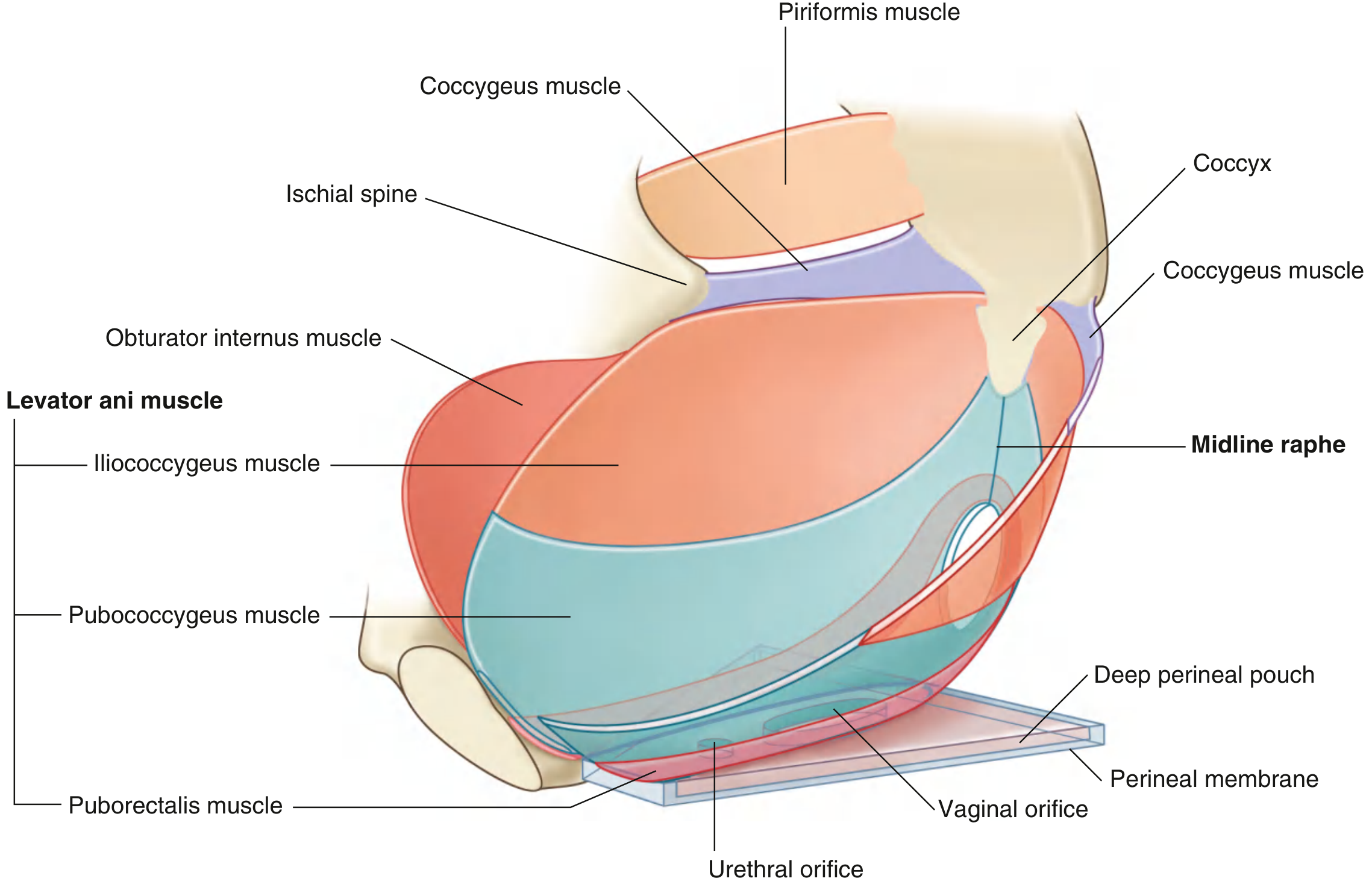
Pelvic floor in a woman - the deep perineal pouch sits as a thin region just above the perineal membrane, below the levator ani.
Boundaries
| Wall | Structure |
|---|---|
| Floor (inferior) | Perineal membrane (thick triangular fibrous sheet) |
| Roof (superior) | Open - merges with pelvic floor above |
| Lateral walls | Ischiopubic rami |
| Posterior border | Free - anchored to perineal body |
| Anterior gap | Small gap between perineal membrane and inferior pubic ligament |
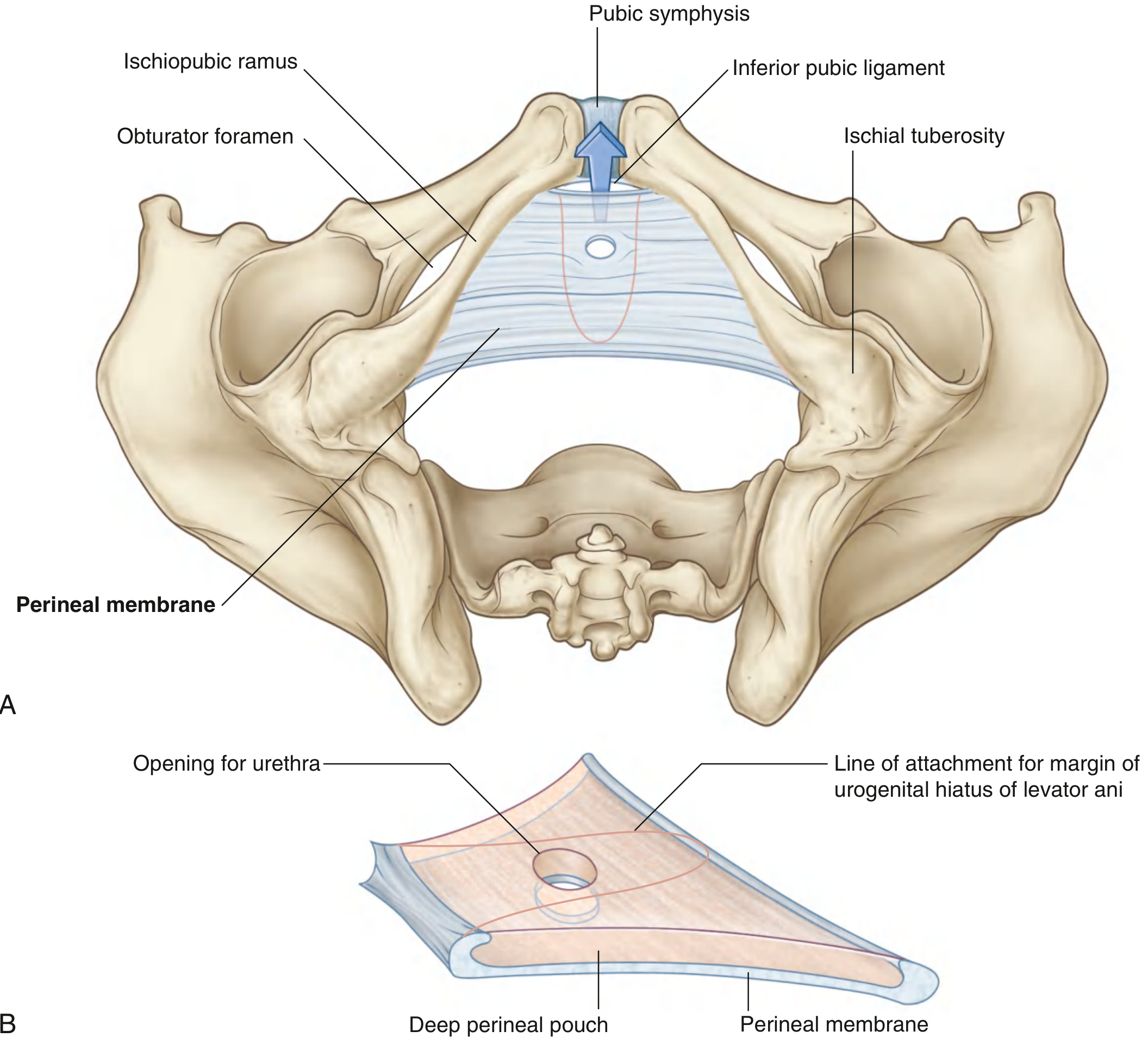
Contents - "2 Muscles + 2 in Women + Vessels + Nerves"
Muscles (Table 5.3)
| Muscle | Origin | Insertion | Nerve | Function |
|---|---|---|---|---|
| External urethral sphincter | Inferior ramus of pubis + walls of deep perineal pouch | Surrounds membranous urethra | Pudendal nerve (S2-S4) | Compresses membranous urethra; relaxes during micturition (voluntary control) |
| Deep transverse perineal | Medial ischial ramus | Perineal body | Pudendal nerve (S2-S4) | Stabilizes position of perineal body |
| Compressor urethrae (women only) | Ischiopubic ramus (each side) | Blends with partner anterior to urethra | Pudendal nerve (S2-S4) | Accessory sphincter of urethra |
| Sphincter urethrovaginalis (women only) | Perineal body | Passes forward lateral to vagina, blends anterior to urethra | Pudendal nerve (S2-S4) | Accessory sphincter (also may help close vagina) |
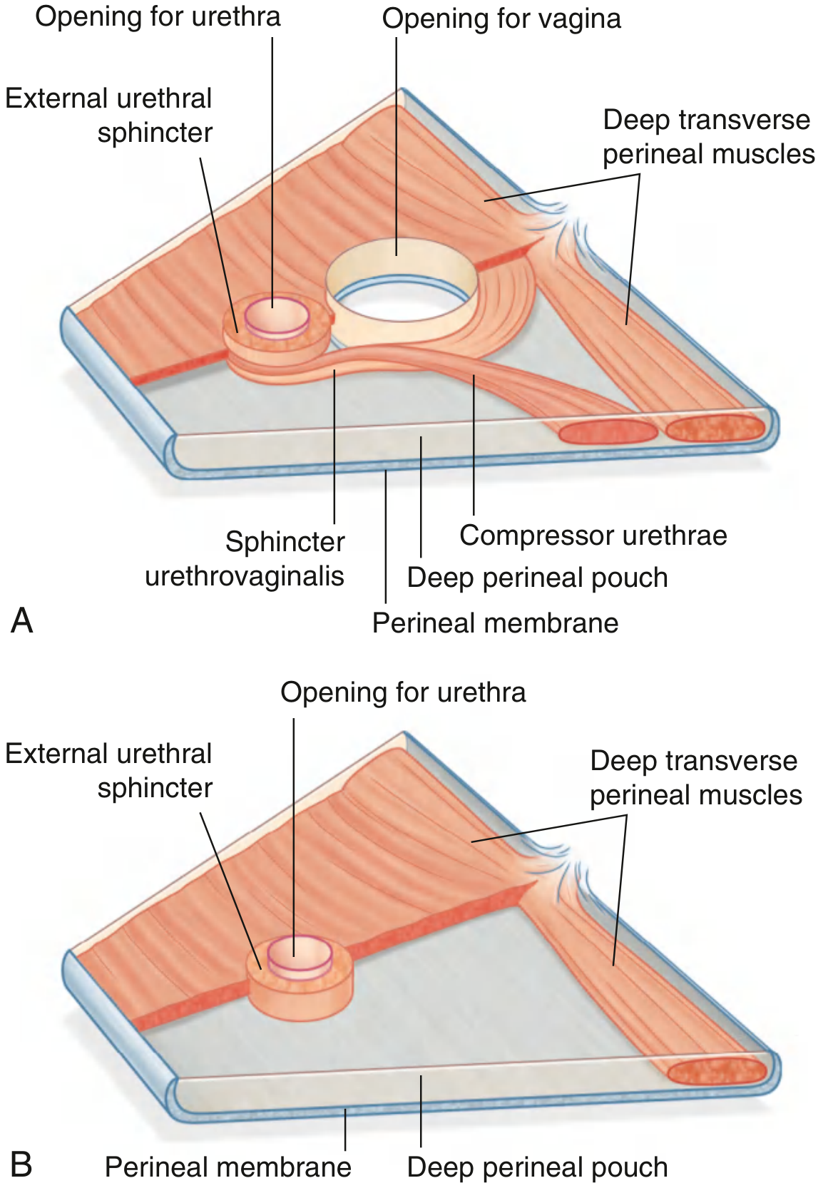
Neurovascular Contents
- Dorsal nerve of penis/clitoris - enters and traverses the lateral margin of the deep perineal pouch, exits anteriorly to reach the dorsum of the penis/clitoris; sensation to glans
- Terminal part of internal pudendal artery - enters pouch, gives off: artery to bulb of penis, urethral artery, deep artery of penis, and dorsal artery of penis (or equivalents in women)
- Bulbo-urethral glands (Cowper's glands) (in men) - small paired glands in the deep perineal pouch; ducts pierce perineal membrane to drain into penile urethra
Structures Passing Through
- Urethra - passes vertically through a hiatus in the perineal membrane going from pelvic cavity to perineum; surrounded here by the external urethral sphincter
- Vagina (in women) - passes through a hiatus just posterior to the urethral opening
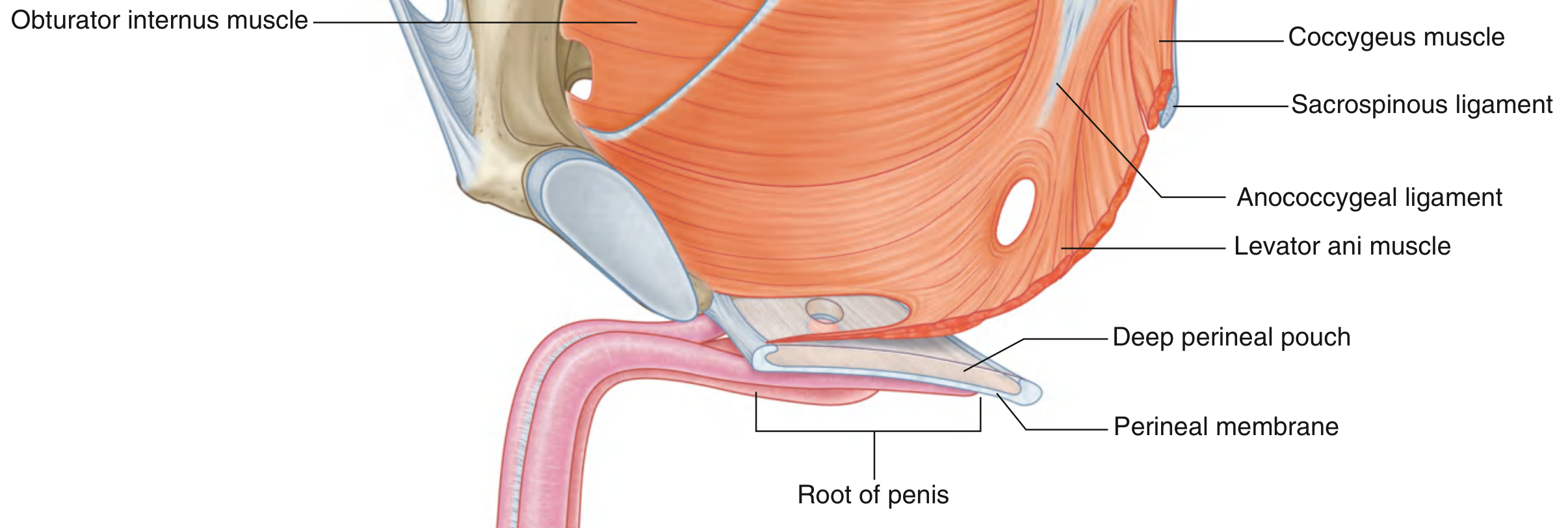
Medial View - Relationship to Penis and Pelvic Floor
Key Functions (Easy Summary)
- Voluntary urinary control - the external urethral sphincter here is the voluntary sphincter; when damaged (e.g., in pelvic fracture or prostatectomy), you get stress incontinence
- Pelvic floor support - the deep perineal pouch + perineal membrane together support pelvic viscera from below, spanning the urogenital hiatus
- Penile/clitoral innervation and blood supply - the dorsal nerve and internal pudendal artery pass through here to supply erectile tissues
- Stabilization of perineal body - the deep transverse perineal muscles anchor the perineal body, which is the junction point for multiple pelvic floor muscles
Clinical Significance
| Scenario | Mechanism |
|---|---|
| Urinary incontinence after prostatectomy | Damage to external urethral sphincter in deep perineal pouch; no voluntary urethral closure |
| Pelvic fracture urethral injury | The membranous urethra (passing through deep perineal pouch) is the most common site of rupture in pelvic fractures - because it is relatively fixed here |
| Perineal laceration/episiotomy repair | Deep transverse perineal muscle and perineal body are key structures to reconstruct to restore pelvic floor integrity |
| Pudendal nerve block | Anesthetic injected near ischial spine to block pudendal nerve before it branches; anesthetizes all structures of the deep perineal pouch |
| Bulbo-urethral gland cyst/abscess | Glands in deep perineal pouch can become infected; present as a perineal swelling between scrotum and anus |
| Female stress urinary incontinence | Weakness of compressor urethrae and sphincter urethrovaginalis (women-specific deep pouch muscles) contributes to urethral incompetence |
Compare: Deep vs. Superficial Perineal Pouch
| Feature | Superficial Perineal Pouch | Deep Perineal Pouch |
|---|---|---|
| Location | Below perineal membrane | Above perineal membrane |
| Contents | Crura/bulb of penis (erectile tissue), ischiocavernosus, bulbospongiosus, superficial transverse perineal | External urethral sphincter, deep transverse perineal, bulbo-urethral glands, dorsal nerve/artery of penis |
| Roof | Perineal membrane | Open (no superior fascia) |
| Floor | Membranous (Colles') fascia | Perineal membrane |
| Clinical injury | Straddle injury → blood/urine in superficial pouch | Pelvic fracture → membranous urethral rupture |
Gray's Anatomy for Students, Table 5.3 and Figs. 5.36-5.37, pp. 531-533
This is a shared conversation. Sign in to Orris to start your own chat.