
Apoptosis from Robbins second year flowchart
Reading File
Searching sources...
Finding Sources
Finding Sources
Reading File
Reading File
Reading File
Reading File
Loading Image
Loading Image
Reading File
Reading File
Here is the complete Robbins apoptosis flowchart with both diagrams and full mechanistic breakdown:
Apoptosis - Robbins Pathology
Definition
Apoptosis is a programmed cell death pathway where cells activate intrinsic enzymes (caspases) that degrade their own nuclear DNA and cytoplasmic proteins. The cell breaks into membrane-bound apoptotic bodies that are rapidly phagocytosed - no inflammatory reaction is elicited.
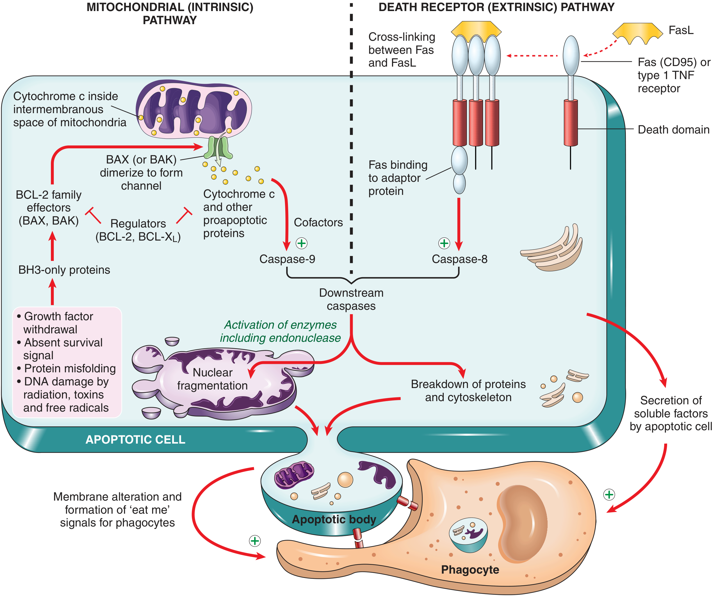
The Two Pathways - Flowchart (Robbins Basic Pathology, Fig. 1.12)
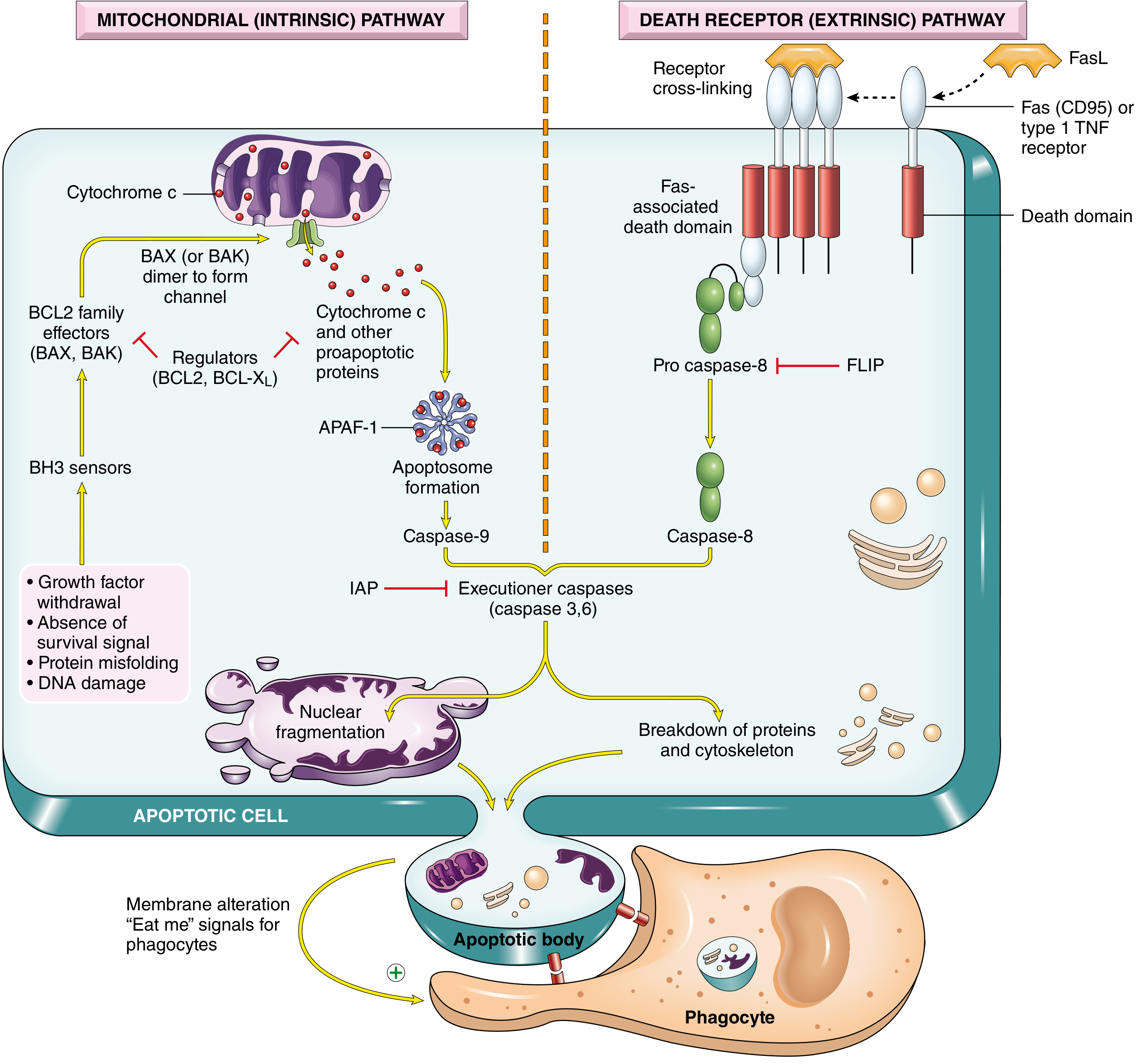
The More Detailed Version (Robbins & Cotran - Pathologic Basis of Disease, Fig. 2.16)
Step-by-Step Breakdown
MITOCHONDRIAL (INTRINSIC) PATHWAY
Triggers:
- Growth factor withdrawal / absence of survival signal
- DNA damage (radiation, toxins, free radicals)
- Protein misfolding (ER stress)
Sequence:
- Stress activates BH3-only sensor proteins (BAD, BIM, BID, PUMA, NOXA)
- BH3-only proteins antagonize anti-apoptotic BCL-2 / BCL-XL (which normally keep mitochondrial outer membrane intact)
- Pro-apoptotic BAX / BAK are activated - they dimerize and insert into the outer mitochondrial membrane, forming channels
- Cytochrome c (+ other pro-apoptotic proteins like Smac/DIABLO) leaks from the intermembranous space into the cytosol
- Cytochrome c binds APAF-1 → forms the apoptosome (wheel-shaped complex)
- Apoptosome recruits and activates Caspase-9 (initiator caspase)
- Caspase-9 activates executioner caspases (Caspase-3, -6)
- IAPs (Inhibitors of Apoptosis Proteins) normally block executioner caspases - Smac/DIABLO neutralizes IAPs, releasing the brake
- → Nuclear fragmentation + breakdown of proteins and cytoskeleton → Apoptotic body formation
DEATH RECEPTOR (EXTRINSIC) PATHWAY
Triggers:
- FasL binds Fas (CD95) on cell surface
- TNF binds Type 1 TNF receptor
Sequence:
- Receptor cross-linking occurs
- Death domain on receptor recruits adaptor protein FADD (Fas-associated death domain)
- FADD recruits Pro-caspase-8 → forms the DISC (Death-Inducing Signaling Complex)
- FLIP (anti-apoptotic protein) can block this step by competing with Pro-caspase-8
- Pro-caspase-8 undergoes autocatalytic cleavage → active Caspase-8 (initiator caspase)
- Caspase-8 activates executioner caspases (Caspase-3, -6)
- → Same final execution phase as intrinsic pathway
BCL-2 Family Summary
| Group | Members | Function |
|---|---|---|
| Anti-apoptotic | BCL-2, BCL-XL, MCL-1 | Keep outer mitochondrial membrane impermeable (4 BH domains: BH1-4) |
| Pro-apoptotic effectors | BAX, BAK | Form channels in outer mitochondrial membrane (BH1-3) |
| BH3-only sensors | BAD, BIM, BID, PUMA, NOXA | Sense cellular stress; activate BAX/BAK and inhibit BCL-2 (only BH3 domain) |
Morphologic Features of Apoptosis (vs. Necrosis)
| Feature | Apoptosis | Necrosis |
|---|---|---|
| Cell size | Shrinkage | Swelling |
| Nucleus | Chromatin condensation (pyknosis), fragmentation | Karyolysis or karyorrhexis |
| Membrane | Intact until late | Early rupture |
| Inflammation | None | Yes |
| Apoptotic bodies | Yes, rapidly phagocytosed | No |
| H&E appearance | Round, intensely eosinophilic mass with dense nuclear fragments | Ghost cells, loss of architecture |
Causes Summary Table
| Condition | Mechanism |
|---|---|
| During embryogenesis | Loss of growth factor signaling |
| Tissue turnover (gut epithelium, lymphocytes) | Loss of survival signals |
| Hormone-dependent involution (endometrium, lactating breast) | Reduced survival signals from hormone withdrawal |
| End of immune/inflammatory response | Loss of activation-induced survival signals |
| Self-reactive lymphocyte elimination | Both intrinsic and extrinsic pathways |
| DNA damage (radiation, cytotoxic drugs) | Activation of BH3-only proteins (p53-mediated) |
| Misfolded protein accumulation | BH3-only protein activation; direct caspase activation |
| Viral infections | Viral proteins activate pro-apoptotic proteins; CTLs activate caspases |
Sources: Robbins & Kumar Basic Pathology (9780323790185), p. 24-25 | Robbins, Cotran & Kumar Pathologic Basis of Disease (9780443264528), p. 56-60
This is a shared conversation. Sign in to Orris to start your own chat.