
NT scan short notes
Reading File
Finding Sources
Finding Sources
Searching PubMed
"nuchal translucency" screening
Reading File
Reading File
Nuchal Translucency (NT) Scan – Short Notes
Definition
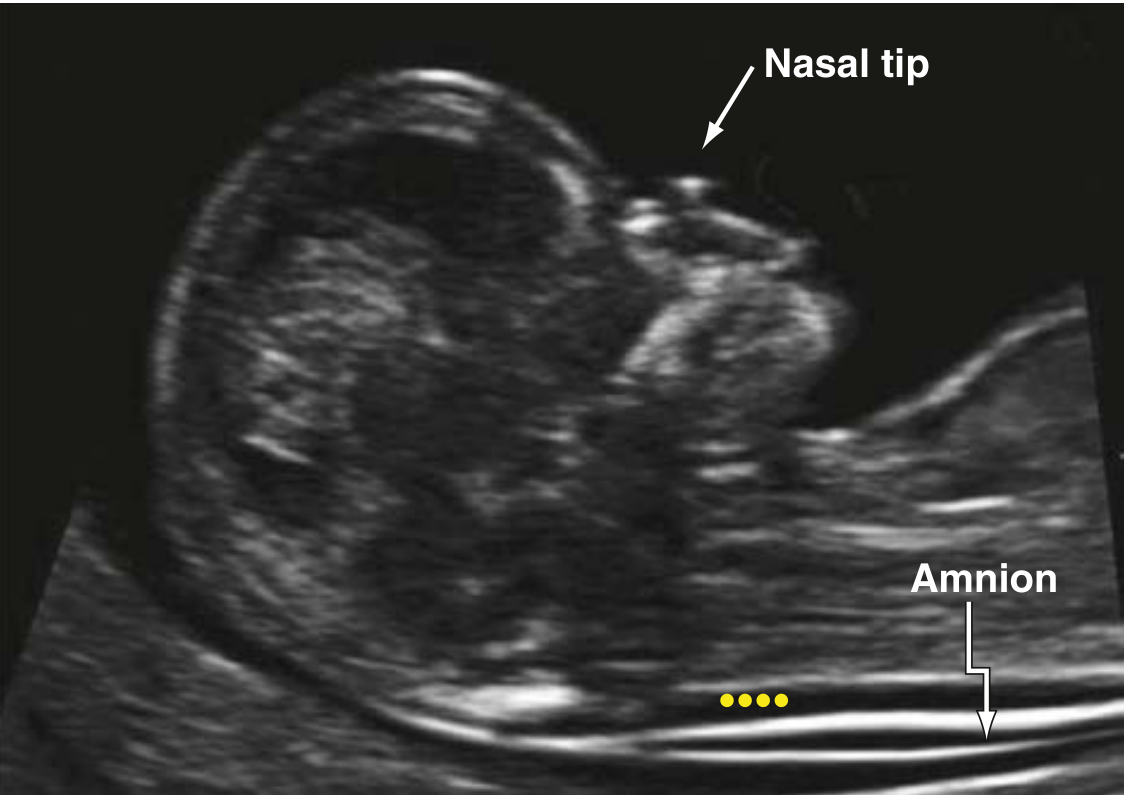
NT is the sonographic measurement of the subcutaneous fluid collection between the fetal cervical spine soft tissue and the overlying skin, seen as an anechoic (translucent) space at the back of the fetal neck on a midsagittal view - Creasy & Resnik's Maternal-Fetal Medicine, p. 635.
Timing / Technique
- Performed at 11-13+6 weeks gestation, when fetal crown-rump length (CRL) is 45-84 mm.
- Midsagittal section of the fetus, neck in neutral position, image magnified so head/neck/upper thorax fill most of the screen.
- Calipers placed "on-to-on" on the inner borders of the nuchal line (measuring the maximum thickness of the translucency, not the nuchal skin fold).
- Must distinguish NT from the amnion (a false membrane can mimic the NT edge).
Purpose
- Used as part of first-trimester aneuploidy screening, typically combined with maternal serum analytes (free beta-hCG and PAPP-A) plus maternal age -> patient-specific risk for trisomy 21, 18, 13.
- NT and PAPP-A are the most efficient individual markers between 9-12 weeks; combined screening performs best around 11 weeks.
Screening performance (combined NT + biochemistry)
- At a 5% screen-positive rate: Down syndrome detection rate ~88% (95% CI 84-89.4%).
- In women >35 years: 90-92% detection with 16-22% false-positive rate.
-
90% of trisomy 18 pregnancies are screen-positive with combined NT + biochemical screening.
Increased NT (above the 95th-99th percentile for CRL, roughly >3 mm) is associated with:
- Chromosomal aneuploidy - trisomy 21, 18, 13, monosomy X (Turner syndrome, often with cystic hygroma), triploidy.
- Structural anomalies, especially congenital cardiac defects - NT screening at 10-14 weeks has been used to flag major cardiac defects even with a normal karyotype.
- Skeletal dysplasias, diaphragmatic hernia, abdominal wall defects.
- Genetic syndromes / RASopathies (e.g., Noonan syndrome) - additional testing (microarray, RASopathy panel, exome sequencing) is recommended when karyotype is normal but NT remains elevated.
- Adverse pregnancy outcome even when karyotype and anatomy are normal - larger NT correlates with higher risk of miscarriage/fetal loss.
Additional first-trimester ultrasound marker - Nasal Bone (NB)
- Absent or hypoplastic NB increases suspicion for trisomy 21 (absent in ~60-70% of Down syndrome fetuses at 11-13 weeks vs a small percentage of euploid fetuses).
- Reflects the flat nasal bridge phenotype of Down syndrome.
Management of increased NT with normal karyotype
- Genetic counseling.
- Consider microarray / expanded genetic testing (cell-free DNA can be falsely reassuring if a structural defect is present).
- Detailed fetal anatomy scan and fetal echocardiography later in pregnancy (18-22 weeks) to look for structural anomalies not evident in the first trimester.
- Serial growth and anatomy surveillance given increased risk of adverse outcome even if initial workup is normal.
Key teaching points
- NT alone is a screening tool, not diagnostic - an elevated NT raises risk but does not confirm a chromosomal or structural abnormality.
- Quality matters: measurement technique is operator-dependent; ongoing epidemiologic monitoring of individual sonographer/sonologist performance (rather than ad hoc image review) is the preferred quality-control method - Tietz Textbook of Laboratory Medicine.
Recent evidence check (2023-2025 systematic reviews/meta-analyses):
- PMID 40519157 (2025) - outcomes of fetuses with early increased NT: systematic review/meta-analysis, reinforcing that increased NT carries risk beyond aneuploidy alone.
- PMID 37019452 (2023) - whole exome sequencing in fetuses with isolated increased NT and normal karyotype: meta-analysis supports added diagnostic yield of exome sequencing, consistent with the textbook recommendation above.
- PMID 40190084 (2025) - explores link between cystic hygroma/NT and congenital heart disease/lymphatic abnormalities.
These don't contradict the textbook material, they support and update the management recommendations (favoring expanded genetic testing, including exome sequencing, in isolated increased NT with normal karyotype).
Verbal autopsy short notes
Finding Sources
Finding Sources
Searching PubMed
"verbal autopsy" cause of death
Reading File
Reading File
Verbal Autopsy - Short Notes
Definition
Verbal autopsy (VA) is a method used to assign the probable cause of death by interviewing family members or community members/caregivers about the signs, symptoms, and circumstances preceding a death, used in settings where medical certification of cause of death is not available - Park's Textbook of Preventive and Social Medicine, p. 650.
Rationale / When used
- In most low- and middle-income countries (LMICs), civil (vital) registration systems are incomplete, so most deaths occur without a physician-certified cause of death.
- VA fills this gap by systematically collecting information (structured questionnaire) from next-of-kin about the terminal illness/event, which is then reviewed (by physicians or algorithm/computer-coded methods) to assign a probable cause of death, usually coded to ICD-10.
Method
- Standardized questionnaires (e.g., WHO Verbal Autopsy Standard instrument) are administered to relatives soon after a death.
- Records of births and deaths are collected periodically among defined populations under demographic surveillance systems run by research institutions in developing countries.
- Cause-of-death assignment can be done by physician review or by automated algorithms (e.g., InterVA, SmartVA) that map reported symptoms to probable causes.
Key applications (from library sources)
-
Maternal mortality measurement - One of the recognized approaches for estimating maternal mortality ratio (MMR) where civil registration is absent, alongside household surveys, sisterhood method, RAMOS (Reproductive Age Mortality Studies), and census methods - Park's PSM, p. 649-650.
-
India - SRS/RHIME - India's Sample Registration System (SRS) uses an enhanced form of verbal autopsy called RHIME (Representative, Re-sampled, Routine Household Interview of Mortality with Medical Evaluation), introduced from 2000 onward as part of a prospective study of ~1 million deaths. RHIME includes random re-sampling of fieldwork by an independent team to maintain data quality, and causes are coded per WHO Global Burden of Disease categories (haemorrhage, sepsis, hypertensive disorders, obstructed labour, abortion, other) - Park's PSM, p. 651.
-
Global Burden of Disease (GBD) studies - GBD estimates (e.g., GBD 2019) combine vital registration, country-level surveillance, disease registries, and verbal autopsy data to model cause-specific mortality worldwide - Fuster and Hurst's The Heart, 15th ed.
-
Cardiovascular disease/stroke burden estimation - In regions with scarce vital registration, VA surveys have been used to estimate CVD burden, e.g., 9-13% of deaths attributable to CVD (especially stroke) in some low-resource settings - Fuster and Hurst's The Heart, 15th ed.
-
Psychiatric epidemiology - VA has revealed underestimation of suicide by official statistics; a VA study in South India found suicide rates roughly 10-fold higher than official national estimates - Kaplan and Sadock's Synopsis of Psychiatry.
-
Standardization - "Verbal autopsy standards" exist for ascertaining and attributing causes of death, listed alongside other health classification systems (ICD, ATC/DDD, ICNP) - Kaplan & Sadock's Comprehensive Textbook of Psychiatry.
-
Forensic/COVID-19 context - WHO-recommended protocols for COVID-19 deaths include general/external examination plus verbal autopsy, alongside criteria of elimination and exclusion, when standard postmortem examination is limited - The Essentials of Forensic Medicine and Toxicology, 36th ed.
Advantages
- Only feasible method for cause-of-death data in areas lacking vital registration or medical certification.
- Useful for community-based, longitudinal demographic surveillance (e.g., HDSS sites) and large national mortality surveys (SRS-India).
- Can capture socially sensitive causes (e.g., suicide, maternal deaths) that are under-reported in official records.
Limitations
- Relies on recall and lay knowledge of the informant, subject to recall bias and misclassification.
- Less accurate for causes with overlapping/nonspecific symptoms (e.g., differentiating some cardiovascular vs. respiratory causes).
- Physician-coded VA can have inter-rater variability; algorithmic methods depend on validation against gold-standard (hospital-certified) causes.
Recent evidence (2023-2024 systematic reviews) - supports and refines library content:
- PMID 39030612 (2024) - systematic review validating malaria-attributed deaths using VA studies, highlighting ongoing efforts to improve VA accuracy for specific causes.
- PMID 37638359 (2023) - systematic review of CVD mortality based on VA in LMICs (Bulletin of WHO), consistent with the textbook's use of VA for CVD/stroke burden estimation.
- PMID 36640854 (2023) - systematic review of global autopsy rates in all-cause and sudden death, contextualizing the gap that VA and other methods aim to fill where formal autopsy rates are low.
No major contradictions with the textbook material; these confirm that VA remains a widely used and actively validated tool for cause-of-death ascertainment in resource-limited settings.
Verbal autopsy short notes in ms obg
Finding Sources
Finding Sources
Finding Sources
Finding Sources
Finding Sources
Verbal Autopsy - Short Notes (MS OBG Perspective)
Verbal autopsy is a favorite short-note topic in Obstetrics under "Maternal Mortality / Measuring Maternal Mortality" - relevant for MS OBG theory and viva.
Definition
Verbal autopsy (VA) is a technique used to assign a probable cause of death by structured interview of family/community members, used where medical certification of cause of death is not available - essential in India where most deaths (especially maternal deaths in rural areas) occur at home without medical attendance - Park's Textbook of Preventive and Social Medicine, p. 650.
Why it matters in Obstetrics
Most maternal deaths in developing countries occur outside health facilities (home deliveries, on the way to hospital), so civil registration/hospital records miss the true cause. VA is one of the key methods to estimate Maternal Mortality Ratio (MMR) when vital registration is incomplete.
Approaches for measuring maternal mortality (know all 6 - common exam list)
- Civil registration systems
- Household surveys
- Sisterhood method (direct/indirect) - interviews respondents about survival of adult sisters
- RAMOS (Reproductive Age Mortality Studies) - investigates all deaths in women of reproductive age using multiple data sources
- Verbal autopsy
- Census (with added questions)
India-specific application - SRS and RHIME
- India's Sample Registration System (SRS) uses RHIME (Representative, Re-sampled, Routine Household Interview of Mortality with Medical Evaluation) - an enhanced verbal autopsy tool.
- Started from year 2000, part of a prospective study covering ~1 million deaths.
- Includes random re-sampling of fieldwork by an independent team for quality control.
- Deaths are coded using WHO Global Burden of Disease categories with ICD-10 codes: haemorrhage, sepsis, hypertensive disorders, obstructed labour, abortion, and other causes.
- This is how India generates nationally representative cause-specific MMR data (EAG states vs southern states vs other states comparison) despite incomplete civil registration in many regions.
Procedure
- Trained field investigator interviews family/attendants soon after death using a structured questionnaire (symptoms, sequence of events, treatment sought, delays).
- Data reviewed/coded by physicians or algorithm (e.g., WHO VA instrument) to assign probable cause of death.
Exam-relevant linking concepts
- Ties into the "Three Delays Model" (delay in decision to seek care, delay in reaching facility, delay in receiving care) often explored through VA narratives.
- Complements Confidential Enquiry into Maternal Deaths and facility-based maternal death review (MDR) - VA specifically covers deaths that never reach a facility.
- Contributes to India's major causes of maternal death list: haemorrhage, sepsis, abortion-related complications, hypertensive disorders, obstructed labour, anaemia (indirect cause) - Park's PSM, p. 651.
Advantages
- Only feasible method in areas without complete civil registration.
- Captures home deaths and community-level cause-of-death patterns missed by hospital data.
- Useful for large-scale surveillance (SRS/RHIME model, HDSS sites).
Limitations (favorite viva point)
- Recall bias / informant may not know exact clinical details.
- Cannot always distinguish between clinically similar causes (e.g., eclampsia vs other hypertensive crisis).
- Coding variability between physician reviewers; requires trained interviewers and standardized instruments.
- Cannot substitute for facility-based clinical/laboratory diagnosis.
One-line answer for viva: "Verbal autopsy is a structured interview-based method to ascertain probable cause of death in the absence of medical certification, used in India's SRS through the RHIME technique to estimate cause-specific maternal mortality."
Note on sourcing: Dedicated obstetrics texts in this library (Creasy & Resnik's Maternal-Fetal Medicine, Berek & Novak's Gynecology) do not carry a specific "verbal autopsy" section - this topic is drawn from Park's Textbook of Preventive and Social Medicine (Community Medicine), which is the standard source MS OBG students use for maternal mortality measurement methods, since it's a shared PSM-Obstetrics exam topic.
Labour analgesia fshort notes ms obg
Finding Sources
Finding Sources
Searching PubMed
labor analgesia epidural
Reading File
Reading File
Labour Analgesia - Short Notes (MS OBG)
Definition
Labour analgesia refers to pain relief methods used during the process of labour and vaginal delivery, ranging from non-pharmacological techniques to neuraxial (epidural/spinal) blockade - a core topic under "Management of Normal Labour" in MS OBG.
Pain pathways in labour (exam favorite)
- First stage (uterine contractions + cervical dilatation): visceral pain via sympathetic afferents entering the spinal cord at T10-L1.
- Second stage (perineal stretching, descent of the head): somatic pain via the pudendal nerve (S2-S4).
- This dermatomal basis explains why a lumbar epidural covering T10-S4 is needed for complete labour analgesia through delivery.
Classification of methods
A. Non-pharmacological
- Psychoprophylaxis (Lamaze technique), breathing exercises, relaxation
- Continuous labour support / doula
- Hydrotherapy (warm water immersion)
- TENS (transcutaneous electrical nerve stimulation)
- Sterile water injections, acupuncture, hypnosis, aromatherapy
- Ambulation and position changes
B. Pharmacological - Systemic
- Parenteral opioids: pethidine (meperidine), fentanyl, remifentanil (PCA)
- Inhalational analgesia: nitrous oxide (Entonox, 50:50 N2O/O2)
- Note: remifentanil PCA is inferior to neuraxial labour analgesia per meta-analyses - Miller's Anesthesia, 10e.
C. Pharmacological - Regional/Neuraxial (gold standard)
- Epidural analgesia - most common and effective; lumbar epidural is the "mainstay of labour analgesia" - Miller's Anesthesia, 10e.
- Combined spinal-epidural (CSE)
- Dural puncture epidural (DPE) - newer technique
- Continuous spinal analgesia
- Programmed intermittent epidural bolus (PIEB) - most effective current technique for labour pain relief along with continuous epidural infusion - Morgan and Mikhail's Clinical Anesthesiology, 7e.
- Pudendal nerve block - for second stage/instrumental delivery
- Paracervical block - rarely used now (risk of fetal bradycardia)
Epidural analgesia - key points
- Drugs: bupivacaine or ropivacaine (low-dose) combined with an opioid (fentanyl/sufentanil) - "walking epidural" - provides excellent analgesia with reduced motor blockade.
- Can be initiated in early labour after obstetric evaluation; timing of initiation (early vs late) does not affect length of labour or mode of delivery (Chestnut et al., cited in Creasy & Resnik's Maternal-Fetal Medicine).
- Provides pain relief for first and second stages without altering maternal consciousness.
Effect on labour progress (important exam point)
- Large natural-experiment data (Zhang et al.) showed epidural analgesia does not increase overall cesarean delivery rate, oxytocin use, or instrumental delivery rate.
- The only consistent effect is prolongation of the second stage of labour (mean ~25 minutes).
- Fear of dystocia/cesarean should not be used to withhold epidural analgesia - Creasy & Resnik's Maternal-Fetal Medicine, p. 968.
Maternal/fetal complications of epidural
- Maternal hypotension (due to sympathetic blockade) - managed with IV prehydration and avoiding supine position (left lateral tilt).
- Late fetal heart rate decelerations from decreased uteroplacental perfusion (more common with bupivacaine than chloroprocaine/lidocaine) - up to 20% of cases.
- Uterine hypertonus with oxytocin augmentation can worsen uteroplacental insufficiency (FHR abnormalities in up to 70% in that setting).
- Post-dural puncture headache (with inadvertent dural puncture).
- Chronic backache - earlier retrospective studies suggested association, but prospective 1-year follow-up studies found no significant difference in persistent back pain between epidural and non-epidural groups.
- Maternal fever, pruritus (with opioids), rare: high spinal block, local anesthetic systemic toxicity, epidural hematoma/abscess.
Contraindications
- Absolute: patient refusal, coagulopathy/anticoagulation, local infection at insertion site, raised ICP, uncorrected hypovolemia/shock.
- Relative: severe cardiac lesions with fixed cardiac output, spinal deformity, sepsis.
Advantages of neuraxial techniques over systemic opioids
- Superior pain relief, doesn't cause maternal/fetal respiratory depression (unlike opioids), maintains maternal alertness, provides ready anesthesia if operative delivery needed.
Instrumental delivery note (related topic often paired in exams)
- Epidural analgesia does not increase rate of operative delivery per current evidence (Morgan & Mikhail, 7e) - contrasts with older teaching.
- Vacuum extraction requires less anesthesia than forceps delivery, relevant when comparing analgesia needs for assisted deliveries.
Recent evidence (2024-2025 meta-analyses) - useful for viva "recent advances"
- PMID 40121178 (2025, Br J Anaesth) - meta-analysis comparing epidural vs dural puncture epidural (DPE) analgesia in labouring women - DPE is an emerging technique combining benefits of CSE and epidural with fewer complications.
- PMID 37855310 (2024) - meta-analysis on dexmedetomidine as an epidural adjunct for labour analgesia, showing improved analgesic quality but noting bradycardia risk.
- PMID 39438806 (2024) - meta-analysis comparing ropivacaine vs levobupivacaine for labour epidural analgesia on maternal/fetal outcomes - both comparable, useful for "choice of local anesthetic" discussion.
These support the textbook material and can be cited as "recent advances" in an MS OBG answer (newer local anesthetic adjuncts, DPE technique) without contradicting core teaching.
One-line summary for viva: "Labour analgesia ranges from non-pharmacological methods to neuraxial techniques, with lumbar epidural analgesia using low-dose local anesthetic-opioid combinations being the gold standard, providing excellent pain relief without increasing cesarean risk, though it modestly prolongs the second stage of labour."
This is a shared conversation. Sign in to Orris to start your own chat.