
Nodular hidradenoma pas positivity
nodular hidradenoma PAS histology clear cells glycogen
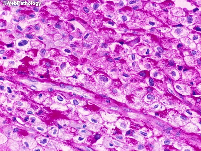
High-fidelity histopathology image captured with brightfield microscopy of a bladder mucosa biopsy stained with Periodic acid–Schiff (PAS). The tumor displays urothelial cells with abundant clear cytoplasm due to glycogen accumulation, characteristic of the clear cell variant of urothelial carcinoma. Cells are arranged in solid sheets and irregular nests within the urothelial/lamina propria region, with nuclear atypia including enlarged, hyperchromatic nuclei and conspicuous nucleoli. The PAS positivity highlights cytoplasmic glycogen; this reaction is typically diastase-labile (glycogen) and may require diastase treatment to confirm glycogen as the staining source (not shown). The tumor cells often border around a fibrovascular stroma, with occasional mitotic figures and occasional inflammatory infiltrates. The differential includes other glycogen-rich clear cell neoplasms and metastasis; immunohistochemical workup (e.g., GATA3, uroplakin, p63) supports urothelial origin while ruling out clear cell renal cell carcinoma. Clinically, this histology correlates with bladder mass lesions in adults and informs prognosis and treatment decisions, including cystectomy or intravesical therapy depending on invasion. This image demonstrates typical features of clear cell urothelial carcinoma, including glycogen-rich cytoplasm, clear cell morphology, and urothelial differentiation markers in adjunct studies. Correlation with imaging and flow cytometry may further refine staging; pathologist interpretation relies on morphological context and corroborative IHC profiles.
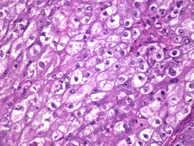
Imaging modality: Hematoxylin and eosin stained histology of breast tissue showing glycogen-rich clear cell carcinoma. The specimen is breast parenchyma with tumor cells arranged in solid sheets and nests; cells have abundant clear to amphophilic cytoplasm and vesicular nuclei with prominent nucleoli. Mitoses are rare, supporting a relatively indolent biology. The cytoplasmic clearing is consistent with glycogen-rich content; PAS staining would be positive and diastase-sensitive. The tumor often displays minimal gland formation and infiltrative growth into surrounding stroma; in this image, neoplastic cells replace normal ducts and lobular architecture. Clinically, such tumors tend to have an excellent prognosis despite a reported 30% axillary lymph node metastasis rate, and five-year survival approaches 100% with appropriate treatment, reflecting a relatively favorable outcome for breast carcinoma variants. Immunophenotype commonly includes hormone receptor positivity (ER/PR) and variable HER2 status, aiding differential diagnosis from metastatic clear cell renal cell carcinoma, which would be negative for breast lineage markers. The pathologic diagnosis highlights clear cell or glycogen-rich clear cell carcinoma of the breast, a rare entity with distinctive cytoplasmic appearance but favorable prognosis. This image is relevant for educational comparisons between histologic patterns of clear cell breast carcinomas and other clear cell neoplasms, and for references in diagnostic radiology-pathology correlation studies and clinical oncology.
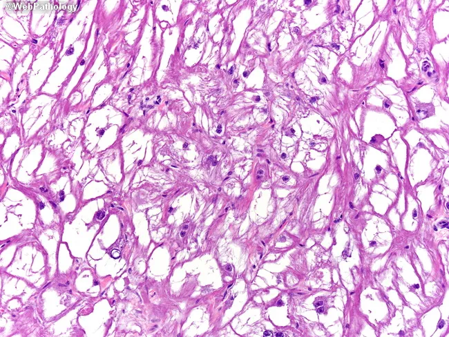
Light microscopy histology image of ventricular myocardial tissue stained with Periodic Acid-Schiff (PAS) highlighting glycogen-rich, vacuolated cardiac myocytes with spider cell morphology. Centrally placed nuclei with radiating, slender myofibrils produce the characteristic spider-like appearance and a distinctive periphery of cytoplasmic processes. PAS stain yields strong magenta coloration because of abundant glycogen, while conventional hematoxylin provides nuclear contrast. Electron microscopy described in the referenced material shows abundant glycogen, few irregular myofibrils, and poorly developed sarcoplasmic reticulum, supporting a benign, hamartomatous rhabdomyoma rather than a malignant sarcoma. Morphologically, tumor cells are large, polygonal, with clear cytoplasm and delicate, sparse myofilaments; cell borders may appear interwoven within the myocardium, reflecting a proliferative but well-differentiated muscular lineage. This constellation—spider cells, glycogen-rich cytoplasm, central nuclei, radiating myofibrils—constitutes a classic histopathologic signature for cardiac rhabdomyoma, especially in pediatric patients and in the context of tuberous sclerosis complex. Clinically, the presence of such tumors can yield arrhythmias or obstruction depending on size and location; prognosis is often favorable with potential spontaneous regression. Diagnostic relevance excludes most malignant cardiac neoplasms; differential diagnoses include rhabdomyosarcoma and myxoma, but immunophenotype and ultrastructure typically resolve the diagnosis. Correlation with clinical imaging and genetics further strengthens the definitive assessment in pediatric care.
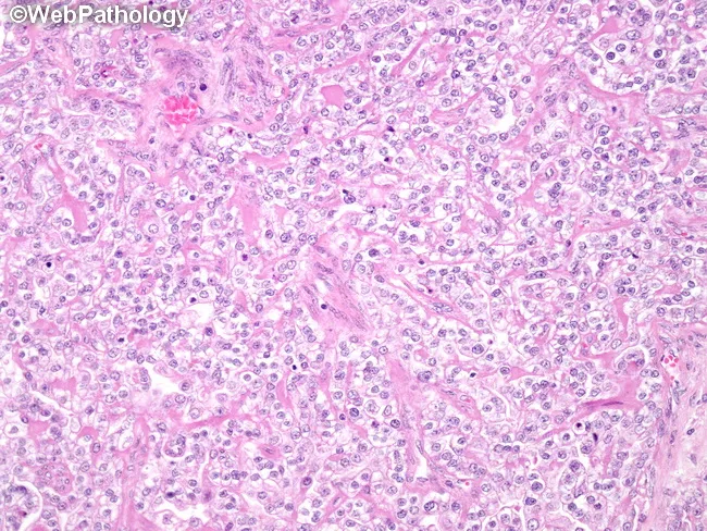
Histology: ovarian clear cell carcinoma, hematoxylin-eosin stained, evaluated at light microscopy. The tissue shows nests and sheets of polygonal cells with distinct cell borders and abundant clear cytoplasm, due to glycogen. Cells are arranged in glandular, tubular or tubulocystic structures; hobnail cells with apically placed nuclei are present, imparting a characteristic appearance. Nuclear enlargement is mild, with minimal pleomorphism; mitotic activity is not appreciably increased. Background stroma is often fibrous; occasional small cystic spaces may be seen. The cytoplasmic clearing is PAS-positive and diastase-sensitive, consistent with glycogen content. Nucleoli are inconspicuous in many cells; cytoplasmic borders crisp. The overall pattern demonstrates a combination of tubulocystic, papillary, and solid growth, which is typical for ovarian CCC. The slide shows features that help distinguish CCC from other ovarian epithelial tumors, such as endometrioid, serous, and mucinous carcinomas. Clinically, ovarian CCC tends to be associated with endometriosis and can show chemoresistance; prognostic implications depend on stage and grade. The presence of hobnail cells is a supportive diagnostic feature. This image likely represents typical example of clear cell histology and may be used for educational purposes to illustrate cellular morphology, glycogen-rich cytoplasm, and glandular architecture in CCC.
nodular hidradenoma PAS positivity glycogen clear cells histopathology
Nodular Hidradenoma - PAS Positivity
What Stains PAS Positive?
| Component | PAS Result | Diastase Sensitivity | Reason |
|---|---|---|---|
| Clear cells (round, pale cytoplasm) | PAS positive | Diastase-sensitive | Cytoplasmic glycogen accumulation |
| Polyhedral/basophilic cells | Less prominent PAS staining | - | Less glycogen content |
| Mucinous stromal areas (if present) | PAS positive | Diastase-resistant | Mucin (not glycogen) |
| Ductal luminal contents | PAS positive | Variable | Secretory glycoproteins |
Key Histopathological Points
- Contain abundant glycogen - this is the primary substrate for PAS positivity
- PAS+ and diastase-sensitive (i.e., the positivity is abolished after diastase pretreatment, confirming glycogen as the source)
- No lipid content (Oil Red O negative)
- Described as a "metabolic variant of epidermoid cells" rather than true secretory differentiation
- Diastase digests glycogen
- If PAS positivity disappears after diastase treatment → glycogen confirmed
- If PAS positivity persists → mucin or other non-glycogen material (mucin is diastase-resistant)
Tumor Architecture
- Solid areas - varying proportions of clear cells, poroid cells, squamoid cells, and rarely mucinous cells
- Cystic areas - lined by cuboidal cells
- Hyalinized eosinophilic stroma between lobules (a distinctive feature)
- Duct-like structures resembling eccrine ducts
Quick Exam Mnemonic
"Clear cells in hidradenoma = Glycogen = PAS+, Diastase-SENSITIVE"(Compare with mucin = PAS+, Diastase-RESISTANT)
Summary Table
| Feature | Detail |
|---|---|
| Tumor type | Benign eccrine acrospiroma |
| PAS+ substrate | Glycogen in clear cells |
| Diastase test | Positive abolished → glycogen confirmed |
| Lipid content | Absent (Oil Red O negative) |
| Other PAS+ areas | Ductal secretions, occasional mucin in stroma |
| Clinical relevance | PAS with diastase helps distinguish from lipid-laden tumors (sebaceous) and mucin-rich lesions |
- Andrews' Diseases of the Skin, p. 766 - Acrospiromas section
- PMC - Clinicopathological Profile of Nodular Hidradenoma: "clear cells contain glycogen and PAS-positive, diastase-resistant material, but no lipid" (note: this reference says diastase-resistant, while Basicmedicalkey cites diastase-sensitive for glycogen - the glycogen component is diastase-sensitive; diastase-resistant positivity refers to any co-existing mucin)
Important clarification: The clear cell glycogen is PAS+/diastase-sensitive. Some sources say "diastase-resistant" when referring to the total PAS positivity of clear cells because the cells may contain both glycogen AND small amounts of mucin/glycoprotein. The glycogen fraction is always diastase-sensitive; the residual PAS positivity after diastase is due to mucin/glycoprotein.
Image