
Polyp
Which polyp topic should I go into in detail?
"colorectal polyps"[MeSH Terms] AND "surveillance"
colonic adenomatous polyp colonoscopy or histology
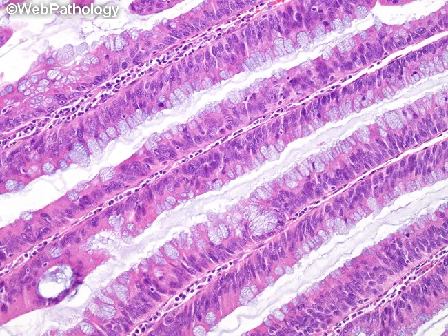
This histology image depicts colonic mucosa displaying villous adenomatous architecture with low-grade dysplasia. Prepared as a routine hematoxylin and eosin stained section, the villous processes are lined by columnar epithelium exhibiting architectural crowding, branching, and cryptal pseudostratification. There is hypercellularity with nuclear overcrowding; nuclei are moderately enlarged and elongated, hyperchromatic yet maintaining orderly polarity. Nucleoli are not prominent. Goblet cell mucin shows partial loss, contributing to a basophilic cytoplasmic appearance on H&E. Mitotic activity is not significantly increased, arguing against high-grade features. The mucosal glands demonstrate lack of maturation from crypt base to luminal surface, with vertical growth and irregular gland size; mucin depletion and basophilia accentuate the neoplastic epithelium. Overall, the lesion is an adenomatous polyp with villous architecture and low-grade dysplasia, a premalignant neoplasm in the colorectal tract. On imaging and clinical context this corresponds to a colon polyp with villous pattern, requiring complete endoscopic resection and histologic subtyping to determine malignant potential. Diagnostic significance includes risk stratification for progression to high-grade dysplasia or invasion; differential diagnoses include tubular adenoma with villous features, mixed adenoma, serrated lesions; clinical correlation: colonoscopy findings, patient age, and polyp burden inform management. Planning for surveillance is essential.
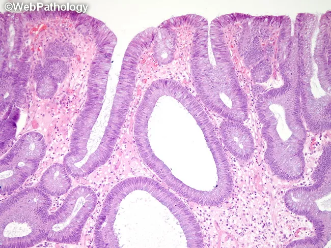
Juvenile polyp from the colon in juvenile polyposis syndrome is illustrated on a hematoxylin and eosin stained slide. The lesion arises from colonic mucosa and shows hamartomatous architecture with distorted, irregular glands embedded in an edematous lamina propria. The surface epithelium is dysplastic, displaying elongated, crowded glands with hyperchromatic, stratified nuclei and mild to moderate atypia, resembling tubular adenoma-type changes rather than a benign juvenile polyp alone. Glandular ducts are variably cystically dilated, and the mucosal stroma is expanded with edema and inflammatory infiltrate. Notable features include gland distortion, cystic dilation, and mitotic activity in dysplastic epithelium. This histology reflects the increased risk of adenocarcinomatous transformation in juvenile polyposis syndrome, either within a juvenile polyp or in concomitant adenomatous lesions. Clinically, these findings emphasize the cancer risk in JPS and the necessity for vigilant surveillance colonoscopy, genetic testing (SMAD4 and BMPR1A mutations), and risk stratification. The image captures the spectrum of neoplastic potential in JPS, where dysplasia signals progression toward colorectal carcinoma and may guide management, including therapeutic polypectomy, colectomy consideration, and targeted screening intervals in affected individuals. Correlation with clinical history, family testing, and endoscopic surveillance improves early cancer detection and informs therapeutic decision-making.
Colorectal polyps
- Neoplastic: tubular, villous and tubulovillous adenomas, and serrated adenomas/polyps.
- Hyperplastic.
- Hamartomatous: juvenile, Peutz-Jeghers, Cronkite-Canada.
- Inflammatory: pseudopolyps and benign lymphoid polyps.
- Adenomas are the neoplastic epithelial polyps. Their hallmark is dysplasia, and they are the precursors of colonic adenocarcinoma.
- Hyperplastic polyps are most common in the left colon and rectum and have no malignant potential. They must be distinguished from sessile serrated adenomas, which are cancer precursors.
- Sessile serrated adenomas lack dysplasia and share some features with hyperplastic polyps.
- Hamartomatous polyps, as in Peutz-Jeghers, carry an increased malignancy risk when they occur in a genetic syndrome.
- FAP is caused by APC mutations. Patients have hundreds of adenomas and develop colon cancer before age 30.
- HNPCC (Lynch syndrome) is caused by mismatch repair gene mutations with microsatellite instability. Patients have far fewer polyps and develop cancer later than in FAP, but earlier than in sporadic cancer.
- Adenomas occur in up to 25% of people over 50 in the US.
- The risk of cancer in an adenoma depends on its type and size:
| Adenoma type | Cancer risk |
|---|---|
| Tubular | about 5% |
| Tubulovillous | about 22% |
| Villous | up to 40% |
- Invasive cancer is rare in polyps under 1 cm. A polyp over 2 cm carries a 35 to 50% risk.
- Large hyperplastic polyps (over 2 cm) may undergo malignant change. Hyperplastic polyposis is a rare condition with multiple large polyps in young adults and an increased cancer risk.
- Hyperplastic polyps are usually under 5 mm and cannot be told apart from adenomas on colonoscopy, so they are often removed.
- Neoplastic polyps should be removed endoscopically.
- Pedunculated polyps are usually removed intact by snare polypectomy.
- Sessile polyps may need saline lift, piecemeal snare excision or endoscopic mucosal resection.
- Rectal sessile polyps are preferably removed by transanal excision. This gives an intact single specimen for staging.
- Tattoo the site of sessile polypectomies to guide later surveillance.
- Colectomy is reserved for lesions that cannot be removed endoscopically, such as large flat lesions, or for confirmed invasive cancer.
- A malignant pedunculated polyp can be treated with endoscopic resection alone if the cancer is confined to the submucosa and has no unfavorable features.
- Unfavorable features that call for prompt surgery are poor differentiation, vascular or lymphatic invasion, and incomplete resection.
- Polypectomy complications are perforation and bleeding. Bleeding can be immediate or delayed and usually stops on its own. Colonoscopy may be needed to clip, re-snare or cauterize the site.
| Finding | Repeat colonoscopy |
|---|---|
| 1 or 2 small tubular adenomas | 5 to 10 years |
| More than 2 adenomas, adenoma of 1 cm or more, villous or high-grade histology, or more than 10 adenomas | Within 3 years |
| Adenoma of 2 cm or more, or piecemeal resection of any adenoma | Within 6 months, to confirm complete removal |
| Sessile serrated polyp under 10 mm without dysplasia | 5 years |
| Serrated polyp with high-grade dysplasia, or traditional serrated adenoma | 3 years |
Recent evidence check
- A 2024 systematic review of surveillance colonoscopy frequency for adenomatous polyps (PMID 37967829). I did not read its full text, so I can't say how its intervals compare with the table above.
- A 2024 meta-analysis of computer-aided diagnosis for leaving polyps in situ (PMID 38768453).
- A 2025 meta-analysis of computer-aided diagnosis versus endoscopists (PMID 40375757).
Endometrial polyp
"endometrial polyp"[Title/Abstract] AND malignancy
endometrial polyp hysteroscopy or histology
endometrial polyp
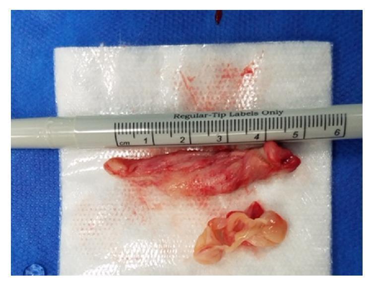
This clinical photograph shows a gross surgical specimen consisting of two tissue fragments excised during a polypectomy and endometrial curettage. The largest fragment is an elongated, pedunculated polyp measuring approximately 5.3 cm in length, as indicated by the adjacent surgical ruler. The polyp exhibits a fleshy, irregular surface with variegated coloring ranging from pale pink to deep red, suggesting vascular congestion or focal hemorrhage. Below the primary polyp is a smaller, fragmented piece of tissue, which appears more yellowish-white, translucent, and edematous, consistent with inactive endometrial curettings. The specimens are placed on a sterile white gauze over a blue surgical drape. This visual provides a gross pathological representation of a large endometrial polyp, a common cause of postmenopausal bleeding. The educational focus is on the morphology of benign endouterine growths and the distinction between a focal polypoid mass and diffuse endometrial tissue obtained through curettage.
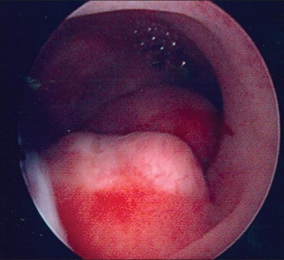
This clinical endoscopic image depicts a hysteroscopic view of the uterine cavity. The primary finding is a large, focal endometrial polyp characterized by a pale, whitish, and raised appearance that contrasts with the surrounding darker, vascularized endometrial tissue. The surface of the polyp shows irregular morphology and areas of focal reddish discoloration, suggestive of localized hemorrhage or hypervascularity. This diagnostic image demonstrates a common benign growth that can cause abnormal uterine bleeding (AUB) or postmenopausal bleeding (PMB). The visualization highlights the high sensitivity of hysteroscopy in detecting focal endometrial pathologies compared to transvaginal ultrasound, particularly in identifying the size, location, and vascular characteristics of endometrial polyps.
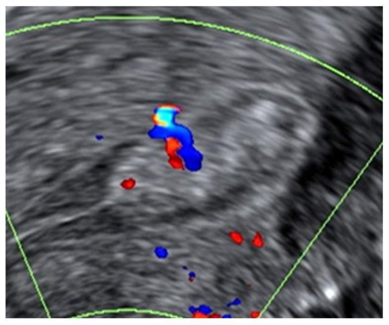
A transvaginal diagnostic ultrasound image demonstrating a benign endometrial polyp in the longitudinal plane of the uterus. The B-mode greyscale imaging shows a well-defined, focal hyperechoic lesion with a regular outline situated within the endometrial cavity. The lesion exhibits homogeneous echogenicity, consistent with a solid polypoid structure. Superimposed color Doppler signals (red and blue) reveal a single prominent feeder vessel entering the lesion, which is a classic vascular pattern for a simple endometrial polyp. The surrounding endometrium appears morphologically normal without evidence of irregularity or multi-vessel vascularity. This image serves as an educational example of typical sonographic features used in gynecological imaging to distinguish benign polyps from suspected endometrial malignancies according to International Endometrial Tumor Analysis (IETA) criteria.
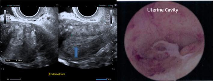
A multi-panel medical image featuring diagnostic imaging of the uterus in a 52-year-old postmenopausal patient. The left panel shows grayscale transvaginal ultrasound (TVUS) images of the uterus in a longitudinal plane. The endometrium is clearly identified, appearing thickened with a heterogeneous echotexture. Electronic calipers indicate an endometrial thickness measurement of 1.14 cm (11.4 mm). A blue arrow highlights a focal hypoechoic area within the thickened endometrial stripe, suggestive of a potential polyp or cystic change. The right panel displays a hysteroscopic view of the uterine cavity. The endoscopic perspective shows a pink, vascular endometrial lining with a prominent, irregular, white-to-tan protruding structure, characteristic of an endometrial polyp. These combined imaging modalities illustrate the evaluation of postmenopausal bleeding and endometrial thickening, which in this clinical case was histologically confirmed as a benign active endometrium.
Endometrial polyps
- They are exophytic masses that project into the endometrial cavity. They may be single or multiple.
- Most are sessile and small, 0.5 to 3 cm, but some are large and pedunculated.
- Cytogenetic studies show acquired chromosomal rearrangements in the stromal cells, similar to other benign mesenchymal tumors. They are best seen as stromal neoplasms.
- Polyps respond to estrogen but show little or no response to progesterone.
- Tamoxifen is associated with polyps because it has weak pro-estrogenic effects in the endometrium.
- Atrophic polyps occur mainly after menopause. They are probably the atrophic remnants of polyps that formed before menopause.
- Adenocarcinoma rarely arises within a polyp.
- Many polyps cause no symptoms. They account for about 25% of abnormal uterine bleeding, usually metrorrhagia (Schwartz's Principles of Surgery, 11th ed, Uterine Corpus).
- Bleeding can be intermenstrual, heavy, irregular or postmenopausal. Polyps are also linked to infertility (Berek & Novak's Gynecology).
- Prevalence is greatest after age 50, and polyps are common in women taking tamoxifen (Grainger & Allison's Diagnostic Radiology, ch. 35).
- A cervical polyp in a postmenopausal woman, especially one on tamoxifen, often comes with an endometrial polyp. Hysteroscopy is advised in that case (Pfenninger and Fowler's Procedures for Primary Care).
- On ultrasound, polyps are typically homogeneous and isoechoic to the endometrium. They are continuous with it and keep the endomyometrial interface.
- They may be centrally cystic, from dilated fluid-filled glands.
- Color Doppler may show a characteristic single feeding vessel.
- Sonohysterography shows them well.
- On MRI they have intermediate T1 and heterogeneous high T2 signal.
- They must be told apart from submucosal fibroids and from malignancy.
- Hysteroscopy with biopsy is needed to exclude malignancy or foci of atypical hyperplasia within a polyp.
- Blind curettage often misses polyps, so hysteroscopic removal is preferred. It can usually be done in the office.
- For small polyps, small-caliber scissors transect the base under hysteroscopic guidance, and grasping forceps remove the polyp.
- Larger polyps may need polyp snares, electrosurgical needles, a resectoscope or an electromechanical tissue removal system to sever the stalk and morcellate the lesion.
- Cut as close to the endometrium as possible to reduce recurrence.
- In infertility, it is unclear whether polyp number or size affects outcome. The textbook advises removing all accessible polyps if this can be done with minimal trauma.
- Send all specimens for histology.
Recent evidence
- A 2026 systematic review of endometrial pathologies, infertility and malignant potential (PMID 41671103).
- A 2026 meta-analysis of the association between endometriosis and endometrial polyps (PMID 41317651).
- A 2025 genome-wide association study of female genital tract polyps (PMID 39986329). It implicates DNA repair and cell proliferation genes.