
Merkel cell carcinoma
Reading File
Now I'll search the library and PubMed in parallel for comprehensive coverage.
Finding Sources
Searching PubMed
"Merkel cell carcinoma"[MeSH Terms]
Excellent - rich results. Let me read the top sources in parallel: Andrews' (most detailed), Robbins Pathology, and Sabiston Surgery.
Reading File
Reading File
Reading File
Searching PubMed
"Merkel cell carcinoma" AND treatment
I now have rich content from multiple textbooks. Let me also read more of the Andrews' section for histology, treatment, and prognosis details, and fetch the clinical image.
Reading File
Loading Image
I now have comprehensive, authoritative content from multiple textbooks. Here is the full clinical overview:
Merkel Cell Carcinoma (MCC)
Also called trabecular carcinoma. First described by Toker in 1972.
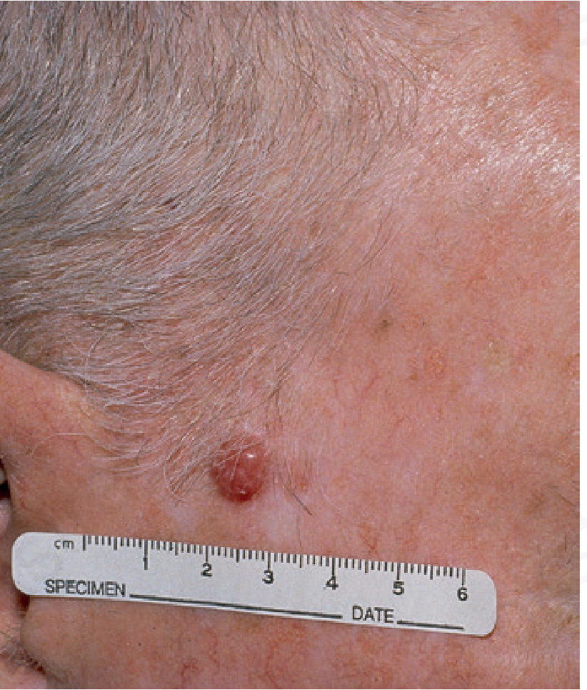
Cell of Origin
MCC arises from Merkel cells - specialized slow-adapting mechanoreceptors in the basal layer of the epidermis, which also have neuroendocrine properties. There is ongoing debate as to whether the progenitor is epidermal or neural crest-derived.
Epidemiology
- Incidence: ~0.44 per 100,000, but rising at 8% per year (faster than melanoma, which rises at 3%/year)
- Over 1,500 new US cases annually
- Demographics: 90% occur in patients >50 years; mean age 74-76. About 60% male, 95% in white individuals
- Location: ~90% on sun-exposed sites - head/neck (36%), extremities (37%), trunk (11%)
- Immunosuppression is a major risk factor: organ transplant recipients, CLL patients, and HIV-infected individuals have substantially elevated risk (8-15% of MCC patients have some form of immune impairment)
- PUVA therapy also increases risk
Etiology: Merkel Cell Polyomavirus (MCPyV)
- ~80% of MCCs in North America are associated with MCPyV (a member of the polyomavirus family); only ~25% in Australia
- The viral DNA is integrated into the cancer cell genome - all progenitor cells share the same viral integration, indicating the infection preceded or coincided with neoplastic development
- MCPyV seroprevalence increases from 30% in children under 5 to ~80% in adults over 50; lymphoid tissue (especially tonsils) is the reservoir
- MCPyV-associated tumors: mainly in immunosuppressed patients; the viral genome carries few acquired mutations and is usually diploid
- MCPyV-negative tumors (UV-induced): seen in immunocompetent individuals; carry a heavy UV mutation burden, with frequent loss-of-function mutations in RB and TP53, plus numerous copy number changes
- Like other transforming DNA viruses, MCPyV encodes proteins that interfere with Rb, p53, PP2A, and MYC
- Patients with MCPyV-positive tumors have a better prognosis (45% vs. 15% 5-year survival)
- MCPyV serology can be used to monitor for tumor recurrence
Clinical Presentation
- Rapidly growing, painless, raised nodule - red to violaceous, shiny surface with overlying telangiectasia
- Most cases are not initially suspected to be malignant
- Mnemonic: AEIOU
- A - Asymptomatic/lack of tenderness
- E - Expanding rapidly
- I - Immune suppression
- O - Older than 50 years
- U - UV-exposed site on a person with fair skin
Behavior and Staging
- Highly aggressive: local recurrence, regional nodal spread, and hematogenous metastases
- At presentation, ~one third have regional lymph node involvement; hematogenous spread occurs in at least one third of patients overall
- Spontaneous remissions have been reported (primarily women with head/neck tumors, often after reducing iatrogenic immunosuppression) - but recurrence after regression can occur
- MCC can present as metastatic disease with no evident primary - these patients paradoxically have a better prognosis than those with a known primary at the same stage
- Staging uses the AJCC system based on maximum tumor diameter (not depth of invasion, unlike melanoma)
Histopathology
- Dermal tumor that may extend into subcutaneous tissue
- Cells ~15 µm in diameter, very scant cytoplasm, hyperchromatic nuclei with a distinctive smudged chromatin pattern
- Numerous mitoses and apoptotic cells; cells arranged in sheets and cords
- Microscopically resembles an aggressive neuroendocrine carcinoma and can be indistinguishable from small cell lung cancer and other small round blue cell tumors
Immunohistochemistry (IHC) - Key Panel
| Marker | MCC | Notes |
|---|---|---|
| CK20 | Positive (perinuclear dot pattern) | Most specific; used in SLNB staining |
| TTF-1 | Negative | Distinguishes from small cell lung cancer (TTF-1+) |
| CK7 | Negative | Tends to stain small cell lung cancer |
| S-100 | Negative | Distinguishes from melanoma |
| LCA (CD45) | Negative | Distinguishes from lymphoma |
Differential diagnosis includes: small cell lung cancer, lymphoma, neuroblastoma, small cell endocrine carcinoma, Ewing sarcoma, melanoma, and BCC.
Workup
- Biopsy: shave or punch biopsy; experienced dermatopathologist often needed
- Sentinel lymph node biopsy (SLNB): recommended for all T stages - up to one third of clinically node-negative patients have a positive SLNB. Biopsy must be stained with CK20 to detect micrometastases
- Imaging: CT, MRI, or PET/CT to evaluate for metastatic disease (CT alone detects metastatic disease in only 20%)
- MCPyV serology at 2-3 months post-diagnosis for baseline, then serial monitoring for recurrence
Treatment
Locoregional Disease
- Surgery: Wide local excision (WLE) with 1-2 cm margins is the primary treatment. Mohs micrographic surgery (MMS) is used where tissue conservation is needed
- Radiation therapy: MCC is a radiosensitive tumor. Adjuvant RT reduces local recurrence from 16% to near 0% even after Mohs surgery. Directed at both the primary site and draining/regional lymph node basins - even when SLNB is negative
- Recurrence is seen in 46-76% of untreated lymph node basins
- Prophylactic lymph node dissection improves local control but not survival - largely being replaced by radiation to the nodal basin
- Adjuvant immunotherapy: Trials support adjuvant anti-PD-1 (pembrolizumab) after resection of high-risk MCC (including SLNB-positive disease). The STAMP/EA6174 trial (n=280) examined adjuvant pembrolizumab after surgery for stage I-IIIB - results were pending at time of publication
Metastatic/Advanced Disease
- Immune checkpoint inhibitors are the treatment of choice:
- Pembrolizumab (PD-1 inhibitor): ~50% response rate; responses appear durable
- Avelumab (PD-L1 inhibitor): also shows good responses in advanced MCC
- Tumors arising in sun-damaged skin (high mutational burden) are especially responsive to checkpoint inhibitors; some patients with distant metastatic disease achieve sustained responses and may be cured
- Combination checkpoint therapy has also shown efficacy
- Chemotherapy: Initially responsive but disease progression occurs; considered palliative in metastatic disease. Traditional adjuvant chemotherapy does not prevent later metastatic spread
- Multikinase inhibitor pazopanib: partial responses seen
Prognosis
- Nodal status is the strongest prognostic factor - node-positive patients have significantly decreased overall survival
- MCPyV-positive patients: 45% 5-year survival vs. 15% in MCPyV-negative (UV-driven) cases
- Natural killer cell response also correlates with prognosis
- Locoregional recurrence is a poor prognostic sign
- Treatment should not be delayed - metastatic disease can develop within weeks of diagnosis
Sources:
- Andrews' Diseases of the Skin, Ch. 29 (Trabecular carcinoma section)
- Robbins, Cotran & Kumar Pathologic Basis of Disease, Ch. 11 (Skin)
- Sabiston Textbook of Surgery, 21st ed., Ch. 63 (Cutaneous malignancies)
This is a shared conversation. Sign in to Orris to start your own chat.