
My mother is facing problem with pain in the knee where she experinces pain when she straighten her leg and special while standing from seated position there is noted huge swelling around the knee and would be the diagnosis what treatment would help to recover better
"knee osteoarthritis" AND "treatment"
Most Likely Diagnosis: Knee Osteoarthritis (OA)
Understanding the Diagnosis
What is Knee Osteoarthritis?
- S. Das: A Manual on Clinical Surgery, 13th Ed.
Why These Specific Symptoms?
| Symptom | Why It Happens |
|---|---|
| Pain when straightening the leg | Bone-on-bone pressure and osteophyte (bone spur) formation cause pain during full extension |
| Pain on rising from seated position | Cartilage-deprived joint surfaces are stressed by the sudden weight-bearing and change in position |
| Large swelling around the knee | Joint effusion (fluid accumulation) from chronic inflammation of the synovial lining |
| Stiffness after rest | Characteristic "gelling" - the joint feels stiff after inactivity and eases with movement |
Other Possibilities to Rule Out
- Chondromalacia patellae (softening of the cartilage behind the kneecap) - more common in younger women
- Rheumatoid arthritis - causes bilateral joint swelling with morning stiffness >1 hour
- Gout / pseudogout - causes sudden, intense swelling (especially in older patients)
- Popliteal (Baker's) cyst - can cause swelling behind the knee that may rupture
How Is It Diagnosed?
- Clinical examination - joint line tenderness, crepitus (crunching sound), reduced range of motion, swelling, possible varus/valgus deformity
- X-ray (weight-bearing) - the most important first investigation. Findings include:
- Loss of joint space (narrowing)
- Osteophytes (bone spurs) at joint margins
- Subchondral sclerosis (bone hardening)
- Subchondral cysts
- MRI - not routinely required; reserved for mechanical problems or surgical planning
- Blood tests - to rule out inflammatory arthritis (rheumatoid factor, ESR, CRP, uric acid)
Treatment Plan
Step 1: Lifestyle & Conservative Measures (First Line)
- Weight loss - if BMI >25; even modest weight reduction significantly slows disease progression and reduces knee loading
- Exercise - low-impact aerobic exercise (walking, swimming, cycling) and quadriceps-strengthening exercises are the most evidence-backed interventions. A 2025 BMJ network meta-analysis confirmed exercise as top-tier treatment
- Cushioned footwear - reduces impact forces on the knee
- Knee brace - improves proprioceptive control and offloads the affected compartment
- Activity modification - avoid prolonged sitting, kneeling, squatting, or stair climbing where possible
Step 2: Medications
- NSAIDs (e.g., ibuprofen, diclofenac) - first-line for pain relief; reduce both pain and inflammation. Use with a stomach-protecting drug (PPI) if she has any gastric issues
- Paracetamol (Acetaminophen) - for mild pain or when NSAIDs are not tolerated
- Topical NSAIDs (e.g., diclofenac gel) - applied directly to the knee; good option for elderly patients with fewer side effects
Step 3: Injections (if medications are insufficient after 4-6 weeks)
- Intraarticular corticosteroid injections - fast-acting relief for swelling and pain; effect lasts weeks to months
- Viscosupplementation (Hyaluronic acid injections) - may provide longer-lasting benefit with repeated courses; results vary by individual
Step 4: Surgical Options (when all else fails)
- Total or Partial Knee Replacement (Arthroplasty) - provides excellent pain relief and functional improvement in advanced OA
- Arthroscopic debridement - limited role in OA; not routinely recommended by AAOS unless there is an unstable meniscal tear or loose body
Immediate Action Steps
- See an orthopedic surgeon or rheumatologist for a proper clinical examination and X-ray
- Do not delay - untreated OA can progress to significant joint deformity
- For now, she can:
- Apply ice packs (15-20 minutes, 3x daily) to reduce swelling
- Elevate the leg when resting
- Avoid activities that worsen pain
- Use a walking aid if needed to reduce knee loading
- Start gentle seated quadriceps exercises (tighten the thigh muscle with the leg straight, hold 5 seconds, repeat 10 times)
Important: This is an educational overview based on authoritative medical textbooks (Campbell's Operative Orthopaedics 15th Ed 2026, Textbook of Family Medicine 9e, S. Das Manual on Clinical Surgery 13th Ed, Firestein & Kelley's Textbook of Rheumatology). Please consult a doctor for a proper physical examination, imaging, and a personalized treatment plan. Do not self-medicate.
It is due to loss of cartilage ?? Iheard a latest news about 15-PGDH Inhibitors where it help to regrow the cartilage faster does this help to this case or not because she experincing pain from so many days and she is using nsaids where there is instant relief but responding well
15-PGDH inhibitor cartilage regeneration osteoarthritis
15-PGDH inhibitor SW033291 cartilage regrowth osteoarthritis 2024 2025 clinical trial
PMID: 41308124
https://med.stanford.edu/news/all-news/2025/11/joint-cartila…
15-PGDH inhibitor human clinical trial Phase 1 cartilage osteoarthritis FDA 2025 2026
Yes - Cartilage Loss is the Core Problem in Knee OA
The 15-PGDH Inhibitor - What the Science Actually Shows
What is 15-PGDH?
The Landmark Study (Science, 2026)
- A small-molecule inhibitor called SW033291 (PGDHi) blocks 15-PGDH activity
- In aged mice: both systemic (body-wide) and local (intra-articular) injections led to genuine articular cartilage regeneration - the cartilage thickened across the entire joint surface
- The mechanism works by reprogramming existing chondrocytes (changing their gene expression from a "degenerative" pattern back to an "extracellular matrix-synthesizing" pattern) - not by growing new stem cells
- It reduced OA-related pain in the mice
- It also reduced inflammatory cytokines in the joint (CCL7, CXCL10, VEGF, IL-2)
- Most importantly for your mother's case: Human OA cartilage tissue taken from knee replacement patients was treated with the inhibitor for just 1 week in the lab. The result - cartilage degradation markers went down, GAG (glycosaminoglycan, the key cartilage building block) content increased, and new articular cartilage formation began
What Stage is This at Right Now (July 2026)?
| Stage | Status |
|---|---|
| Mouse studies (aged + injury models) | Completed - very positive results |
| Human tissue (lab explants) | Completed - positive signals |
| Phase 1 clinical trials | An oral 15-PGDH inhibitor has already completed Phase 1 safety testing in humans - but for muscle weakness, not OA |
| Phase 1/2 trials specifically for OA/cartilage | Not yet started - researchers say they "hope a similar trial will be launched soon" |
| Available for patients | Not yet - this is NOT available as a treatment |
Can Your Mother Use This Right Now?
- Not approved or commercially available - SW033291 is purely a research compound. It cannot be prescribed, purchased, or obtained outside of a clinical trial
- No OA-specific clinical trial is running yet - Phase 1 trials for OA specifically have not yet been announced as of July 2026
- Human safety in joints is unproven - Phase 1 for muscle weakness showed safety, but joint administration (intra-articular injection vs. systemic) for OA has not been tested in humans
What This Means for Her Current Situation
Immediate (available today)
- Continue NSAIDs under doctor's supervision, but discuss stomach protection (a PPI like omeprazole) if she is on them long-term
- Ask her orthopedic doctor about intraarticular corticosteroid injection for the swelling - fast relief, directly at the site
- Start a supervised quadriceps strengthening program - this is the single most evidence-backed intervention to reduce load on the knee and slow progression
- Weight management if relevant
Medium term
- Ask about hyaluronic acid (viscosupplementation) injections - may offer longer-lasting relief than steroids
- Knee brace for offloading
Future (watch this space)
- Monitor news about Phase 1/2 OA trials for 15-PGDH inhibitors - if a clinical trial opens, she could potentially enroll depending on eligibility criteria
- This research genuinely represents the first mechanistic hope for cartilage regeneration - worth following
Give a brief about my mother how can I explain it orthopedic doctor
Patient Summary for Orthopedic Consultation
- Pain on straightening the leg (full extension)
- Pain especially when standing up from a seated position
- Noticeable large swelling around the knee
- Currently taking NSAIDs, which are giving good/instant relief for the pain
- Possible knee osteoarthritis - given the age-related pattern of pain on rising/standing, swelling, and response to anti-inflammatory medication
- "Can you confirm the diagnosis with a physical exam and weight-bearing X-ray of the knee?"
- "Given the swelling, should the joint fluid be drained or tested (to rule out infection, gout, or inflammatory arthritis)?"
- "Since NSAIDs are helping now, is it safe for her to continue them long-term, or should we add stomach protection (like a PPI)?"
- "Would a corticosteroid or hyaluronic acid (viscosupplementation) injection help with the swelling and pain?"
- "Should she start physiotherapy or specific quadriceps-strengthening exercises, and is a knee brace advisable?"
- "What is the current severity/stage of the joint damage, and are we looking at conservative management or should we discuss surgical options (like partial/total knee replacement) down the line?"
- "Is there any dietary or weight management advice specific to her condition?"
- "I read about a new 15-PGDH inhibitor drug from a 2026 Stanford study (published in Science) that showed cartilage regeneration in animal and human tissue studies. I understand it's still in early research and not yet available as a treatment - just wanted to check if you're aware of it or expect any relevant trials to open up."
What to look for in xray
knee osteoarthritis X-ray joint space narrowing osteophytes
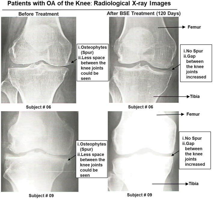
This diagnostic image is a comparison chart featuring anteroposterior (AP) X-ray radiographs of the knee joints from two patients (Subject #06 and Subject #09) diagnosed with osteoarthritis. The figure is organized into two columns: 'Before Treatment' and 'After BSE Treatment (120 Days).' In the 'Before Treatment' images, radiological signs of osteoarthritis are evident, including visible marginal osteophytes (bone spurs) at the joint edges and significant narrowing of the joint space between the femur and tibia. The 'After BSE Treatment' images illustrate the results of Boswellia serrata extract (BSE) therapy, showing a notable reduction or absence of the previously noted osteophytes and a visible increase in the joint space gap. The images are annotated with arrows identifying the femur and tibia, along with descriptive text boxes highlighting the increase in joint gap and the disappearance of spurs post-treatment. This comparison serves as a clinical illustration of potential radiological improvement in osteoarthritic knee conditions following anti-inflammatory pharmacological intervention.
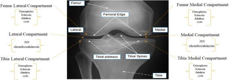
This diagnostic image is an annotated anteroposterior (AP) x-ray radiograph of a human knee joint, functioning as an educational diagram for osteoarthritis (OA) assessment. The central image identifies key anatomical landmarks, including the distal femur, proximal tibia, femoral edges, tibial plateaus, and tibial spines. The joint is divided into lateral and medial compartments. Callout boxes categorize secondary OA-related radiographic features by anatomical location. The femoral and tibial lateral/medial compartments are sites for identifying osteophytes, subchondral sclerosis, bone attrition, and subchondral cysts. The joint space itself is highlighted for the evaluation of joint space narrowing (JSN) and chondrocalcinosis. This diagram serves as a clinical guide for orthopedic and radiological classification of knee osteoarthritis severity, emphasizing the spatial distribution of degenerative findings according to standardized systems like the OARSI grading scale.
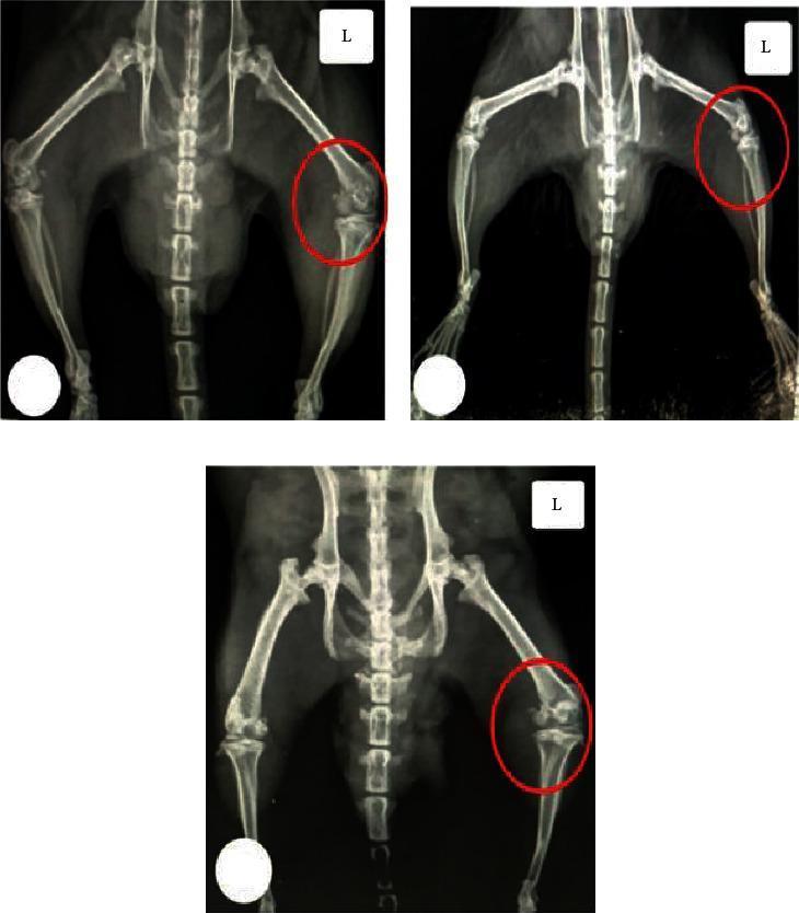
This diagnostic image consists of three X-ray radiographs illustrating a comparative study of knee osteoarthritis (OA) in a rat model. The radiographs focus on the left knee joint (marked 'L' and circled in red) across different experimental conditions. (A) Normal control group: Shows preserved joint space, smooth articular surfaces, and normal bone density. (B) MIA-treated group (monosodium iodoacetate-induced OA): Demonstrates significant radiographic alterations including marked narrowing of the joint space, subchondral bone sclerosis, surface irregularities suggesting cartilage erosion, and the formation of marginal osteophytes (bony spurs). (C) MIA + CUR-loaded PLGA NP-treated group: Shows a joint morphology more similar to the normal control, with improved joint space maintenance and smoother articular surfaces compared to the untreated OA group. This comparison is used to evaluate the therapeutic efficacy of curcumin-loaded nanoparticles in attenuating joint destruction and hindering the progression of osteoarthritis.
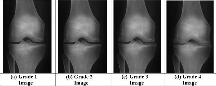
This diagnostic comparison chart displays four anteroposterior (AP) radiographic views of the human knee joint, illustrating the progression of knee osteoarthritis according to Kellgren–Lawrence (KL) grading. The images, labeled (a) through (d), represent Grade 1 to Grade 4 of the disease. In Grade 1 (a), the joint space appears largely preserved with minimal radiographic changes. As the progression moves toward Grade 4 (d), there is a clear chronological worsening of pathological features. Key diagnostic signs demonstrated include progressive joint space narrowing (JSN), particularly in the medial compartment, and the development of osteophytes (bone spurs) along the joint margins. Advanced stages, specifically Grade 3 and Grade 4, exhibit more severe narrowing, subchondral sclerosis (increased bone density appearing as whiter areas on the X-ray), and potential subchondral cysts or bone contour irregularities. This visual is intended for orthopedic and radiological education to assist in the classification and early detection of osteoarthritis using deep learning models (GAO-DCNN).
What to Look For on Her Knee X-ray
1. Joint Space Narrowing (JSN)
- The gap between the femur (thigh bone) and tibia (shin bone) becomes narrower as cartilage wears away
- Usually starts on the medial (inner) side of the knee first, since that compartment bears more weight
- Best seen on a weight-bearing (standing) X-ray - this is important to specifically ask for, since a normal lying-down X-ray can look deceptively better
2. Osteophytes (Bone Spurs)
- Small bony outgrowths that form at the joint margins as the body tries to stabilize the damaged joint
- Seen at the edges of the femur and tibia
3. Subchondral Sclerosis
- Increased bone density/whiteness just beneath the cartilage surface, where bone is rubbing directly against bone
4. Subchondral Cysts
- Small fluid-filled cavities that form in the bone just under the joint surface in advanced disease
The Grading Scale They'll Likely Use: Kellgren-Lawrence (KL)
| Grade | Findings |
|---|---|
| 0 | Normal |
| 1 (Doubtful) | Tiny/questionable osteophyte only |
| 2 (Minimal) | Definite osteophytes, joint space narrowing <50% |
| 3 (Moderate) | Multiple osteophytes, clear joint space narrowing, some sclerosis |
| 4 (Severe) | Large osteophytes, severe joint space narrowing, marked sclerosis, bone deformity |
Practical Tip for the Doctor Visit
-
How advanced the disease is
-
Whether she's a candidate for a brace (varus/valgus offloading braces target specific compartments)
-
Roughly how urgent surgical discussion might become
-
Bailey and Love's Short Practice of Surgery, 28th Ed.
-
Miller's Review of Orthopaedics, 9th Ed.
What are other fifferntials
Differential Diagnosis for Knee Pain with Swelling (Pain on Extension / Rising from Sitting)
Categorized by Pattern (Monoarticular = single joint, like her case)
| Differential | Key Distinguishing Features |
|---|---|
| Osteoarthritis | Gradual onset, worse with activity/standing, morning stiffness <30 min, crepitus, bony swelling, older age |
| Gout | Sudden, intense pain and swelling, often red/hot joint, more common in men, linked to high uric acid |
| Pseudogout (CPPD) | Similar to gout but calcium crystals; common in elderly, can mimic OA flare |
| Septic arthritis | Fever, warmth, severe pain even at rest, cannot bear weight - medical emergency, must be ruled out |
| Trauma / Hemarthrosis | History of fall or injury, rapid swelling (within hours) |
| Rheumatoid arthritis | Usually symmetric, affects multiple small joints too, morning stiffness >1 hour |
| Housemaid's knee (Prepatellar bursitis) | Swelling more localized to front of kneecap, history of kneeling |
| Baker's (popliteal) cyst | Swelling more behind the knee, may cause calf swelling if ruptured |
| Meniscal tear | Often history of twisting injury, locking/catching sensation |
Red Flags That Would Point AWAY from Simple OA (worth mentioning to the doctor)
- Fever or feeling generally unwell → suggests infection (septic arthritis) - urgent
- Redness and warmth over the joint → suggests gout, pseudogout, or infection
- Multiple joints involved symmetrically → suggests rheumatoid arthritis
- Sudden onset overnight → more typical of gout/pseudogout than OA
- Recent fall or twisting injury → suggests trauma, ligament, or meniscal injury
- Unable to bear any weight at all → needs urgent evaluation
How the Doctor Will Differentiate
- Joint aspiration (arthrocentesis) if there's uncertainty - checking the fluid for crystals (gout/pseudogout), infection (septic arthritis), or blood (trauma) is the most definitive test when swelling is significant
- Blood tests - ESR, CRP (inflammation), uric acid (gout), rheumatoid factor/anti-CCP (RA), complete blood count (infection)
- X-ray - as discussed, looks for OA-specific changes (joint space narrowing, osteophytes, sclerosis, cysts)
- Physical exam pattern - whether it's one joint (monoarticular, favors OA/gout/septic/trauma) versus many joints (polyarticular, favors RA/PsA/viral)
- ROSEN's Emergency Medicine, Table 102.1
- Firestein & Kelley's Textbook of Rheumatology