
Why anencephaly cause maternal polyhydraminos
Why Anencephaly Causes Maternal Polyhydramnios
What is Anencephaly?
The Core Mechanism: Absent Swallowing Reflex
- The cerebral cortex and higher brain centers are absent - the neural circuitry required to initiate and coordinate swallowing does not exist
- The fetus therefore cannot swallow amniotic fluid
- Fluid continues to be produced (fetal urine, lung fluid secretions) but is not being removed by swallowing
- Over the last 2 months of pregnancy, this accumulation leads to polyhydramnios (excess amniotic fluid, typically 1,500-2,000 mL or more)
Normal Amniotic Fluid Regulation (for context)
| Source of fluid IN | Source of fluid OUT |
|---|---|
| Fetal urine (main source, 2nd/3rd trimester) | Fetal swallowing (main route) |
| Lung fluid secretions | Intramembranous absorption |
| Oral/tracheal secretions | Transmembranous flow |
Clinical Points
- Polyhydramnios from anencephaly is most prominent in the last 2 months of pregnancy
- Anencephaly can be detected on ultrasound (absent skull vault is the hallmark sign)
- Elevated maternal serum and amniotic fluid AFP (alpha-fetoprotein) is also a marker, as neural tissue is exposed to amniotic fluid
- Anencephaly occurs in ~1 per 5,000 births, is more common in females, and is always fatal
- It falls under the category of neural tube defects, most of which can be prevented by maternal folic acid supplementation (400 µg daily) before and during pregnancy
Summary in One Sentence
Spina bifida
Spina Bifida
Definition and Embryological Basis
- More common in girls than boys
- Incidence of spina bifida cystica: ~1 in 5,000 births (considerable geographic variation)
- Strongly linked to folic acid deficiency - maternal supplementation (400 µg/day) significantly reduces incidence
- Most commonly affects the lumbar vertebrae
Classification: Four Types
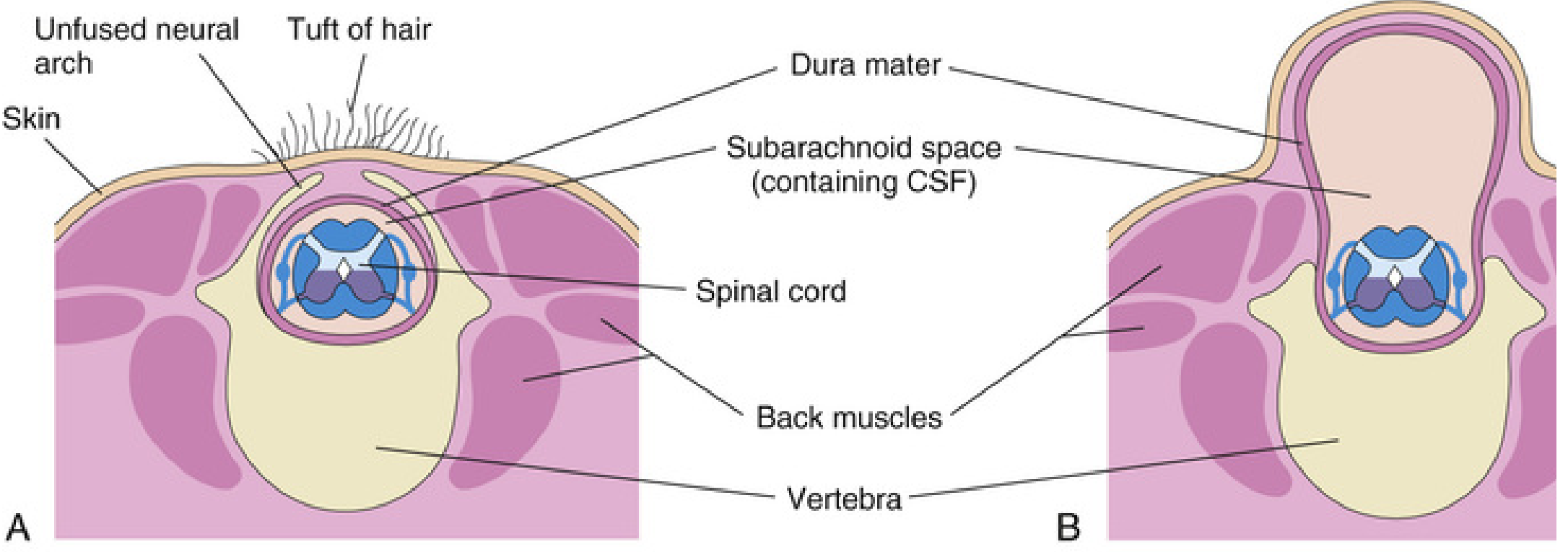
1. Spina Bifida Occulta
- Bony defect only - the neural arches of L5 or S1 fail to fuse in the midline, but there is no herniation of neural tissue
- Found in ~10% of otherwise normal people - usually an incidental finding
- May be marked externally by a small dimple, tuft of hair (hypertrichosis), lipoma, dermal sinus, or birthmark over the lumbosacral region
- Usually asymptomatic; rarely causes significant spinal cord or nerve root deficits
- The dimple marks the site where the caudal neuropore closed at the end of week 4
2. Spina Bifida with Meningocele
- Meninges (and CSF) herniate through the bony defect forming a cyst-like sac; spinal cord and nerve roots remain in their normal position
- The sac is covered by skin
- Neurological deficits may be absent or mild
- If skin cover is adequate, may be managed conservatively; a large sac can be surgically excised at 3-6 months
3. Spina Bifida with Meningomyelocele (the most common severe form)
- Both meninges and spinal cord/nerve roots herniate into the sac
- The roof of the defect is formed by exposed neural tissue (not covered by normal skin)
- Causes motor, sensory, and autonomic deficits below the level of the lesion
- Dermatomal loss of sensation, skeletal muscle paralysis, and sphincter dysfunction (bladder/bowel) are common
- 75-90% of cases develop hydrocephalus, nearly always due to the associated Arnold-Chiari type II malformation
- Most lesions are lumbosacral
4. Spina Bifida with Myeloschisis (Rachischisis)
- The most severe form - the neural tube lies completely open; the spinal cord is a flat, exposed plate of neural tissue
- No cyst; the cord is directly exposed to amniotic fluid
- Severe neurological deficits; poor prognosis
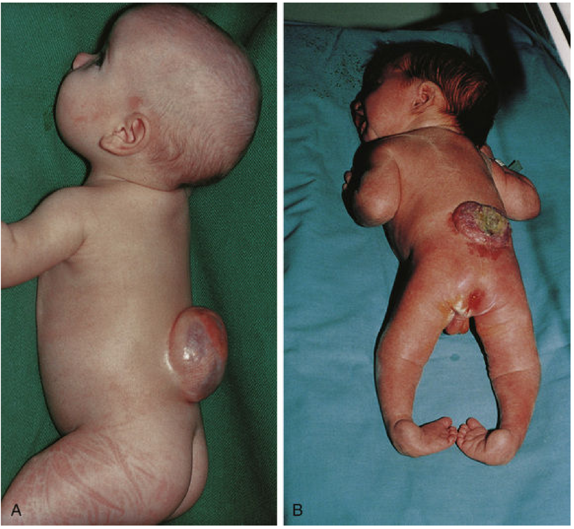
Types Summary Table
| Type | Bony defect | Meninges | Spinal cord/roots | Skin cover | Neurological deficit |
|---|---|---|---|---|---|
| Occulta | Yes | No herniation | Normal | Intact | Usually none |
| Meningocele | Yes | Herniated (CSF) | Normal position | Thin/intact | Mild or none |
| Meningomyelocele | Yes | Herniated | Herniated into sac | Absent/incomplete | Moderate-severe |
| Myeloschisis | Yes | Open | Exposed plate | Absent | Severe |
Associated Abnormalities
Arnold-Chiari Type II Malformation
- Present in almost all infants with spina bifida (myelomeningocele)
- The medulla oblongata and cerebellar tonsils herniate downward through the foramen magnum into the cervical spinal canal
- Causes pressure on the lower medulla
- Strongly associated with obstructive hydrocephalus (blockage of CSF flow)
- Features: headache, vomiting, visual disturbances, cerebellar ataxia, sensory disturbances, or paralysis
Hydrocephalus
- Occurs in 75% of meningomyelocele cases
- Mostly due to the Arnold-Chiari malformation blocking CSF outflow
- Requires surgical CSF diversion (ventriculoperitoneal shunt)
Tethered Cord Syndrome
- Progressive neurological deficit due to spinal cord tethering and traction as the child grows
- Part of the broader group of spinal dysraphism disorders
Neuropathic Bladder
- Common long-term complication of lumbosacral meningomyelocele
- Leads to urinary incontinence, recurrent UTIs, and upper tract damage
Neurological Deficits by Level
| Level | Motor deficit | Sensory loss | Bladder/bowel |
|---|---|---|---|
| Thoracic | Paraplegia | Trunk/legs | Yes |
| Lumbar (L1-L3) | Hip flexion/adduction weakness | Thigh/leg | Yes |
| Lumbar (L4-L5) | Foot drop, knee instability | Below knee | Common |
| Sacral | Foot/ankle weakness | Perianal (saddle) | Common - sphincter paralysis |
Prenatal Diagnosis
- Ultrasound: absence of posterior vertebral arches; banana sign (cerebellum pulled caudally) and lemon sign (frontal bone scalloping) due to associated Chiari malformation
- Elevated maternal serum AFP: leaked from exposed neural tissue into amniotic fluid and then maternal blood. Used in routine antenatal screening (15-20 weeks)
- Amniotic fluid AFP and acetylcholinesterase: confirmatory if serum AFP elevated
Management
Immediate (Neonatal)
- Cover the defect with sterile moist dressings to protect neural tissue
- Early surgical closure within 24-48 hours to prevent meningitis and CSF contamination
- Treat hydrocephalus with ventriculoperitoneal (VP) shunt
Selective Approach for Myelomeningocele
- Previously, all defects were closed surgically; now a selective approach is used
- Surgery is considered inappropriate in infants with severe hydrocephalus, large open defect, and no distal neurological function - majority of these infants die in the first year without closure
- With antibiotics, early surgical closure, and shunting: ~50% of children who survive the first 24 hours reach school age
Fetal Surgery
- In utero repair of myelomeningocele (before 26 weeks gestation) reduces the need for shunting and improves motor outcomes (MOMS trial)
Long-term Multidisciplinary Care
- Neuropathic bladder - clean intermittent catheterization (CIC), urodynamics
- Neuromuscular scoliosis - bracing or surgical correction
- Bone and joint deformity
- Skin care (pressure sores due to sensory loss)
- Cognitive and developmental support
Prevention
Why and how meningomyelocele causes hydrocepwhuksu
How Myelomeningocele Causes Hydrocephalus
Step 1 - The Root Cause: Why Does Myelomeningocele Lead to Chiari II?
"Failure of closure of the neural tube results in CSF leakage through the open spinal defect, leading to lack of development of the posterior fossa and abnormalities of neural and calvarial development."
- Creasy & Resnik's Maternal-Fetal Medicine
- The neural tube fails to close in the lumbosacral region at ~week 4 of gestation
- The open myelomeningocele defect allows CSF to leak freely out of the spinal canal into the amniotic fluid throughout fetal development
- Because CSF is continuously lost, there is insufficient hydraulic pressure inside the developing neural tube
- This low pressure means the posterior fossa (the bony space at the back of the skull housing the brainstem and cerebellum) does not expand properly - it remains abnormally small
- The cerebellar vermis, tonsils, fourth ventricle, and medulla are forced downward through the enlarged foramen magnum into the cervical spinal canal - this is the Arnold-Chiari type II malformation
Step 2 - How Chiari II Produces Hydrocephalus
Obstruction at the Foramen Magnum
- The herniated medulla, cerebellar vermis and tonsils physically plug the foramen magnum
- CSF flowing down from the ventricular system cannot pass freely around the craniocervical junction
- This creates a non-communicating (obstructive) hydrocephalus
Obstruction of the Fourth Ventricle
- The fourth ventricle is dragged downward and becomes elongated and slit-like
- Its outlets (foramina of Luschka and Magendie) are displaced and compressed
- CSF cannot exit the fourth ventricle normally
Aqueductal Stenosis (secondary)
- The midbrain shows "beaking" of the tectum (the posterior midbrain appears elongated on sagittal MRI)
- This distortion can narrow the cerebral aqueduct (of Sylvius), adding another level of obstruction between the third and fourth ventricles
The Full Pathway (Summary)
Myelomeningocele (open neural tube defect)
↓
CSF leaks out of spinal canal throughout fetal life
↓
Posterior fossa remains small / underdeveloped
↓
Cerebellum + brainstem + 4th ventricle herniate
downward through foramen magnum
↓
= Arnold-Chiari Type II Malformation
↓
Obstructs CSF outflow at:
• Foramen magnum (physical plug)
• 4th ventricle outlets (Luschka, Magendie)
• Cerebral aqueduct (beaking/kinking)
↓
Non-communicating (obstructive) hydrocephalus
(entire ventricular system dilates)
Key Numbers
| Fact | Figure |
|---|---|
| Myelomeningocele cases with Chiari II | ~100% |
| Myelomeningocele cases developing hydrocephalus | 80-90% |
| Hydrocephalus present at birth | ~25% |
| Hydrocephalus developing after postnatal MMC repair | ~80% of remaining cases |
| Cases with symptomatic brainstem dysfunction | ~20% (but high mortality) |
MRI Appearances of Chiari II
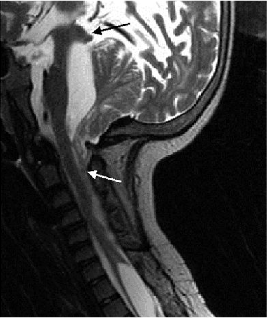
- Small, shield-shaped posterior fossa
- Downward displacement of cerebellar vermis, tonsils, 4th ventricle, medulla
- "Beaking" of the midbrain tectum
- Slit-like fourth ventricle (normal/enlarged = suggests isolated 4th ventricle or hydrocephalus)
- Medullary kinking
- Enlarged massa intermedia
- On antenatal ultrasound: "banana sign" (compressed, curved cerebellum) and "lemon sign" (frontal bone scalloping due to caudal traction on the brain)
Treatment of the Hydrocephalus
- Ventriculoperitoneal (VP) shunt - traditional standard; diverts CSF from the lateral ventricles to the peritoneal cavity
- Endoscopic Third Ventriculostomy (ETV) + Choroid Plexus Cauterization (CPC) - avoids a permanent shunt; effective in >70% of MMC patients
- Fetal surgery (in utero MMC repair before 26 weeks) - reduces the need for shunting by allowing the posterior fossa to develop more normally before birth; 40% still require shunting despite prenatal repair (MOMS trial data)
- Posterior fossa decompression - for symptomatic brainstem compression from the Chiari herniation itself
Why there is dilated 4 ventricle with absent cerebellum in dandy walker malformation
Dandy-Walker Malformation: Why the 4th Ventricle is Dilated and the Cerebellum is Absent
What is Dandy-Walker Malformation?
- Cystic dilatation of the 4th ventricle (the 4th ventricle balloons into a large posterior fossa cyst)
- Aplasia or hypoplasia of the cerebellar vermis (the midline portion of the cerebellum is absent or rudimentary)
- Enlarged posterior fossa with upward displacement of the tentorium, torcula, and straight sinus above the lambdoid suture
The Core Developmental Problem
Normal Development
- This membrane perforates at around week 26 of gestation to form the foramen of Magendie (median aperture) in the midline
- The foramina of Luschka (lateral apertures) also open
- These three openings allow CSF produced inside the 4th ventricle to exit freely into the subarachnoid space
- With free CSF egress, the 4th ventricle remains normally sized
- Simultaneously, CSF pressure inside the developing posterior fossa drives normal expansion of the cerebellar vermis and hemispheres
What Goes Wrong in Dandy-Walker
Foramina of Luschka & Magendie fail to open
↓
CSF cannot exit the 4th ventricle
↓
CSF accumulates inside the 4th ventricle
↓
4th ventricle progressively expands into a large cyst
(= "roofless 4th ventricle" lined by ependyma)
↓
The ballooning cyst DISPLACES and COMPRESSES the
developing cerebellar vermis rather than allowing
it to grow → vermis is absent or hypoplastic
↓
Posterior fossa enlarges to accommodate the cyst
Tentorium and torcula are pushed upward
↓
Hydrocephalus (obstructive) from blocked CSF outflow
"This cyst represents the expanded, roofless fourth ventricle in the absence of a normally formed vermis."
- Robbins & Kumar Pathologic Basis of Disease
Why Specifically is the Vermis Absent?
1. Primary Developmental Failure
2. Mechanical Compression/Displacement by the Expanding Cyst
The MRI - What You See
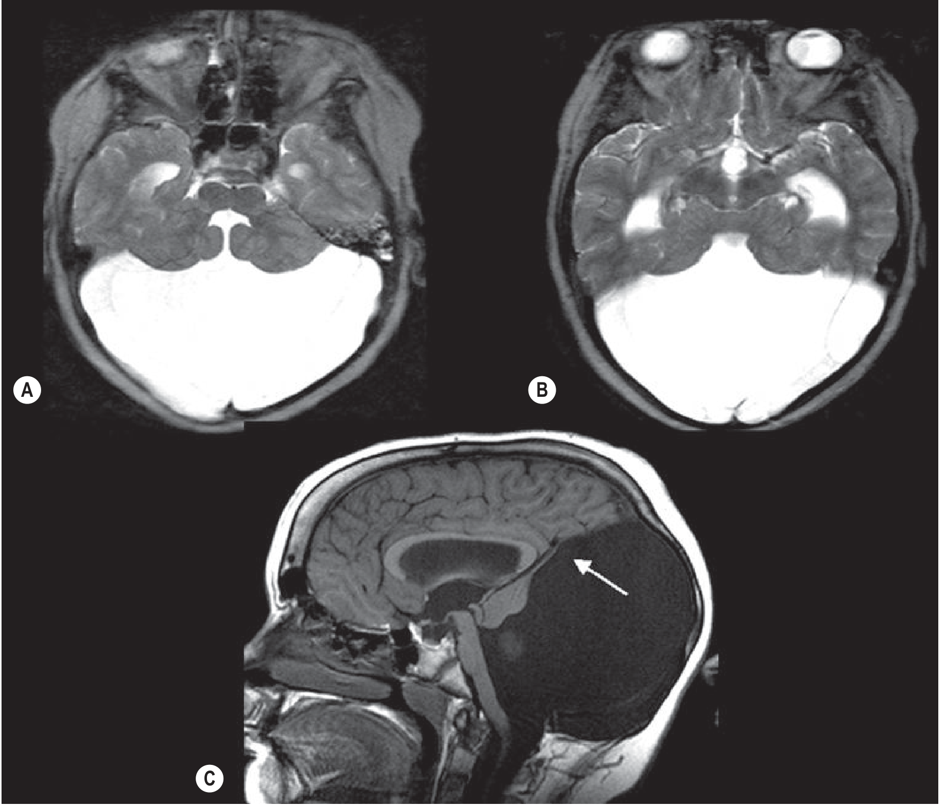
| Feature | Explanation |
|---|---|
| Large posterior fossa cyst | Expanded, roofless 4th ventricle |
| Absent/hypoplastic cerebellar vermis | Primary failure of vermis formation + compression |
| Elevated tentorium/torcula above lambdoid suture | Posterior fossa expansion pushes tentorium up |
| Hydrocephalus (dilated lateral and 3rd ventricles) | Obstruction of CSF outflow from 4th ventricle |
| Thin rim of cerebellar tissue at cyst wall | Compressed, displaced cerebellar remnant |
| Dilated cerebral aqueduct + 3rd ventricle | Retrograde obstruction from blocked 4th ventricle outflow |
Associated Features and Anomalies
- Corpus callosum agenesis/hypoplasia - in ~20-25% of cases
- Heterotopia of inferior olivary nuclei
- Pachygyria of the cerebral cortex
- Hydrocephalus - develops in the majority due to obstruction; may not be present at birth but emerges in infancy
- Brainstem nuclei dysplasias - often found on pathology
- Other midline anomalies (septal, hypothalamic)
- Can be associated with chromosomal abnormalities and genetic syndromes
Dandy-Walker Spectrum (from severe to mild)
| Variant | 4th Ventricle | Vermis | Posterior Fossa | Hydrocephalus |
|---|---|---|---|---|
| Classic DWM | Massively dilated cyst | Absent/aplastic | Greatly enlarged | Common |
| DW Variant | Mildly enlarged | Hypoplastic (partial) | Mildly enlarged | Variable |
| Blake's Pouch Cyst | Mildly enlarged | Normal | Normal/mildly enlarged | Possible |
| Mega Cisterna Magna | Normal | Normal | Enlarged cisterna only | Absent |
Contrast with Chiari II
| Dandy-Walker | Chiari II | |
|---|---|---|
| 4th ventricle | Massively dilated/cystic | Small, slit-like, displaced downward |
| Cerebellum | Vermis absent, cyst fills posterior fossa | Herniated downward through foramen magnum |
| Posterior fossa | Enlarged | Small |
| Tentorium | Elevated | Low |
| Association | Isolated or genetic syndromes | Myelomeningocele (nearly 100%) |
| Mechanism of hydrocephalus | Blocked 4th ventricle outlets (foramina) | Foramen magnum obstruction by herniated tissue |
Clinical Presentation
- Macrocephaly (enlarging head circumference) - often the first sign in infancy
- Bulging posterior fontanelle
- Signs of raised intracranial pressure (irritability, vomiting, sunset sign)
- Developmental delay - more related to associated brain anomalies than hydrocephalus alone
- Cerebellar signs - ataxia, hypotonia (if cerebellar tissue remains)
- Seizures - from associated cortical dysplasias
Treatment
- VP shunt for hydrocephalus - drains the cyst directly (cystoperitoneal shunt) or lateral ventricles
- Management of associated anomalies
- Prognosis depends heavily on associated brain malformations, not just the posterior fossa cyst
Why these Forman fail to perforate
Dandy-Walker malformation why foramina Luschka Magendie fail to open causes genetics pathogenesis
https://en.wikipedia.org/wiki/Dandy%E2%80%93Walker_malformat…
Why Do the Foramina of Luschka and Magendie Fail to Perforate in Dandy-Walker?
The Honest Answer First
"Taggart and Walker... ascribed the potential cause as underdevelopment of the foramina of Luschka and Magendie - now no longer believed to be significant." - Wikipedia (citing updated literature)
What Actually Causes the Developmental Arrest?
1. Genetic Causes (most important)
| Gene/Chromosome | Mechanism |
|---|---|
| ZIC1 and ZIC4 (chromosome 3q) | Zinc finger transcription factors critical for cerebellar vermis development; deletion causes DWM |
| ZIC2 and ZIC5 (chromosome 13q) | Involved in Blake's pouch cyst variant (haploinsufficiency from 13q deletion) |
| FOXC1 | Forkhead transcription factor; mutations linked to DWM, particularly involving mesenchymal development of the posterior fossa |
| Trisomy 18 (most common trisomy association) | Extra chromosome 18 - DWM is the most frequent trisomy association |
| Trisomy 13, 21, 9 | Also associated with DWM |
| Chromosomal deletions/duplications | Various partial chromosomal imbalances |
| Autosomal recessive/dominant inheritance | Rare familial forms exist |
2. The Blake's Pouch Theory (Developmental Timing Failure)
- The roof of the 4th ventricle passes through a stage called Blake's Pouch - a blind-ended invagination
- Normally at ~week 26 of gestation, this pouch ruptures/perforates to form the foramen of Magendie
- If the posterior membranous area fails to regress at the programmed time, the pouch remains intact as a cyst
- In classic DWM, the primary failure is of cerebellar vermis formation (not foraminal atresia) - the vermis simply never develops, so the posterior wall of the 4th ventricle is never properly formed by neural tissue
- In Blake's Pouch Cyst (a milder variant), the vermis IS normal but the membrane just fails to perforate - this is the purer "foraminal failure" form
3. Environmental / Teratogenic Causes
| Teratogen | Evidence |
|---|---|
| Warfarin (during pregnancy) | Linked to DWM in 1985; also causes corpus callosum agenesis and ocular dysgenesis |
| Maternal diabetes | Increased risk of DWM |
| Alcohol | Suggested but uncertain evidence |
| Viral infections (congenital) | Cytomegalovirus, rubella - disrupt hindbrain development |
| Isotretinoin (vitamin A derivative) | Hindbrain teratogen |
The Underlying Mechanism - Putting It Together
Primary defect: Genetic mutation OR teratogen
(weeks 4-6 of gestation)
↓
Abnormal development of:
• Rhombic lip (germinal zone → cerebellar vermis)
• Posterior fossa mesenchyme
• Posterior membranous area (Blake's pouch) timing
↓
Cerebellar vermis fails to form (aplasia/hypoplasia)
↓
The posterior wall of the 4th ventricle is never
properly "closed" by vermian tissue
↓
Blake's pouch / posterior membranous area fails to
perforate (secondary to the primary malformation)
↓
4th ventricle communicates with an expanding
posterior fossa cyst instead of the subarachnoid space
↓
Progressive cyst enlargement → hydrocephalus
(in ~70-80% of cases)
Key Point: Foraminal Atresia vs. Primary Vermian Failure
| Classical teaching | Modern understanding |
|---|---|
| Foramina fail to open → CSF trapped → 4th ventricle dilates → compresses vermis | Primary failure of vermis/posterior fossa development → 4th ventricle has no posterior wall → acts as an open cyst |
| Foraminal atresia is the PRIMARY cause | Foraminal non-patency is SECONDARY to primary mesenchymal/cerebellar maldevelopment |
| Analogous to a "blocked drain" | More like a "drain that was never properly built" |
Why is the Cause Frequently Unknown?
- Many responsible gene mutations have not yet been discovered
- The condition is genetically heterogeneous (many different genes can produce the same phenotype)
- Environmental contributions are hard to confirm retrospectively
- The event occurs very early (weeks 4-6), often before a woman knows she is pregnant
AC malformation type ll how occur in ass with syringomyelia
Arnold-Chiari Type II and Syringomyelia: How They Are Connected
What is a Syrinx?
The Fundamental Problem: Blocked CSF Flow at the Foramen Magnum
- Partially or completely occludes the subarachnoid space at the craniocervical junction
- Disrupts the normal two-way pulsatile flow of CSF between the cranial and spinal compartments
- Systolic arterial pulsation causes a brief rise in intracranial pressure
- CSF surges smoothly downward through the foramen magnum into the spinal subarachnoid space
- It then returns during diastole
- This pulsatile flow is free and symmetric
The Three Theories of How the Syrinx Forms
Theory 1 - Gardner's Hydrodynamic Theory ("Water Hammer" / Piston Theory)
- The herniated tonsils block the foramina of Luschka and Magendie (or the foramen magnum)
- CSF cannot exit the 4th ventricle normally
- With each systolic pulse of the choroid plexus, a pressure wave ("water hammer effect") is transmitted down the central canal of the spinal cord
- This repeated pulsatile pressure progressively dilates and then ruptures the central canal wall, forming a syrinx
- The syrinx is therefore essentially a dilated, diverticulated central canal
"A pulse wave of CSF pressure generated by systolic pulsations of the choroid plexuses is transmitted into the cord from the fourth ventricle through the central canal... the syrinx consists essentially of a greatly dilated central canal with a diverticulum that ramifies from the central canal."
- Adams & Victor's Principles of Neurology
Theory 2 - Oldfield/Heiss "Piston Effect" / Subarachnoid Pressure Theory
- The herniated cerebellar tonsils act as a piston in a cylinder (the spinal canal)
- With each heartbeat, the tonsils are pushed slightly downward by the intracranial pulsation
- This compresses the spinal subarachnoid CSF from outside the cord
- The pressure wave drives CSF into the spinal cord parenchyma through Virchow-Robin perivascular spaces and other subpial channels (NOT through the central canal)
- Over time, these small fluid collections coalesce to form a syrinx cavity
- The syrinx initially forms outside the central canal, but eventually connects to it (hydromyelia ex vacuo)
"The compressive effect of the cerebellar tonsils partially occludes the subarachnoid space at the foramen magnum and creates pressure waves that compress the spinal cord from without and not from within; the pressure waves propagate syrinx fluid caudally with each heartbeat."
- Adams & Victor's Principles of Neurology (Heiss et al.)
- The syrinx can form without a direct 4th ventricle-to-central canal connection
- Decompression of the foramen magnum (removing the "piston") often causes the syrinx to shrink or disappear
Theory 3 - Virchow-Robin Perivascular Tracking
- Subarachnoid obstruction at the craniocervical junction increases spinal CSF pressure during straining, coughing, or physical effort
- Under this elevated pressure, CSF tracks inward along Virchow-Robin (perivascular) spaces into the cord parenchyma
- Small fluid pools accumulate and coalesce over years into a syrinx cavity
- This also explains why coughing or straining worsens symptoms acutely in syringomyelia
Why Chiari II Specifically (vs. Chiari I)?
| Chiari I | Chiari II | |
|---|---|---|
| Structures herniated | Cerebellar tonsils only | Tonsils + vermis + 4th ventricle + medulla |
| Foramen magnum occlusion | Partial | More complete |
| Association with syrinx | Common (majority of syringomyelia cases) | Present but often overshadowed by hydrocephalus |
| Timing of symptoms | Adolescence/adulthood | Infancy (due to MMC/hydrocephalus) |
The Full Sequence - Visual Summary
Arnold-Chiari II Malformation
(herniated tonsils, vermis, medulla through foramen magnum)
↓
Foramen magnum partially/fully occluded
↓
BLOCKED pulsatile CSF flow at craniocervical junction
↓
With each heartbeat...
↓
┌─────────────────────────────────┐
│ Theory 1 (Gardner) │ Theory 2 (Heiss/Oldfield)
│ Pressure wave travels DOWN │ Tonsils act as PISTON
│ central canal of cord │ Compress cord from OUTSIDE
│ → dilates central canal │ → CSF enters via
│ → syrinx (dilated canal) │ Virchow-Robin spaces
└─────────────────────────────────┘
↓
Progressive fluid accumulation inside cord parenchyma
↓
SYRINX (fluid-filled cavity, usually cervical cord)
↓
May extend upward into brainstem = SYRINGOBULBIA
Clinical Features of Syringomyelia (when it complicates Chiari II)
| Symptom | Mechanism |
|---|---|
| Dissociated sensory loss - loss of pain & temperature, preserved touch/vibration | Central cord destruction at anterior commissure (crossing spinothalamic fibers) |
| Cape/shawl distribution of sensory loss (neck, shoulders, arms) | Cervical syrinx |
| Painless injuries and burns of the hands | Loss of pain sensation |
| Hand/arm weakness and wasting (lower motor neuron) | Anterior horn cell destruction |
| Absent upper limb reflexes | Arc disrupted at cervical level |
| Spastic paraplegia (if large syrinx) | Corticospinal tract involvement |
| Scoliosis | Asymmetric muscle weakness |
| Syringobulbia (if extends upward) | Nystagmus, tongue weakness, dysphagia, facial sensory loss |
Treatment Implication
- Posterior fossa decompression (craniectomy + C1 laminectomy ± duraplasty) - restores normal CSF flow at the craniocervical junction
- The syrinx often collapses or shrinks after decompression without directly treating the syrinx
- In Chiari II with hydrocephalus, VP shunting reduces intracranial pressure and can indirectly resolve the syrinx
- Direct syrinx shunting (syringoperitoneal shunt) is reserved for cases that fail decompression
Can you simplify me the spinal tract and their damage lead to which disorder
Spinal Tracts and What Damage to Each Causes
The Spinal Cord Cross-Section - A Map
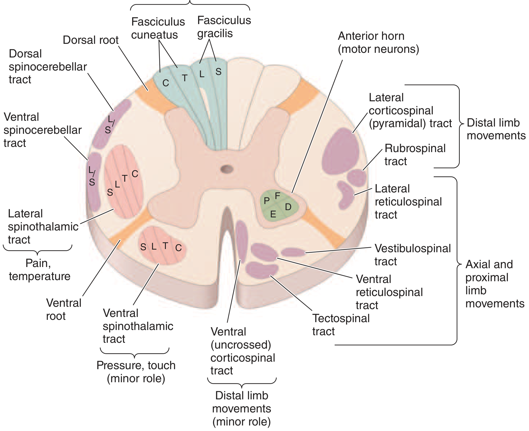
- Posterior (dorsal) = sensory (fine sensation)
- Lateral = motor (corticospinal) + pain/temperature (spinothalamic)
- Anterior (ventral) = motor neurons + crude touch
THE ASCENDING (SENSORY) TRACTS
1. Posterior Columns (Dorsal Columns)
| What they carry | Where they cross | Side of cord |
|---|---|---|
| Vibration, proprioception (joint position), fine touch, pressure | Cross in the medulla (high up) | Travel ipsilateral (same side) in the cord |
- Ipsilateral loss of vibration sense and joint position below the lesion
- Sensory ataxia - patient cannot feel where their limbs are → wide-based unsteady gait
- Positive Romberg sign (falls when eyes closed - relies on vision to compensate)
- Classic diseases: Subacute combined degeneration (B12 deficiency), tabes dorsalis (syphilis), Friedreich's ataxia, posterior cord syndrome
2. Lateral Spinothalamic Tract
| What it carries | Where it crosses | Side of cord |
|---|---|---|
| Pain and temperature | Crosses at the anterior commissure (within spinal cord) | Travels contralateral (opposite side) |
- Contralateral loss of pain and temperature 1-2 levels below the lesion
- Touch is preserved (carried by both posterior column and ventral spinothalamic)
- Classic scenario: Syringomyelia destroys the fibers as they cross → "cape/shawl" distribution loss
- Somatotopy: sacral fibers are outermost → deep lesions spare sacral ("sacral sparing")
3. Ventral (Anterior) Spinothalamic Tract
| What it carries | Side |
|---|---|
| Crude touch and pressure (minor role) | Contralateral |
4. Spinocerebellar Tracts (Dorsal + Ventral)
| What they carry | Function |
|---|---|
| Unconscious proprioception from muscles/joints | Coordination and balance via cerebellum |
- Ipsilateral limb ataxia (clumsy, uncoordinated movements)
- Loss of smooth, coordinated gait
- Seen in: Friedreich's ataxia, multiple sclerosis
THE DESCENDING (MOTOR) TRACTS
5. Lateral Corticospinal Tract (Pyramidal Tract) - THE MOST IMPORTANT
| Origin | Crosses | Side in cord |
|---|---|---|
| Motor cortex | At the pyramidal decussation in the medulla | Travels ipsilateral (already crossed) |
- Ipsilateral spastic weakness below the lesion
- Increased tone (spasticity)
- Hyperreflexia (exaggerated deep tendon reflexes)
- Positive Babinski sign (extensor plantar response)
- No muscle wasting (unlike LMN)
6. Anterior Horn Cells (not a tract, but critical)
- Ipsilateral segmental flaccid weakness
- Decreased/absent reflexes
- Muscle wasting and fasciculations
- No Babinski
- Seen in: polio, motor neuron disease (at level of lesion in ALS)
SUMMARY TABLE - TRACTS AT A GLANCE
| Tract | Location | Carries | Crosses where | Side of deficit |
|---|---|---|---|---|
| Posterior column | Dorsal | Vibration, proprioception, fine touch | Medulla | Ipsilateral |
| Lateral spinothalamic | Lateral | Pain, temperature | Anterior commissure (spinal cord) | Contralateral |
| Ventral spinothalamic | Anterior | Crude touch | Spinal cord | Contralateral |
| Spinocerebellar | Lateral | Unconscious proprioception | Mostly ipsilateral | Ipsilateral |
| Lateral corticospinal | Lateral | Voluntary movement | Medulla (already crossed) | Ipsilateral |
| Anterior horn cell | Anterior grey | LMN to muscle | None | Ipsilateral, segmental |
CLINICAL SYNDROMES FROM COMBINED TRACT DAMAGE
Brown-Séquard Syndrome (Hemicord - one side cut)
| Side of lesion | Finding |
|---|---|
| Ipsilateral | UMN weakness (corticospinal) + loss of vibration/proprioception (dorsal column) |
| Contralateral | Loss of pain and temperature 1-2 levels below (spinothalamic crossed) |
Central Cord Syndrome (Central canal damaged)
- Pain and temperature fibers as they cross are destroyed
- Arms worse than legs (cervical crossing fibers affected first)
- "Cape/shawl" dissociated sensory loss - pain/temperature lost, touch/vibration preserved
- Causes: Syringomyelia, hyperextension injury in elderly, intrinsic tumour
Anterior Cord Syndrome (Anterior spinal artery occlusion)
| Lost | Preserved |
|---|---|
| Motor (bilateral UMN weakness) | Vibration and proprioception |
| Pain and temperature (bilateral) | (posterior columns spared) |
| Bladder/bowel dysfunction |
Posterior Cord Syndrome
| Lost | Preserved |
|---|---|
| Vibration, proprioception (bilateral) | Motor power |
| Sensory ataxia, Romberg positive | Pain and temperature |
Complete Cord Transection
- Bilateral UMN weakness → spastic paraplegia/quadriplegia
- All sensation lost below level
- Bladder, bowel, sexual dysfunction
- Initially: spinal shock (flaccid, areflexic) → after days/weeks: spasticity and hyperreflexia return
Conus Medullaris Syndrome (sacral cord)
- Saddle anaesthesia (S3-S5 - perianal, perineal area)
- Bladder/bowel dysfunction (urinary retention + incontinence, lax anal tone)
- Impotence
- Bulbocavernosus and anal reflexes absent
- Leg strength largely preserved (distinguishes from cauda equina)
Cauda Equina Syndrome (nerve roots, not cord)
- Low back + radicular pain (prominent)
- Asymmetric leg weakness and sensory loss
- Variable areflexia
- Relative sparing of bladder/bowel (vs. conus)
- LMN signs (flaccid, wasted)
Quick Memory Aid
Posterior columns = "Same side, Fine sensation, Falls with eyes closed"
Spinothalamic = "Opposite side, Pain & Temp, Crosses in cord"
Corticospinal = "Same side, Motor, UMN signs"
BACK of cord = feel where you are
SIDE of cord = pain/temp (opposite) + movement (same)
FRONT of cord = motor neurons (LMN)
CENTRE = crossing pain fibers → cape distribution
Why umn and lmn damage cause ipsilateral loss and why lmn not cause babinski sign
Why UMN and LMN Damage Both Cause Ipsilateral Weakness - and Why Only UMN Causes Babinski
Part 1: Why Does Both UMN and LMN Damage Cause Ipsilateral Weakness in the Spinal Cord?
The Motor Pathway - Two Neurons
BRAIN (Motor cortex)
↓
Upper Motor Neuron (UMN)
= corticospinal tract
[crosses at the MEDULLA - pyramidal decussation]
↓
SPINAL CORD (Anterior horn)
↓
Lower Motor Neuron (LMN)
= alpha motor neuron
[exits via ventral root - NEVER crosses]
↓
MUSCLE
The Key Point: The Tract Has ALREADY Crossed Before It Enters the Cord
| Location of damage | Side of weakness |
|---|---|
| Above the decussation (motor cortex, internal capsule, cerebral peduncle, upper medulla) | Contralateral (opposite side to lesion) |
| Below the decussation (spinal cord - UMN damage in cord) | Ipsilateral (same side as lesion) |
| LMN (anterior horn, ventral root, nerve) | Ipsilateral (same side, same segment) |
"Motor cortex or medulla (above the pyramidal decussation): contralateral to weakness. Cervical spinal cord (below the pyramidal decussation): ipsilateral to weakness."
- Neuroanatomy through Clinical Cases 3rd Ed.
Diagram
LEFT Motor Cortex
|
| [UMN descends in LEFT internal capsule]
|
MEDULLA ←—— PYRAMIDAL DECUSSATION ——→
| (fibres CROSS here)
|
RIGHT lateral corticospinal tract
| [now on the RIGHT side of the cord]
|
RIGHT anterior horn cell (LMN)
|
RIGHT limb muscles
Why Does LMN Damage Also Cause Ipsilateral Weakness?
Part 2: Why Does UMN Damage Cause Babinski Sign, But LMN Damage Does NOT?
What Is the Babinski Sign?
- Normal adult response (LMN intact, UMN intact): All toes flex downward (plantar flexion) - negative Babinski
- UMN lesion response: The big toe dorsiflexes (extends upward) and the other toes fan out - positive Babinski
"The Babinski sign is dorsiflexion of the great toe and fanning of the other toes when the lateral aspect of the sole of the foot is scratched. In adults, the normal response to this stimulation is plantar flexion in all the toes."
- Ganong's Review of Medical Physiology
What the Babinski Sign Actually Represents
- Present normally in infants under 1 year (corticospinal tract is not yet myelinated)
- Suppressed in normal adults by the descending inhibitory influence of the corticospinal tract
"The Babinski sign... a disinhibited flexion withdrawal reflex, characterized by dorsiflexion (extension) of the large toe... Such response is considered normal until the age of 1 year."
- Localization in Clinical Neurology 8e
Why UMN Damage Releases the Babinski
- This inhibitory control is removed
- The primitive spinal reflex arc is released (disinhibited)
- The spinal cord reverts to its primitive withdrawal pattern
- The result: big toe extends upward → positive Babinski
Normal adult:
Corticospinal tract ——INHIBITS——→ Primitive withdrawal reflex
(kept suppressed)
Result: Normal plantar flexion (toes curl down)
UMN lesion:
Corticospinal tract DAMAGED → inhibition REMOVED
↓
Primitive reflex RELEASED
Result: Babinski sign (big toe up, toes fan)
Why LMN Damage Does NOT Cause Babinski
- Intact sensory nerve (afferent signal from sole of foot)
- Intact spinal cord interneurons
- Intact LMN (alpha motor neuron) + intact neuromuscular junction + intact muscle
- UMN must be absent/damaged (to remove inhibition)
LMN lesion:
Sole stimulus → sensory nerve → spinal cord interneuron
↓
Alpha motor neuron DAMAGED
↓
No signal reaches muscle
↓
NO REFLEX AT ALL (areflexia)
NO Babinski possible
Complete Comparison Table: UMN vs LMN
| Feature | UMN Lesion | LMN Lesion |
|---|---|---|
| Location | Motor cortex, internal capsule, brainstem (above decussation) OR corticospinal tract in spinal cord (below decussation) | Anterior horn cell, ventral root, peripheral nerve, NMJ |
| Tone | Increased (spasticity) | Decreased (flaccidity) |
| Weakness | Present | Present |
| Reflexes | Hyperreflexia (exaggerated) | Areflexia (absent) |
| Babinski sign | Positive (big toe up) | Absent (reflex arc broken) |
| Muscle wasting | Late/minimal | Early and prominent |
| Fasciculations | Absent | Present (dying LMN fires randomly) |
| Clonus | Present (in severe cases) | Absent |
| Side of deficit | Ipsilateral in cord; contralateral if above medullary decussation | Ipsilateral always |
| Example diseases | Stroke, MS, spinal cord compression, MND (UMN component) | Polio, Guillain-Barré, disc herniation, MND (LMN component) |
Why Reflexes Are Hyperactive in UMN But Absent in LMN
Tendon tap → muscle stretch → muscle spindle (Ia fibre)
→ enters dorsal horn → DIRECTLY synapses on alpha motor neuron (LMN)
→ LMN fires → muscle contracts (reflex)
-
UMN damage: The arc is intact. But the descending inhibitory signals that normally dampen the sensitivity of this arc are gone. The arc becomes overactive → hyperreflexia, clonus. The muscle spindle is now set at higher sensitivity.
-
LMN damage: The alpha motor neuron (the LMN) IS the reflex arc. Damage to it means the arc is physically broken - no matter how strongly the spindle fires, there is no motor neuron to respond → areflexia.
Relation between lateral horn of spinal tract and Horner syndrome
The Lateral Horn, the Sympathetic Pathway, and Horner Syndrome
The Lateral Horn - What Is It?
| Horn | Location | Contains |
|---|---|---|
| Dorsal (posterior) horn | Back | Sensory interneurons |
| Ventral (anterior) horn | Front | Lower motor neurons (alpha motor neurons) |
| Lateral horn | Side | Preganglionic autonomic neurons |
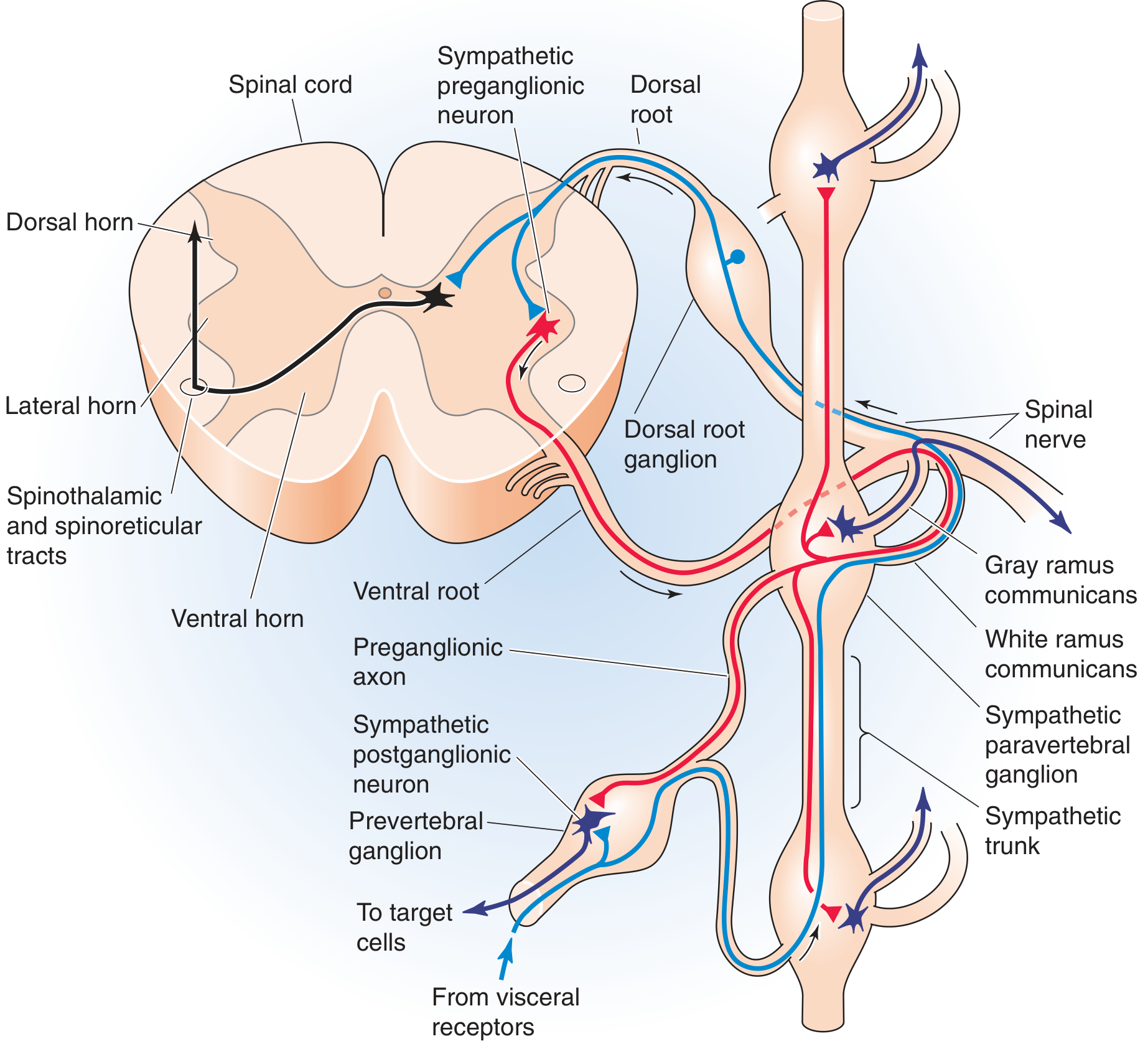
"The cell bodies of preganglionic sympathetic motor neurons are located in the thoracic and upper lumbar spinal cord between levels T1 and L3. At these spinal levels, autonomic neurons lie in the intermediolateral cell column, or lateral horn, between the dorsal and ventral horns."
- Medical Physiology (Boron & Boulpaep)
The Three-Neuron Sympathetic Pathway to the Eye
Neuron 1 - Central Neuron (First-Order)
- Origin: Hypothalamus (posterior hypothalamus)
- Path: Descends through the brainstem (tegmentum) → lateral reticulospinal tract of the spinal cord
- Destination: Synapse in the lateral horn at C8-T2 (the ciliospinal centre of Budge)
- Never crosses - stays ipsilateral throughout
Neuron 2 - Preganglionic Neuron (Second-Order) - THE LATERAL HORN NEURON
- Origin: Lateral horn at C8-T1-T2 (ciliospinal centre)
- Path: Exits via ventral root → enters white ramus communicans → travels over the apex of the lung → over the subclavian artery → ascends the neck alongside the carotid artery
- Destination: Synapses in the superior cervical ganglion (in the neck, at level of C2-C3)
- This is where the T1 lateral horn neuron is most relevant to Horner syndrome
Neuron 3 - Postganglionic Neuron (Third-Order)
- Origin: Superior cervical ganglion
- Path: Travels along the internal carotid artery → into the cavernous sinus → joins the ophthalmic branch of the trigeminal nerve (V1) → enters the orbit
- Destination: Innervates:
- Pupillary dilator muscle (iris)
- Müller's muscle (upper eyelid elevator - smooth muscle)
- Lower lid retractor (inferior tarsal muscle)
- Sweat glands of the face (via external carotid → facial branches)
The Connection: How Lateral Horn Damage Causes Horner Syndrome
- Dilates the pupil (radial/dilator muscle of iris)
- Elevates the upper eyelid (Müller's smooth muscle)
- Elevates the lower lid (inferior tarsal muscle)
- Stimulates sweating on same side of face
- Causes slight protrusion of the eyeball
Horner Syndrome - The Classic Triad
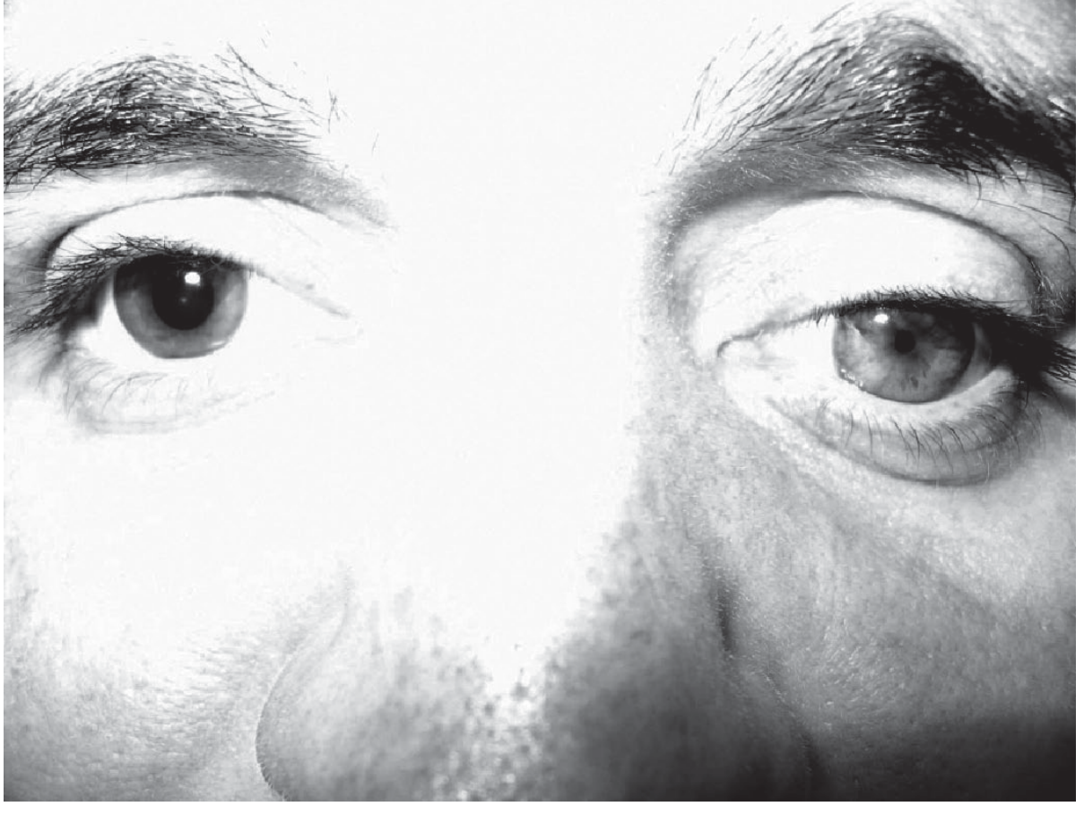
| Sign | Mechanism | Structure lost |
|---|---|---|
| Miosis (small pupil) | Dilator pupillae muscle paralysed; constrictor (parasympathetic) unopposed | Sympathetic → dilator |
| Ptosis (drooping upper lid) | Müller's smooth muscle (upper lid) paralysed | Sympathetic → Müller's muscle |
| Anhidrosis (no sweating) | Loss of sympathetic sudomotor supply to ipsilateral face | Sympathetic → sweat glands |
| (Enophthalmos) | Apparent sunken eye - actually an illusion from narrowed palpebral fissure | - |
| (Lower lid elevation) | "Reverse ptosis" - inferior tarsal muscle paralysed, lower lid rises slightly | Sympathetic → inferior tarsal |
Where Along the 3-Neuron Chain Can Damage Occur?
HYPOTHALAMUS
↓ (Neuron 1 - Central)
BRAINSTEM → LATERAL RETICULOSPINAL TRACT
↓
LATERAL HORN at C8-T1-T2
↓ (Neuron 2 - Preganglionic - exits lateral horn)
Over apex of lung → over subclavian artery
↓
SUPERIOR CERVICAL GANGLION (neck)
↓ (Neuron 3 - Postganglionic)
Along internal carotid → cavernous sinus → orbit
↓
PUPIL DILATOR + MÜLLER'S MUSCLE + SWEAT GLANDS
| Level of Damage | Neuron | Common Causes |
|---|---|---|
| Hypothalamus or brainstem | 1st order (central) | Stroke, MS, lateral medullary infarct (Wallenberg syndrome), tumour |
| Spinal cord (C8-T2 lateral horn) | 2nd order (preganglionic) | Syringomyelia, spinal cord trauma, MS, Brown-Séquard |
| Apex of lung / T1 root | 2nd order | Pancoast tumour (apical lung cancer - most classic cause), cervical rib |
| Neck (sympathetic chain) | 2nd order | Neck surgery, carotid dissection, lymph nodes, trauma |
| Along internal carotid | 3rd order | Carotid artery dissection, cavernous sinus thrombosis |
| Cavernous sinus | 3rd order | Cavernous sinus lesion, aneurysm |
Why Spinal Cord Lesions Cause Horner Syndrome
- Syringomyelia: the expanding central cavity erodes outward to the lateral horn
- Spinal cord trauma at cervicothoracic level
- Brown-Séquard syndrome: the hemicord lesion also damages lateral horn + descending sympathetics
- Multiple sclerosis plaques in the lateral cord
- Hemisection of the cord at any level above T2 disrupts the descending first-order sympathetic pathway
Anhidrosis Pattern Helps Localize the Lesion
| Level of lesion | Pattern of anhidrosis |
|---|---|
| Central (1st order - hypothalamus/brainstem/spinal cord) | Entire ipsilateral face, arm, and trunk |
| Pre-ganglionic (2nd order - T1 to superior cervical ganglion) | Entire ipsilateral face |
| Post-ganglionic (3rd order - along carotid) | No anhidrosis (or only medial forehead/side of nose) - because sudomotor fibres diverge before the superior cervical ganglion |
Quick Summary
Lateral horn (T1-T2) = home of the 2nd-order preganglionic sympathetic neuron
Damage to lateral horn (or anywhere along 3-neuron chain)
↓
Loss of sympathetic supply to ipsilateral eye and face
↓
HORNER SYNDROME:
• Miosis (small pupil) - dilator paralysed
• Ptosis (drooping lid) - Müller's muscle paralysed
• Anhidrosis (no sweat) - sweat glands denervated
Werding Hoffman disease
Werdnig-Hoffmann Disease (SMA Type I)
Definition and Classification
- The most common lethal autosomal recessive disease of childhood after cystic fibrosis
- A pure LMN disease - the corticospinal tract and upper motor neurons are completely spared
- Named after Guido Werdnig and Johann Hoffmann who described it in the 1890s
Molecular Genetics - The SMN Gene
Two copies of the gene exist side by side:
| Gene | Function | Result |
|---|---|---|
| SMN1 | Produces full-length, stable, functional SMN protein | The "working" gene |
| SMN2 | Similar sequence but a key base-pair difference alters mRNA splicing → produces mostly truncated, unstable SMN protein (rapidly degraded) | Backup gene - only ~10-15% functional protein |
What goes wrong in SMA:
"All forms of 5q-SMA are caused by loss-of-function mutations in SMN1 (usually deletions), with the differences in clinical phenotype being determined by the number of SMN2 gene copies."
- Robbins, Cotran & Kumar Pathologic Basis of Disease
- SMN1 is deleted or mutated on both chromosomes (homozygous loss)
- Motor neurons are highly dependent on SMN protein for survival (it is involved in assembly of the spliceosome - the RNA processing machinery)
- Without adequate SMN protein, anterior horn cells progressively die
- The more copies of SMN2 a patient has, the more backup functional protein is made → milder disease
Why Werdnig-Hoffmann (SMA I) is the Worst:
SMA Classification - All Types at a Glance
| Type | Name | SMN2 copies | Age of onset | Clinical features | Prognosis |
|---|---|---|---|---|---|
| SMA I | Werdnig-Hoffmann | 2 | Preterm to 6 months | Floppy baby, unable to sit, weak suck/swallow, may have arthrogryposis | Few survive 1 year |
| SMA II | Dubowitz disease | ≥3 | 6-15 months | Proximal weakness, fasciculation, fine hand tremor, unable to stand | Death from respiratory complications |
| SMA III | Kugelberg-Welander | ≥3 | 1 yr to adolescence | Delayed motor development, proximal leg weakness | Slowly progressive, variable |
| SMA IV | Adult onset | ≥4 | After age 30 | Proximal limb + diaphragm weakness | Slowly progressive; wheelchair eventually |
| Kennedy syndrome | Bulbospinal atrophy | - | Early adulthood | Scapuloperoneal/distal atrophy, bulbar weakness, gynecomastia | Slowly progressive |
Pathology
- Selective degeneration of anterior horn cells of the entire spinal cord (especially cervical and lumbar enlargements)
- Motor nuclei of the brainstem (especially CN IX, X, XII) also affected
- Spinal cord looks shrunken; ventral roots are thin
- Denervation atrophy of skeletal muscles - groups of atrophic fibres next to normal "re-innervated" fibres (grouped atrophy)
- The corticospinal tracts, dorsal columns, and sensory neurons are entirely spared - this is a pure LMN disease
- Arthrogryposis (fixed joint contractures) may occur in utero because of absent fetal movement
Clinical Features of SMA I (Werdnig-Hoffmann)
Onset: Birth to 6 months (most before 3 months; some noted in utero as reduced fetal movements)
The "Floppy Baby"
- Baby lies in a characteristic "frog-leg" posture - hips abducted, knees flexed, arms limp at sides
- Profound generalised flaccid weakness - limbs feel like "wet rags"
- Head lag - head drops back completely on pull-to-sit
LMN Signs (all are LMN - NO UMN features)
| Sign | Finding |
|---|---|
| Tone | Severely reduced (hypotonia, flaccidity) |
| Reflexes | Absent (areflexia) - deep tendon reflexes cannot be elicited |
| Babinski | Absent - no Babinski sign (LMN arc broken) |
| Fasciculations | Present - visible in tongue (characteristic), sometimes limb muscles |
| Muscle wasting | Severe, progressive |
| Weakness | Proximal > distal; legs > arms |
Bulbar Involvement (brainstem motor nuclei)
- Weak suck - poor feeding, difficulty latching
- Weak swallow - aspiration risk
- Tongue fasciculations - highly characteristic finding, nearly diagnostic
- Weak cry - feeble, high-pitched cry
- Facial expression relatively preserved (facial muscles less involved)
Respiratory
- Intercostal muscle weakness → paradoxical breathing (belly breathes in while chest sinks in - "chest caving on inspiration")
- Diaphragm relatively spared early → characteristic bell-shaped chest (narrow rib cage, protruding abdomen)
- Progressive respiratory failure is the cause of death
- Recurrent aspiration pneumonia
Sensory and Cognitive - NORMAL
- Sensation is completely intact (sensory neurons not affected)
- Intelligence is normal - these children are bright and alert, which makes the condition heartbreaking
- Eye movements are preserved
What the Child CANNOT Do
- Cannot sit unsupported (defining feature of SMA I)
- Cannot roll
- Cannot hold head up
- Cannot bear weight on legs
Diagnosis
| Investigation | Finding |
|---|---|
| Genetic testing (SMN1 deletion) | Gold standard - homozygous deletion of SMN1 exon 7 confirms diagnosis |
| EMG | Fibrillations, positive sharp waves, fasciculation potentials (denervation); reduced interference pattern |
| Nerve conduction studies | Motor conduction velocities normal or near-normal (axons intact; it is cell bodies dying) |
| Muscle biopsy | Grouped atrophy - large groups of atrophic fibres (denervated) adjacent to hypertrophied fibres (reinnervated) |
| CK (creatine kinase) | Normal or mildly elevated (unlike muscular dystrophy where CK is markedly elevated) |
| Antenatal diagnosis | Possible via amniocentesis/CVS if family history known |
Differential Diagnosis of the "Floppy Baby"
| Condition | Key distinguishing feature |
|---|---|
| SMA I | Tongue fasciculations, areflexia, normal CK, SMN1 deletion |
| Congenital myotonic dystrophy | Mother has myotonia on exam, face diplegia prominent |
| Congenital myopathy (nemaline, central core) | Muscle biopsy shows structural abnormalities |
| Duchenne muscular dystrophy | Markedly elevated CK; presents later (3-5 yrs) |
| Prader-Willi syndrome | Hypotonia + hypogonadism + feeding difficulties; genetic (chromosome 15) |
| Hypothyroidism | Treatable; thyroid function tests diagnostic |
| Birth asphyxia | History, UMN signs may emerge later |
| Hypotonic cerebral palsy | UMN signs emerge; developmental regression with time |
| Guillain-Barré | Ascending weakness, CSF protein elevated |
| Botulism (infant) | Descending paralysis, dilated fixed pupils, exposure to honey |
Treatment
Previously: No effective treatment - supportive only
- Nutritional support (nasogastric/PEG feeding)
- Physiotherapy, splinting to prevent contractures
- Respiratory support (BiPAP, eventually ventilation)
- Palliative care discussion
Now: Three Disease-Modifying Treatments (Revolutionary)
"Two novel treatments have been remarkable developments in this otherwise dismal field."
- Adams & Victor's Principles of Neurology
| Treatment | Mechanism | Route |
|---|---|---|
| Nusinersen (Spinraza) | Antisense oligonucleotide - modifies SMN2 mRNA splicing to produce more full-length functional SMN protein | Intrathecal injection (repeated) |
| Onasemnogene abeparvovec (Zolgensma) | Gene therapy - AAV9 vector delivers a functional copy of SMN1 directly to motor neurons | Single intravenous infusion (~$2 million per dose) |
| Risdiplam (Evrysdi) | Small molecule - modifies SMN2 splicing (similar to nusinersen but oral) | Oral daily |
Prognosis of SMA I (Without Treatment)
- Most infants die within the first year of life
- Death is from respiratory failure - typically due to recurrent pneumonia and inability to clear secretions
- Virtually none survive beyond 2 years without ventilatory support
- With maximal supportive care (ventilation), survival can be extended
Why It Is a Pure LMN Disease - Summary
SMN1 gene deleted (chromosome 5q13)
↓
Insufficient SMN protein
↓
Anterior horn cells (and brainstem motor nuclei) degenerate
↓
No signal reaches muscles (final common pathway dead)
↓
Flaccid paralysis + areflexia + fasciculations + wasting
(ALL LMN signs - NO UMN signs whatsoever)
↓
Respiratory muscles fail → respiratory failure → death
Sporadic vs mutant
What context are you asking about sporadic vs mutant in?
Generally
Sporadic vs Familial (Mutant/Hereditary) Disease - General Concept
The Core Question
"Where did the disease-causing mutation come from - was it inherited from a parent, or did it arise fresh in this individual?"
1. Familial (Hereditary / Mutant) Disease
Definition
Key features:
- Positive family history - other relatives are affected or carry the gene
- Follows a predictable Mendelian inheritance pattern:
- Autosomal dominant (AD) - one mutant copy sufficient → 50% chance each child affected
- Autosomal recessive (AR) - two mutant copies needed → both parents are carriers
- X-linked - carried on the X chromosome
- The same mutation can be found in parent(s) and affected offspring
- Risk of recurrence in future children is calculable
Examples:
| Disease | Inheritance | Mutation |
|---|---|---|
| Huntington's disease | AD | CAG repeat expansion in HTT gene |
| SMA (Werdnig-Hoffmann) | AR | SMN1 deletion (both parents carriers) |
| Duchenne muscular dystrophy (familial) | X-linked recessive | Dystrophin gene deletion |
| Familial ALS | AD (mainly) | SOD1, C9orf72, FUS, TDP-43 mutations |
| Familial Parkinson's | AD or AR | LRRK2, PINK1, Parkin mutations |
| Cystic fibrosis | AR | CFTR mutation |
| Familial breast cancer | AD | BRCA1/BRCA2 mutations |
2. Sporadic Disease
Definition
Mechanism A - De Novo (New) Germline Mutation
- The child is affected
- Parents are genetically normal
- Risk to siblings: very low (the mutation was a one-off event)
- Risk to the child's own future children: 50% (if autosomal dominant) - the mutation will now be heritable
- Achondroplasia - ~80% of cases are de novo mutations in FGFR3; parents are normal height
- Neurofibromatosis type 1 - ~50% are de novo mutations in NF1
- Duchenne muscular dystrophy - ~1/3 are de novo mutations in the dystrophin gene
- Marfan syndrome - ~25% de novo in FBN1
Mechanism B - Acquired (Somatic) Mutation
- Most cancers are sporadic in this way
- Exposure to mutagens (radiation, chemicals, viruses) damages somatic cell DNA
- The mutation affects only the cells descended from that original mutated cell (a clone)
- Sporadic colon cancer - acquired APC, KRAS, TP53 mutations
- Sporadic leukaemia - acquired chromosomal translocations (e.g., BCR-ABL in CML)
- Sporadic Parkinson's disease - environmental + somatic mitochondrial damage
Mechanism C - Multifactorial / Complex Disease (No Single Mutation)
- No clear Mendelian pattern
- Runs in families but doesn't follow strict dominant/recessive ratios
- Examples: Type 2 diabetes, hypertension, schizophrenia, most common diseases
- Called "sporadic" because individual cases don't have a single identifiable mutation
Mechanism D - Unknown / Idiopathic
- An undiscovered gene
- Epigenetic changes
- Environmental insult
- Truly random biological error
Side-by-Side Comparison
| Feature | Familial (Hereditary/Mutant) | Sporadic |
|---|---|---|
| Origin of mutation | Inherited from parent | New (de novo) or acquired |
| Family history | Positive - relatives affected | Negative - no affected relatives |
| Parents | One or both carry mutation | Genetically normal (usually) |
| Recurrence risk in siblings | High (depends on pattern) | Low (for de novo germline) |
| Recurrence risk in patient's children | High (depends on pattern) | High if de novo germline (50% if AD) |
| Inheritance pattern | Follows Mendelian rules | No family pattern |
| Example (ALS) | Familial ALS - SOD1 mutation inherited | Sporadic ALS - no mutation found, environmental/unknown |
| Example (Parkinson's) | LRRK2 mutation passed through family | Most Parkinson's - no known inherited mutation |
Practical Example: SMA / ALS
- Nearly ALL cases are familial AR - both parents are carriers of SMN1 deletion
- "Sporadic" SMA would be a de novo SMN1 deletion (very rare)
- ~90-95% of ALS cases are sporadic - no family history, no identifiable inherited mutation
- ~5-10% are familial - an inherited mutation (SOD1 is the most common, causing ~20% of familial cases; C9orf72 repeat expansion is now recognized as the most common overall)
- Sporadic ALS likely involves a combination of acquired somatic mutations, oxidative stress, environmental factors, and undiscovered genetic risk variants
Simple Summary
Did it come from the parents?
↓
YES → FAMILIAL (mutant/hereditary)
Inherited mutation, family history positive,
Mendelian pattern, high recurrence risk
NO → SPORADIC
├── De novo germline mutation (new, heritable)
├── Acquired somatic mutation (not heritable)
├── Multifactorial (many genes + environment)
└── Unknown cause (idiopathic)
Can you explain the reason behind freiederich ataxia symptoms
How friedreish ataxia is associated to hypertrophic cardiomyopathy
What are your subscription plan