
Neuroraphy in orthopaedics 10 marks pg answer with flow chart and images
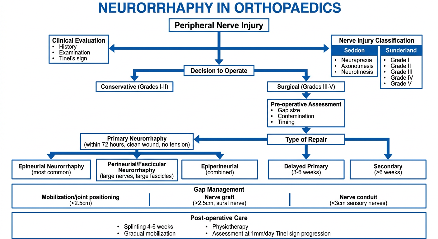
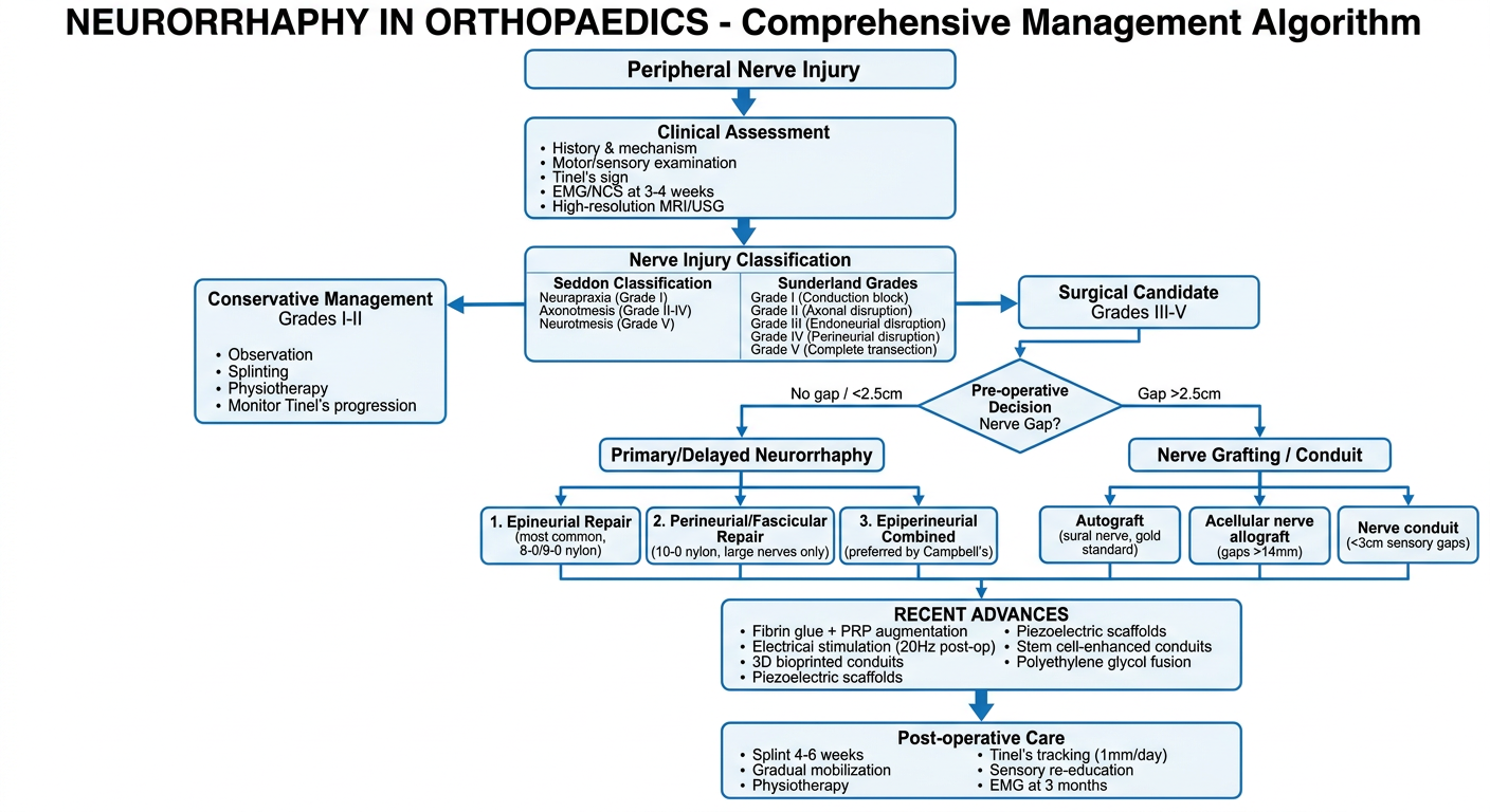
A detailed medical flowchart titled "NEURORRHAPHY IN ORTHOPAEDICS" showing the decision-making algorithm for peripheral nerve repair. The flowchart starts at the top with "Peripheral Nerve Injury" which branches into assessment steps: "Clinical Evaluation (History, Examination, Tinel's sign)" then "Nerve Injury Classification" showing Seddon (Neurapraxia, Axonotmesis, Neurotmesis) and Sunderland (Grade I-V). This flows to "Decision to Operate" with branches for "Conservative (Grades I-II)" and "Surgical (Grades III-V)". The surgical branch leads to "Pre-operative Assessment: Gap size, Contamination, Timing". Then to "Type of Repair" with three branches: "Primary Neurorrhaphy (within 72 hours, clean wound, no tension)", "Delayed Primary (3-6 weeks)", and "Secondary (>6 weeks)". The primary neurorrhaphy branch splits into "Epineurial Neurorrhaphy (most common)" and "Perineurial/Fascicular Neurorrhaphy (large nerves, large fascicles)" and "Epiperineurial (combined)". Below this shows "Gap Management" with options: "Mobilization/joint positioning (<2.5cm)", "Nerve graft (>2.5cm, sural nerve)", "Nerve conduit (<3cm sensory nerves)". Final box shows "Post-operative Care: Splinting 4-6 weeks, Gradual mobilization, Physiotherapy, Assessment at 1mm/day Tinel sign progression". Clean medical diagram style with blue and white color scheme.
NEURORRHAPHY IN ORTHOPAEDICS
DEFINITION
FLOWCHART: DECISION ALGORITHM FOR NEURORRHAPHY
CLASSIFICATION OF NERVE INJURIES (Prerequisite to Repair Decision)
| Seddon | Sunderland | Pathology | Spontaneous Recovery |
|---|---|---|---|
| Neurapraxia | Grade I | Conduction block only | Complete, weeks |
| Axonotmesis | Grade II | Axon disrupted, endoneurium intact | Yes, 1 mm/day |
| Axonotmesis | Grade III | Endoneurium disrupted | Partial |
| Axonotmesis | Grade IV | Perineurium disrupted | None without surgery |
| Neurotmesis | Grade V | Complete nerve division | None without surgery |
TIMING OF REPAIR
| Type | Timing | Indication |
|---|---|---|
| Primary | Within 6-8 hours | Clean sharp wounds, no tension, healthy tissue |
| Delayed Primary | 3-6 weeks | Contaminated or crush wounds; allows demarcation of viable tissue |
| Secondary | >3-6 months | Missed injuries, failed primaries |
PREREQUISITES FOR NEURORRHAPHY
- Clean, well-vascularized wound bed
- No tension at the repair site
- Healthy nerve ends (confirmed by serial cuts until normal fasciculi are visible under microscope)
- Use of magnification (operating microscope or loupes)
- Meticulous hemostasis
- Non-reactive, non-absorbable suture material (monofilament nylon)
TYPES OF NEURORRHAPHY
1. EPINEURIAL NEURORRHAPHY (Most Common)
- Expose and dissect the nerve ends; confirm the gap can be closed without tension
- Resect the neuroma/glioma with a sharp razor blade or diamond-bladed knife against a sterile wooden tongue depressor (nerve miter box)
- Make serial 1-mm cuts until normal fasciculi are visible under the operating microscope
- Control bleeding with thrombin or gelatin sponges
- Determine rotational alignment using surface vessels and fascicular patterns; place epineurial orientation sutures 1 cm from each cut edge
- Place a rubber/plastic background under the nerve for contrast
- Place first suture on the deep (posterior) surface of the epineurium and leave it long for easier rotation
- Place sutures in all four quadrants, then add sufficient interrupted 8-0 or 9-0 monofilament nylon to complete the repair
- Before closure, assess tension through range of motion to guide postoperative mobilization
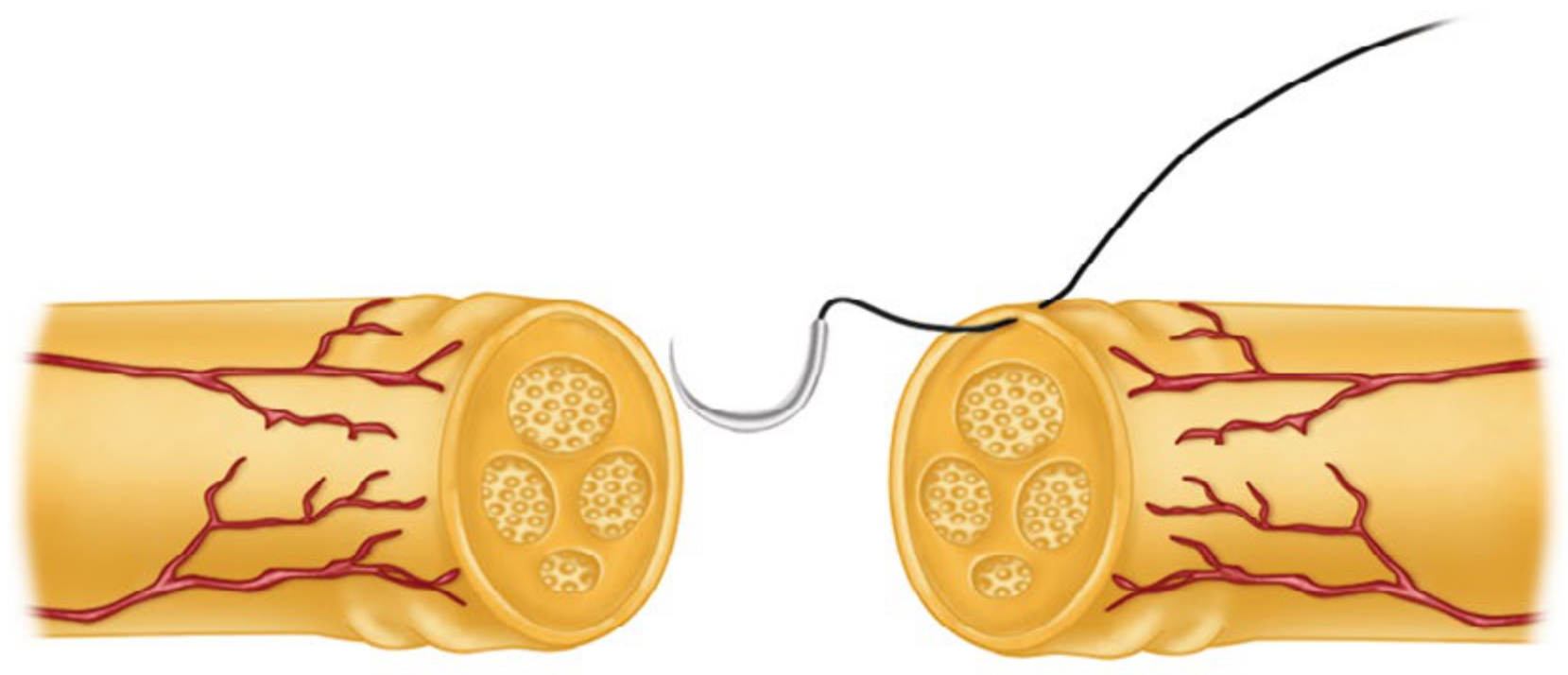
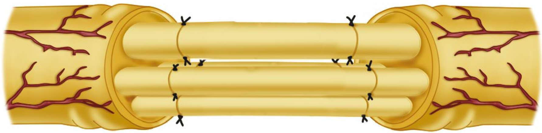
A: Nerve ends trimmed, fascicles identified. B: Epineurial suture placed through matching fascicular site. C: Repair completed.
2. PERINEURIAL (FASCICULAR) NEURORRHAPHY
- Surgeon must be proficient with the operating microscope and 10-0 suture
- Resect nerve ends as for epineurial repair
- Place nerve ends in proper rotational alignment
- Under magnification, identify corresponding fascicular groups in proximal and distal stumps (diagram the arrangement on sterile paper)
- Incise the epineurium longitudinally to expose fasciculi
- Approximate corresponding fasciculi individually with interrupted 9-0 or 10-0 nylon sutures
- Typically 2 sutures per fascicle are sufficient
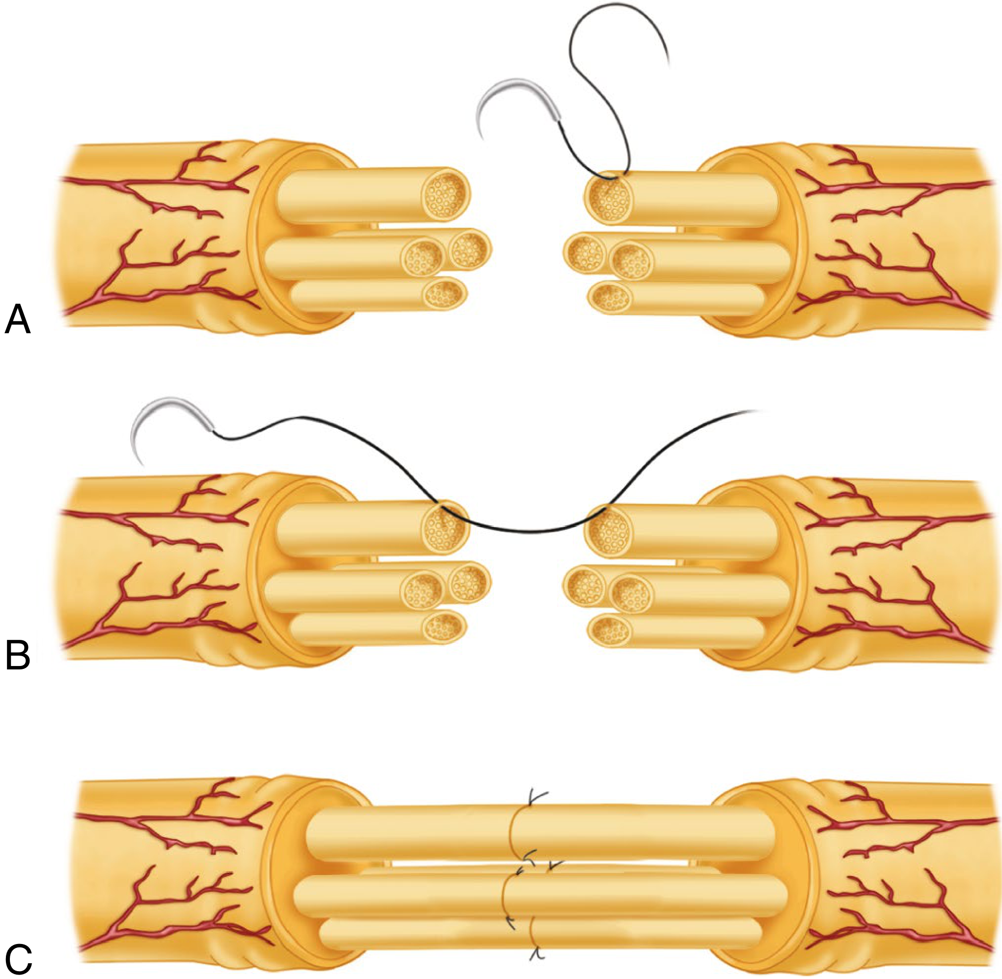
A: Epineurium excised, fascicles exposed. B: Suture passed through corresponding fascicles on either side. C: Neurorrhaphy completed, usually with two 10-0 nylon sutures per fascicle.
3. EPIPERINEURIAL (COMBINED) NEURORRHAPHY
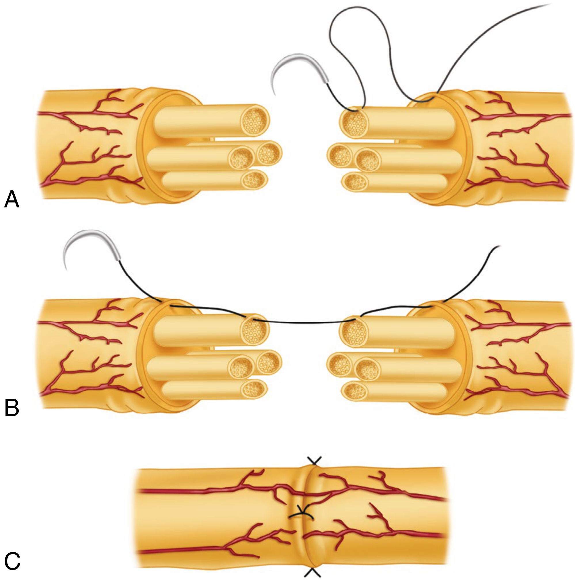
A: Epineurium excised/retracted; sutures placed through epineurium near large peripheral fascicle and through its perineurium. B: Suture passed through matching fascicle on opposite cut surface. C: Repair completed.
4. PARTIAL NEURORRHAPHY
- If >50% of a large nerve (especially sciatic or brachial plexus) is disrupted - partial neurorrhaphy is advisable
- If stimulation of remaining fascicles shows good motor response, do NOT risk injuring intact fascicles (e.g., peroneal or ulnar) just to restore sensation
GAP MANAGEMENT
| Gap Size | Method |
|---|---|
| <2.5 cm | Nerve mobilization, joint flexion positioning |
| >2.5 cm (motor nerves) | Nerve grafting (interfascicular - sural nerve donor) |
| <3 cm (sensory nerves) | Nerve conduit (collagen tube) |
| >14 mm | Acellular nerve allograft outperforms conduits |
SUTURE MATERIALS
| Technique | Suture Size |
|---|---|
| Epineurial (large nerves) | 8-0 monofilament nylon |
| Epineurial (wrist/hand level) | 9-0 monofilament nylon (optimal tension) |
| Perineurial (fascicular) | 9-0 or 10-0 monofilament nylon |
POSTOPERATIVE CARE
- Immobilization: Plaster splint/cast for 4-6 weeks
- Upper extremity: posterior molded plaster splint x 4 weeks
- Shoulder involvement: Velpeau dressing + plaster
- Lower extremity (sciatic/peroneal): spica cast x 6 weeks minimum
- Wound review: Day 7-10, suture removal
- Mobilization: Gradual extension over 2-3 weeks after cast removal
- Lower limb: Long leg brace controlling knee extension after cast removal
- Physiotherapy: Active and passive range of motion; sensory re-education
FACTORS AFFECTING RESULTS
| Favorable | Unfavorable |
|---|---|
| Young age | Delayed repair (>3 months) |
| Distal nerve level | Proximal injury (long regeneration distance) |
| Sharp clean injury | Avulsion/crush injury |
| Early repair | Contaminated wound |
| Good fascicular matching | Poor rotational alignment |
| Tension-free repair | Tension at repair site |
KEY PRINCIPLES SUMMARY
- Tension-free repair is the single most important technical principle
- Fascicular alignment prevents wasteful misdirection of regenerating axons (around 50% of axons fail to cross even a well-performed repair site)
- Magnification (operating microscope) is mandatory for all neurorrhaphy
- Monofilament non-absorbable nylon remains the gold standard suture
- Literature has NOT shown perineurial repair to be superior to epineurial repair; at most institutions, epineurial repair is preferred to limit suture bulk trauma
- Sunderland's conditions for fascicular repair: (1) large enough fascicular groups, (2) wasteful regeneration likely with epineurial repair, (3) fascicular groups occupy constant positions (median/ulnar at wrist, radial at elbow)
Include recent advances and generate new one
neurorrhaphy peripheral nerve repair recent advances
nerve repair conduit scaffold regeneration orthopaedic
recent advances neurorrhaphy nerve repair 2023 2024 2025 fibrin glue electrical stimulation polyethylene glycol
PMID: 41948775
https://link.springer.com/article/10.1186/s12967-025-07567-z
PMID: 39025114
A detailed professional medical flowchart titled "NEURORRHAPHY IN ORTHOPAEDICS - Comprehensive Management Algorithm" with clean clinical design. TOP: "Peripheral Nerve Injury" box ↓ "Clinical Assessment" box with bullet points: History & mechanism, Motor/sensory examination, Tinel's sign, EMG/NCS at 3-4 weeks, High-resolution MRI/USG ↓ "Nerve Injury Classification" box showing two columns: LEFT: Seddon Classification - Neurapraxia (Grade I), Axonotmesis (Grade II-IV), Neurotmesis (Grade V). RIGHT: Sunderland Grades I through V with brief description ↓ splits into two branches: LEFT branch: "Conservative Management" for Grades I-II: Observation, Splinting, Physiotherapy, Monitor Tinel's progression RIGHT branch: "Surgical Candidate" for Grades III-V RIGHT branch continues ↓ "Pre-operative Decision" diamond: "Nerve Gap?" - "No gap / <2.5cm" → "Primary/Delayed Neurorrhaphy" - "Gap >2.5cm" → "Nerve Grafting / Conduit" "Primary/Delayed Neurorrhaphy" splits into 3 boxes: 1. Epineurial Repair (most common, 8-0/9-0 nylon) 2. Perineurial/Fascicular Repair (10-0 nylon, large nerves only) 3. Epiperineurial Combined (preferred by Campbell's) "Nerve Grafting/Conduit" splits into: - Autograft (sural nerve, gold standard) - Acellular nerve allograft (gaps >14mm) - Nerve conduit (<3cm sensory gaps) Both merge into "RECENT ADVANCES" box containing: Fibrin glue + PRP augmentation, Electrical stimulation (20Hz post-op), 3D bioprinted conduits, Piezoelectric scaffolds, Stem cell-enhanced conduits, Polyethylene glycol fusion ↓ Final box: "Post-operative Care" - Splint 4-6 weeks, Gradual mobilization, Physiotherapy, Tinel's tracking (1mm/day), Sensory re-education, EMG at 3 months Blue and white color scheme, medical diagram style, clear arrows, professional typography
NEURORRHAPHY IN ORTHOPAEDICS
10-Mark PG Answer with Recent Advances
COMPREHENSIVE MANAGEMENT FLOWCHART
1. DEFINITION
2. NERVE INJURY CLASSIFICATION
| Seddon | Sunderland | Pathology | Recovery |
|---|---|---|---|
| Neurapraxia | Grade I | Conduction block; no structural damage | Spontaneous, weeks |
| Axonotmesis | Grade II | Axon disrupted; endoneurium intact | Spontaneous, 1 mm/day |
| Axonotmesis | Grade III | Endoneurium disrupted | Partial, may need surgery |
| Axonotmesis | Grade IV | Perineurium disrupted | No spontaneous; surgery needed |
| Neurotmesis | Grade V | Complete division | No spontaneous; surgery needed |
Neurorrhaphy is indicated for Sunderland Grade III-V injuries.
3. DIAGNOSIS AND ASSESSMENT
- Clinical: Motor/sensory deficit, Tinel's sign progression (1 mm/day = healthy regeneration)
- EMG/NCS: At 3-4 weeks to confirm denervation and assess prognosis
- Imaging: High-resolution MRI neurography and ultrasound - can identify neuroma-in-continuity, scarring, and gap length
4. TIMING OF NEURORRHAPHY
| Type | Timing | Indication |
|---|---|---|
| Primary | Within 6-8 hours | Clean sharp wound, tension-free, healthy tissue |
| Delayed primary | 3-6 weeks | Contaminated/crush wounds; allows tissue demarcation |
| Secondary | 3-6 months | Missed injuries, failed primary repairs |
5. PREREQUISITES FOR NEURORRHAPHY
- Clean, well-vascularized wound bed
- No tension at repair site
- Healthy nerve ends confirmed by serial 1-mm cuts under operating microscope
- Magnification (operating microscope or minimum 3.5x loupes)
- Non-reactive, non-absorbable monofilament nylon suture
- Meticulous hemostasis
6. TYPES OF NEURORRHAPHY (Surgical Techniques)
A. EPINEURIAL NEURORRHAPHY (Most Common)
- Expose nerve, confirm tension-free closure is possible
- Resect neuroma/glioma: serial 1-mm cuts with razor blade or diamond knife in a nerve miter box until normal fasciculi are visible
- Achieve rotational alignment using surface vessels and fascicular patterns; place epineurial orientation sutures 1 cm proximal and distal to cut
- Place rubber/plastic background beneath nerve for suture contrast
- Begin with a deep (posterior) epineurial suture (8-0 or 9-0 nylon), leave long for rotation
- Place sutures in all 4 quadrants, then add sufficient interrupted sutures for a watertight repair
- Assess repair tension through range of motion before wound closure
B. PERINEURIAL (FASCICULAR) NEURORRHAPHY
- Requires operating microscope proficiency and ease with 10-0 suture
- Resect nerve ends as above
- Diagram fascicular arrangement on sterile paper
- Incise epineurium longitudinally to expose fasciculi
- Approximate corresponding fasciculi with interrupted 9-0 or 10-0 nylon (2 sutures per fascicle)
C. EPIPERINEURIAL (COMBINED) NEURORRHAPHY
Literature has not shown perineurial repair to be superior to epineurial repair. Epineurial repair is preferred at most centers to limit foreign material inside the nerve.
D. PARTIAL NEURORRHAPHY
7. GAP MANAGEMENT
| Gap | Method |
|---|---|
| < 2.5 cm | Nerve mobilization + joint flexion positioning |
| > 2.5 cm (motor nerves) | Interfascicular nerve grafting (sural nerve) |
| < 3 cm (sensory nerves) | Nerve conduit (collagen or synthetic tube) |
| Gaps < 14 mm | Conduit achieves MRC S3+ in 67% of digital nerve reconstructions |
| Gaps > 14 mm | Acellular nerve allograft outperforms synthetic conduits |
8. SUTURE MATERIALS AND SIZE
| Level | Suture |
|---|---|
| Large nerve trunks (epineurial) | 8-0 monofilament nylon |
| Wrist and hand level | 9-0 monofilament nylon (optimal tension setting) |
| Fascicular repair | 9-0 or 10-0 monofilament nylon |
9. POSTOPERATIVE CARE
- Immobilization: 4 weeks for upper extremity; 6 weeks spica cast for sciatic/peroneal repairs
- Wound review: Day 7-10; suture removal
- Mobilization: Gradual extension over 2-3 weeks after cast/splint removal; long leg brace for lower limb
- Physiotherapy: Active-passive ROM, sensory re-education, desensitization
- Monitoring: Tinel's progression monthly (1 mm/day), EMG at 3 months
10. RECENT ADVANCES (2023-2026)
A. FIBRIN GLUE NEURORRHAPHY + PRP AUGMENTATION
- Fibrin glue neurorrhaphy (FGN) achieves comparable motor and sensory recovery to microsuture neurorrhaphy
- Shorter operative time and simpler technical execution
- When FGN is combined with platelet-rich plasma (PRP): faster functional recovery with no major complications
- PRP provides neuroprotective, anti-inflammatory, and neurotrophic support for axonal regeneration
B. ELECTRICAL STIMULATION (ES) - MOST PROMISING ADVANCE
- Brief intraoperative electrical stimulation (20 Hz, 1 hour, immediately post-neurorrhaphy) accelerates axonal sprouting and Schwann cell activity
- Stimulation promotes: increased Schwann cell proliferation, neurotrophic factor secretion, myelin production, and vascular growth
- Wireless piezoelectric scaffolds (PVDF-TrFE nanofibers): deliver localized electrical stimulation non-invasively without implanted batteries
- Graphene-based conductive scaffolds combined with ES show superior regeneration in 15-mm defect models
- Bordett et al. 2024: Ultrasound, laser, and magnetic stimulation are also being explored as adjuncts
C. ADVANCED NERVE CONDUITS AND SCAFFOLDS
| Generation | Material | Advantage |
|---|---|---|
| 1st gen | Silicone tubes | Mechanical protection only |
| 2nd gen | Collagen conduits (current standard) | Biodegradable, FDA-approved for <3 cm sensory gaps |
| 3rd gen (emerging) | Hyaluronic acid scaffolds | Aligned channels, anti-scarring properties |
| Cutting-edge | 3D bioprinted patient-specific conduits | Custom geometry, controlled porosity, dual electroactive properties |
- Hyaluronic acid-based conduits (2025): mechanistic advances in mimicking native extracellular matrix to guide axons
- Auxilium NeuroSpan Bridge trial (enrolled May 2025): scaffold-based peripheral nerve regeneration for long-gap injuries
D. STEM CELL-ENHANCED REPAIR
- Adipose-derived stem cells (ADSCs) embedded in polycaprolactone conduits: prevents muscle atrophy and restores innervation in 6-mm sciatic nerve defects
- iPSC-derived mesenchymal stem cells in 3D bioengineered conduits: superior morphology, function, and neurotrophic factor expression vs. silicone tubes
- Stem cell exosomes (cell-free approach): reduce risks of live cell transplantation while preserving neurotrophic and immunomodulatory effects
E. POLYETHYLENE GLYCOL (PEG) AXONAL FUSION
- PEG applied within 24 hours of sharp nerve transection can fuse divided axons directly, preventing Wallerian degeneration
- Ghergherehchi et al. (2023): PEG-fused hydrogel on viable transected nerve ends showed encouraging early results
- Less scarring than fibrin glue; comparable tensile strength
- Limitation: degrades slowly (up to 20 months at implant site); pressure on surrounding vasculature due to swelling
F. GENE THERAPY
- Targeted viral vector delivery of BDNF, GDNF, and NT-3 genes to Schwann cells to sustain neurotrophic support during long regeneration periods
- Genetic modification of Schwann cell phenotype to remain in a regeneration-supportive state for longer
- Currently pre-clinical only
G. IMPROVED IMAGING FOR DIAGNOSIS AND SURGICAL PLANNING
- High-resolution MRI neurography: identifies neuroma-in-continuity, fascicular injury patterns, and gap length preoperatively
- Intraoperative ultrasound: real-time fascicular mapping during repair
- AI-assisted image analysis: early identification of reinnervation patterns and treatment response monitoring
11. FACTORS AFFECTING OUTCOME
| Favorable | Unfavorable |
|---|---|
| Young age | Delayed repair (>3 months) |
| Distal level (hand/wrist) | Proximal injury (long regeneration to target) |
| Sharp clean transection | Avulsion / crush mechanism |
| Tension-free repair | Tension at repair site |
| Good fascicular alignment | Poor rotational orientation |
| Early physiotherapy | Muscle denervation >12-18 months |
| Use of PRP/ES adjuncts (emerging) | Heavy contamination |
12. SUMMARY TABLE - NEURORRHAPHY TYPES AT A GLANCE
| Type | Suture Layer | Suture Size | Best For |
|---|---|---|---|
| Epineurial | Epineurium only | 8-0 / 9-0 | Most peripheral nerves |
| Perineurial | Perineurium of fascicles | 10-0 | Large fascicular nerves (median/ulnar at wrist) |
| Epiperineurial | Both layers | 8-0 + 10-0 | Campbell's preferred technique |
| Partial | Partial nerve | As above | Partial nerve injuries (sciatic, brachial plexus) |
Key take-home points for PG exam:
- Tension-free repair is the single most important technical factor
- ~50% of regenerating axons fail to cross even a technically perfect repair site - this is why fascicular alignment matters
- Literature does NOT show fascicular repair superior to epineurial repair in clinical outcomes
- Fibrin glue + PRP = comparable outcomes to microsuture with shorter OR time (2026 systematic review)
- Brief intraoperative electrical stimulation (20 Hz, 1 hr) is the most promising emerging adjunct
- Axonal regeneration = 1 mm/day; track with Tinel's sign progression
Neurotization in orthopaedics 10 marks pg answer with flow chart and images
neurotization nerve transfer peripheral nerve orthopaedic
PMID: 38296247
neurotization nerve transfer recent advances 2024 2025 upper limb brachial plexus spinal cord injury
distal nerve transfer spinal cord injury tetraplegia upper extremity
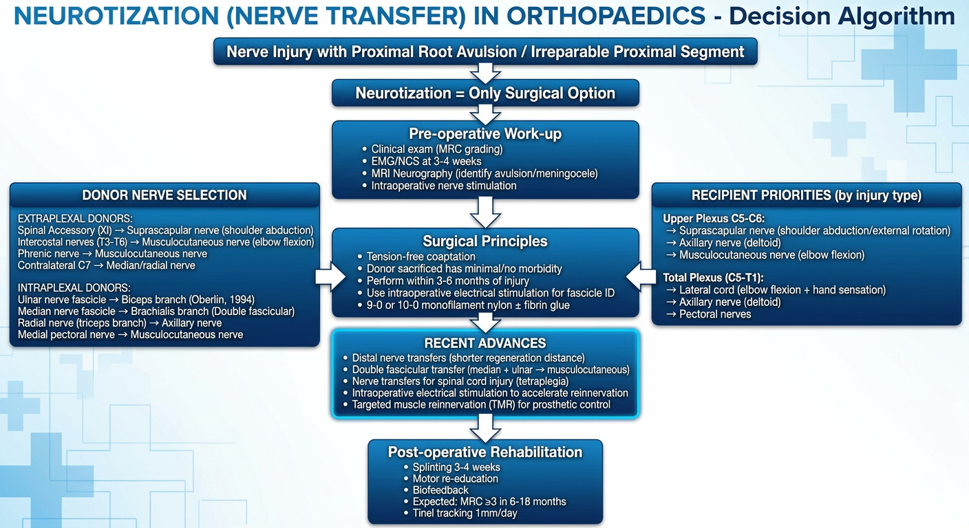
A comprehensive professional medical flowchart titled "NEUROTIZATION (NERVE TRANSFER) IN ORTHOPAEDICS - Decision Algorithm" in blue and white clinical style. TOP BOX: "Nerve Injury with Proximal Root Avulsion / Irreparable Proximal Segment" → "Neurotization = Only Surgical Option" ↓ "Pre-operative Work-up" box: Clinical exam (MRC grading), EMG/NCS at 3-4 weeks, MRI Neurography (identify avulsion/meningocele), Intraoperative nerve stimulation ↓ splits into two columns: LEFT COLUMN titled "DONOR NERVE SELECTION" listing: EXTRAPLEXAL DONORS: - Spinal Accessory (XI) → Suprascapular nerve (shoulder abduction) - Intercostal nerves (T3-T6) → Musculocutaneous nerve (elbow flexion) - Phrenic nerve → Musculocutaneous nerve - Contralateral C7 → Median/radial nerve INTRAPLEXAL DONORS: - Ulnar nerve fascicle → Biceps branch (Oberlin, 1994) - Median nerve fascicle → Brachialis branch (Double fascicular) - Radial nerve (triceps branch) → Axillary nerve - Medial pectoral nerve → Musculocutaneous nerve RIGHT COLUMN titled "RECIPIENT PRIORITIES (by injury type)": Upper Plexus C5-C6: → Suprascapular nerve (shoulder abduction/external rotation) → Axillary nerve (deltoid) → Musculocutaneous nerve (elbow flexion) Total Plexus (C5-T1): → Lateral cord (elbow flexion + hand sensation) → Axillary nerve (deltoid) → Pectoral nerves ↓ MERGE TO CENTRAL BOX: "Surgical Principles" • Tension-free coaptation • Donor sacrificed has minimal/no morbidity • Perform within 3-6 months of injury • Use intraoperative electrical stimulation for fascicle ID • 9-0 or 10-0 monofilament nylon ± fibrin glue ↓ "RECENT ADVANCES" highlighted box: • Distal nerve transfers (shorter regeneration distance) • Double fascicular transfer (median + ulnar → musculocutaneous) • Nerve transfers for spinal cord injury (tetraplegia) • Intraoperative electrical stimulation to accelerate reinnervation • Targeted muscle reinnervation (TMR) for prosthetic control ↓ FINAL BOX: "Post-operative Rehabilitation" Splinting 3-4 weeks, Motor re-education, Biofeedback, Expected: MRC ≥3 in 6-18 months, Tinel tracking 1mm/day Clean medical diagram, professional typography, blue gradient boxes, white arrows
NEUROTIZATION IN ORTHOPAEDICS
10-Mark PG Answer with Flowchart, Images & Recent Advances
COMPREHENSIVE DECISION FLOWCHART
1. DEFINITION
- The term "neurotization" was introduced to describe reinnervation of a denervated muscle by a functional nerve source
- It is the procedure of choice when the proximal nerve stump is unavailable (root avulsion) or too damaged for direct repair or grafting
| Nerve Grafting | Neurotization | |
|---|---|---|
| Proximal stump | Available | Unavailable (avulsion) |
| Continuity | Restores original nerve | Creates new nerve pathway |
| Donor | Expendable sensory nerve (sural) | Functional motor/mixed nerve |
| Gap | Bridges gap | No gap - direct coaptation |
2. INDICATIONS
- Brachial plexus root avulsion - the most common indication; roots torn from spinal cord (preganglionic injury), making nerve repair impossible
- Irreparable proximal nerve injury with long gap not amenable to grafting
- Proximal nerve injury where regeneration distance would be too long for meaningful recovery
- Spinal cord injury (tetraplegia) - nerve transfers to restore hand function (emerging indication)
- Obstetric brachial plexus palsy in children not responding to conservative management
3. PRE-OPERATIVE EVALUATION
| Investigation | Purpose |
|---|---|
| Clinical exam (MRC grading) | Baseline motor/sensory mapping |
| EMG/NCS at 3-4 weeks | Confirm denervation; identify intact donor fascicles |
| MRI cervical spine (T2 coronal/axial) | Identify root avulsion, pseudomeningocele, rootlet absence (sensitivity 81-93%) |
| CT myelography | Gold standard for root avulsion if MRI equivocal |
| Intraoperative nerve stimulation | Identify functional fascicles in donor nerve for selective harvest |
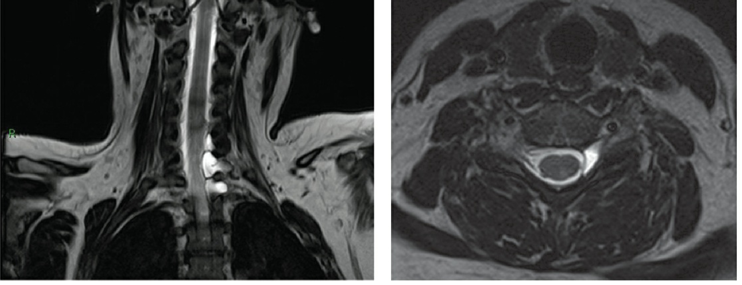
Coronal (left) and axial (right) T2 MRI: ventral/dorsal rootlets visible on normal side; absent on avulsed side with meningoceles in foramina - pathognomonic of preganglionic root avulsion.
4. CLASSIFICATION OF DONOR NERVES
A. EXTRAPLEXAL DONORS
| Donor Nerve | Recipient | Function Restored | Notes |
|---|---|---|---|
| Spinal accessory (CN XI) | Suprascapular nerve | Shoulder abduction + external rotation | Most reliable extraplexal donor; 95% improvement in some series |
| Intercostal nerves (T3-T6) | Musculocutaneous nerve | Elbow flexion | Direct suture to musculocutaneous restores grade 3 flexion in 80% within 6 months |
| Phrenic nerve | Musculocutaneous nerve | Elbow flexion | Significant respiratory morbidity; now rarely used |
| Contralateral C7 | Median or radial nerve | Wrist/finger function | Long regeneration distance; used for total plexus injuries in select centers |
B. INTRAPLEXAL DONORS
| Donor | Recipient | Procedure | Function Restored |
|---|---|---|---|
| Ulnar nerve fascicle (FCU branch) | Nerve to biceps | Oberlin transfer (1994) | Elbow flexion |
| Median nerve fascicle (FDS branch) | Nerve to brachialis | Double fascicular transfer | Elbow flexion (combined with Oberlin) |
| Radial nerve (triceps branch, long head) | Axillary nerve | Leechavengvongs transfer | Shoulder abduction (deltoid) |
| Medial pectoral nerve | Musculocutaneous nerve | Pectoral to MCN transfer | Elbow flexion |
| Thoracodorsal nerve | Musculocutaneous nerve | Alternative for C5-C6 injuries | Elbow flexion |
5. SPECIFIC TECHNIQUES
THE OBERLIN TRANSFER (Technique 67.4 - Campbell's)
- Anterior arm incision, 8-10 cm, 4 cm distal to pectoralis major insertion
- Incise fascia; retract biceps laterally to expose musculocutaneous nerve and its branch to biceps
- Identify ulnar nerve in the brachial canal
- Under electrical stimulation, identify and isolate the fascicle innervating flexor carpi ulnaris (FCU) from the ulnar nerve - FCU function is sacrificed but compensated by other wrist flexors
- Divide selected ulnar fascicle; coapt directly to the nerve to biceps with 9-0 nylon + fibrin glue
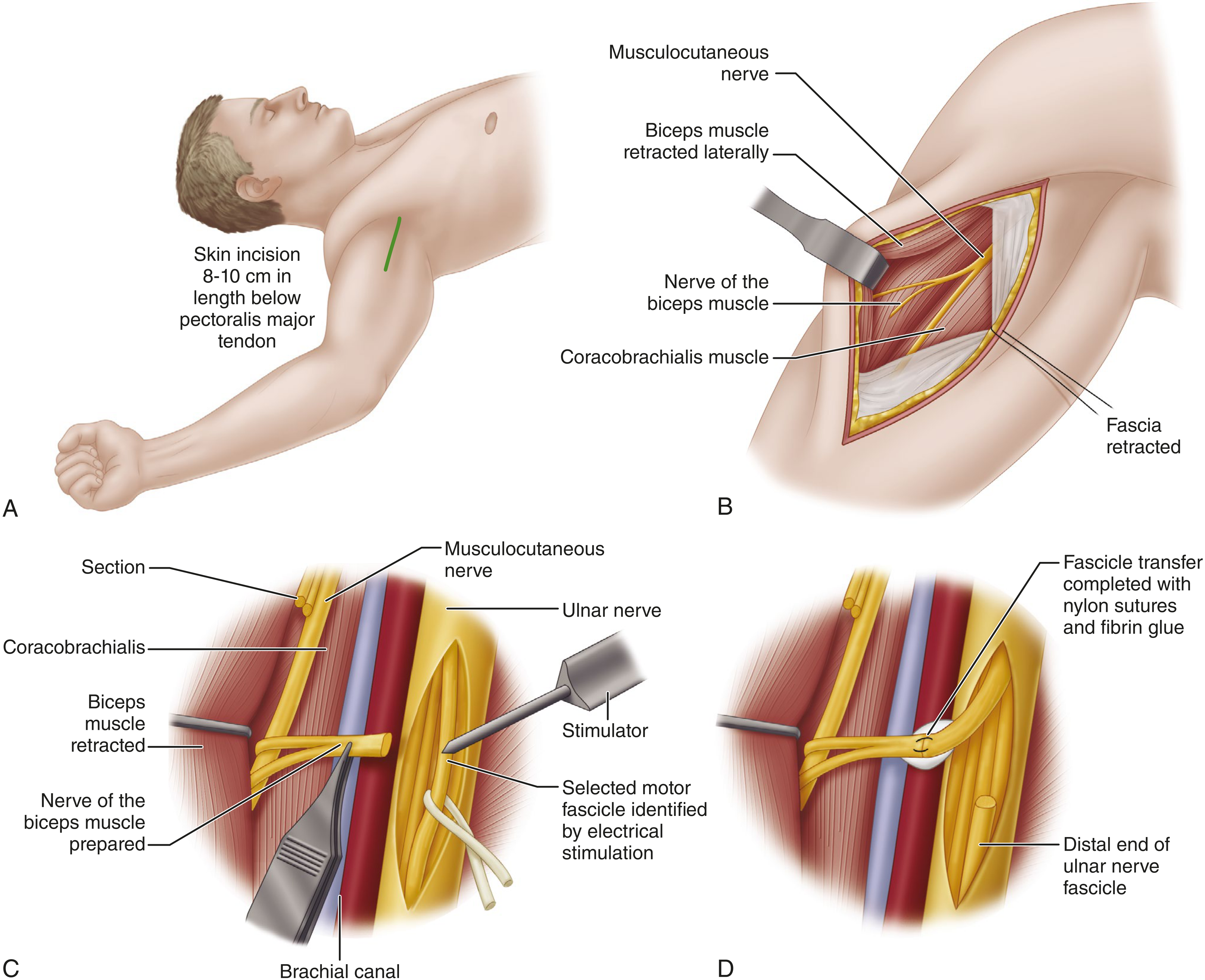
A: Skin incision 8-10 cm below pectoralis major. B: Musculocutaneous nerve + nerve to biceps exposed with biceps retracted laterally. C: Ulnar nerve isolated; selected motor fascicle identified by electrical stimulator. D: Fascicle transfer completed with nylon sutures + fibrin glue.
DOUBLE FASCICULAR TRANSFER
- Combines Oberlin transfer (ulnar fascicle → nerve to biceps) with median nerve fascicle (FDS branch → nerve to brachialis)
- Provides dual innervation to elbow flexors
- Produces stronger and more reliable elbow flexion than single fascicular transfer
SPINAL ACCESSORY → SUPRASCAPULAR TRANSFER
- Spinal accessory nerve divided at its entry into trapezius and sutured to suprascapular nerve
- Mean shoulder abduction of 115 degrees after combined accessory to suprascapular + long head of triceps to axillary nerve transfers (Leechavengvongs et al.)
INTERCOSTAL → MUSCULOCUTANEOUS TRANSFER
- T3-T6 intercostal nerves harvested at the midaxillary line
- Direct suture to musculocutaneous nerve
- At least MRC grade 3 elbow flexion in 80% of patients operated within 6 months of injury
6. SURGICAL PRINCIPLES
- Timing: Ideally within 3-6 months of injury; denervation changes become irreversible beyond 12-18 months
- Tension-free coaptation: Direct end-to-end suture; no grafts required as donor is brought directly to recipient
- Donor morbidity: Must be minimal - function lost from donor is compensated by overlapping muscles or sensory territories
- Fascicular selectivity: Use intraoperative electrical stimulation to identify pure motor fascicles in donor nerve
- Magnification: Operating microscope mandatory; 9-0 or 10-0 nylon suture ± fibrin glue
- Proximity: Donor-to-target muscle distance must allow reinnervation before irreversible atrophy - this is the core advantage of distal nerve transfers
7. PRIORITY OF RECONSTRUCTION (Brachial Plexus)
| Injury Level | Priority 1 | Priority 2 | Priority 3 |
|---|---|---|---|
| Upper (C5-C6) | Elbow flexion (MCN) | Shoulder abduction (suprascapular) | External rotation (axillary) |
| Upper + C7 | Elbow flexion | Shoulder | Wrist/finger extension |
| Total (C5-T1) | Elbow flexion (lateral cord) | Sensation to hand | Deltoid / axillary nerve |
8. RESULTS
| Transfer | Outcome | % achieving MRC ≥3 |
|---|---|---|
| Ulnar fascicle → Biceps (Oberlin) | Elbow flexion | ~75-80% |
| Medial pectoral → MCN | Elbow flexion | ~85-90% |
| Accessory → Suprascapular | Shoulder abduction | ~95% |
| Intercostal → MCN | Elbow flexion | ~80% (within 6 months) |
| Triceps branch → Axillary | Deltoid | ~70-80% |
9. RECENT ADVANCES (2023-2026)
A. DISTAL NERVE TRANSFERS
- Coaptation near the muscle means reinnervation in weeks to months rather than years
- Bertelli et al. J Hand Surg Eur 2024: Notable progress in distal nerve transfer techniques markedly enhancing outcomes for peripheral nerve, brachial plexus, and spinal cord injuries
- Examples: PIN branch → AIN, radial sensory → median sensory, motor branches within the forearm
B. NERVE TRANSFERS FOR SPINAL CORD INJURY / TETRAPLEGIA
- Nerve transfers are now being used to restore upper limb function in C5-C6 tetraplegics who retain some intact motor axons below the injury
- Technique: transfer of paralyzed but re-educable muscles (e.g., posterior interosseous nerve branch) to restore wrist extension or hand grip
- van Zyl et al. (Lancet 2019, updated series 2024): expanding traditional tendon transfer techniques with nerve transfers in tetraplegia shows significant functional gains
- Combination of multiple nerve transfer procedures has proven to improve upper limb function in SCI (Nature Communications 2025)
C. INTRAOPERATIVE ELECTRICAL STIMULATION
- Brief intraoperative ES (20 Hz, 1 hour) immediately after neurotization accelerates axonal sprouting from donor stumps
- Promotes Schwann cell proliferation, BDNF release, and faster reinnervation of recipient muscle
- Now incorporated into brachial plexus programs at leading centers (WashU Medicine/Barnes-Jewish Hospital, 2025)
D. TARGETED MUSCLE REINNERVATION (TMR)
- Residual nerve stumps after amputation are transferred to nearby motor nerve branches
- Creates additional EMG signal sources to control myoelectric prostheses with greater degrees of freedom
- Neurotization here serves a dual purpose: eliminates neuroma pain AND enables intuitive prosthetic control
E. REGENERATIVE PERIPHERAL NERVE INTERFACE (RPNI)
- Residual nerve stumps embedded in free muscle grafts
- Provides an amplified EMG signal for prosthetic control
- Eliminates neuroma formation in amputees
F. AI-GUIDED SURGICAL PLANNING
- Machine learning analysis of MRI neurography and EMG data to predict which donor-recipient combinations will yield the best outcomes
- Intraoperative ultrasound for real-time fascicle identification and coaptation assessment
10. COMPLICATIONS
| Complication | Prevention/Management |
|---|---|
| Donor function loss | Select purely expendable fascicles; intraoperative stimulation |
| Co-contraction | Motor re-education, biofeedback therapy |
| Neuroma at coaptation | Tension-free repair, fibrin glue |
| Failure of reinnervation | Confirm diagnosis, re-explore if no Tinel's at 3 months |
| Phrenic nerve sacrifice (respiratory) | Avoid in bilateral phrenic use; check pulmonary function first |
11. FACTORS AFFECTING OUTCOME
| Favorable | Unfavorable |
|---|---|
| Surgery within 3-6 months | Delayed repair >12 months |
| Young age (<25 years) | Advanced age (poor CNS plasticity) |
| Distal coaptation (near target muscle) | Proximal coaptation (long regeneration) |
| Pure motor donor to motor recipient | Mixed fascicles, type mismatch |
| Good motor re-education post-op | Poor compliance with rehabilitation |
| Partial plexus injury | Total avulsion injury |
KEY TAKE-HOME POINTS FOR PG EXAM
- Neurotization = nerve transfer: donor nerve is functionally intact and sacrificable; recipient nerve has no available proximal stump (root avulsion)
- Oberlin transfer (1994) = ulnar nerve fascicle (FCU branch) → nerve to biceps = prototype modern neurotization
- Spinal accessory → suprascapular = most reliable for shoulder abduction (95% achieve MRC 3+)
- Intercostal → musculocutaneous = 80% achieve MRC 3+ elbow flexion if done within 6 months
- Key advantage of neurotization over grafting: no proximal stump needed; bypasses irreparable zone completely
- Distal nerve transfers are the most important recent advance - shorter regeneration distance = better and faster outcomes
- Nerve transfers are now expanding into spinal cord injury (tetraplegia) as an emerging frontier
Retry
A comprehensive professional medical flowchart titled "NEUROTIZATION (NERVE TRANSFER) IN ORTHOPAEDICS" with clean clinical blue and white design, clear boxes and arrows. START at top: Large box "PERIPHERAL NERVE INJURY WITH UNAVAILABLE PROXIMAL STUMP" with subtitle "Root Avulsion / Irreparable Proximal Segment" Arrow down to: "PRE-OPERATIVE ASSESSMENT" box listing: - Clinical exam + MRC grading - EMG/NCS at 3-4 weeks - MRI Neurography / CT Myelography - Intraoperative nerve stimulation Arrow down to diamond DECISION box: "TYPE OF INJURY?" Three arrows from diamond going to three boxes side by side: LEFT: "PARTIAL PLEXUS (C5-C6 / C5-C7)" MIDDLE: "TOTAL PLEXUS (C5-T1)" RIGHT: "ISOLATED NERVE INJURY" Below LEFT box, arrow to: "INTRAPLEXAL DONORS" box with list: • Ulnar fascicle → Biceps nerve (Oberlin) • Median fascicle → Brachialis (Double fascicular) • Triceps branch (radial) → Axillary nerve • Medial pectoral → Musculocutaneous Below MIDDLE box, arrow to: "EXTRAPLEXAL DONORS" box with list: • Spinal Accessory (XI) → Suprascapular • Intercostal T3-T6 → Musculocutaneous • Phrenic → Musculocutaneous (rarely) • Contralateral C7 → Median/Radial Below RIGHT box, arrow to: "TARGETED TRANSFER" box: • AIN/PIN branches • Distal upper limb transfers All three merge downward to: "SURGICAL PRINCIPLES" box: • Tension-free coaptation • Within 3-6 months of injury • Intraoperative electrical stimulation for fascicle ID • 9-0 / 10-0 nylon ± fibrin glue • Minimum donor morbidity Arrow down to: "RECENT ADVANCES" highlighted yellow/orange box: • Distal nerve transfers (near target = faster reinnervation) • Double fascicular transfer • Nerve transfers in tetraplegia (SCI) • Targeted Muscle Reinnervation (TMR) • Intraoperative electrical stimulation protocol Arrow down to final "POST-OP REHABILITATION" box: • Splinting 3-4 weeks • Motor re-education + biofeedback • Expected MRC ≥3 in 6-18 months • Tinel sign progression 1mm/day Professional medical infographic style, blue gradient header, white background, clean sans-serif font, color-coded sections
A detailed anatomical diagram showing the brachial plexus nerve transfer (neurotization) donor and recipient nerve map for the upper limb. Clean medical illustration style on white background. LEFT SIDE labeled "DONOR NERVES": - Spinal Accessory Nerve (CN XI) shown at top of neck with arrow labeled "→ Suprascapular nerve" - Intercostal nerves T3, T4, T5, T6 shown along ribcage with arrow "→ Musculocutaneous nerve" - Phrenic nerve shown with "→ Musculocutaneous (rarely)" - Contralateral C7 with dashed arrow "→ Median/Radial nerve" CENTER shows brachial plexus anatomy: C5, C6, C7, C8, T1 roots → Upper, Middle, Lower Trunks → Anterior/Posterior Divisions → Lateral, Posterior, Medial Cords → Terminal branches RIGHT SIDE labeled "INTRAPLEXAL TRANSFERS": - Ulnar nerve fascicle (FCU branch) → arrow to Nerve to Biceps "OBERLIN TRANSFER" - Median nerve fascicle (FDS branch) → Nerve to Brachialis "DOUBLE FASCICULAR" - Triceps branch of Radial nerve → Axillary nerve "LEECHAVENGVONGS" - Medial Pectoral → Musculocutaneous nerve Color coding: DONOR nerves in green, RECIPIENT nerves in red, TRANSFER arrows in blue dashed lines. Title at top in bold navy blue. Professional medical textbook illustration quality.
nerve transfer brachial plexus outcomes results recent
PMID: 42324199
PMID: 41855458
NEUROTIZATION IN ORTHOPAEDICS
10-Mark PG Answer — Flowchart, Images & Recent Advances
FLOWCHART
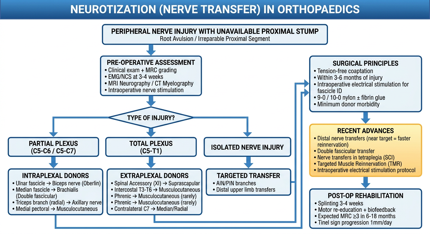
DONOR-RECIPIENT NERVE MAP
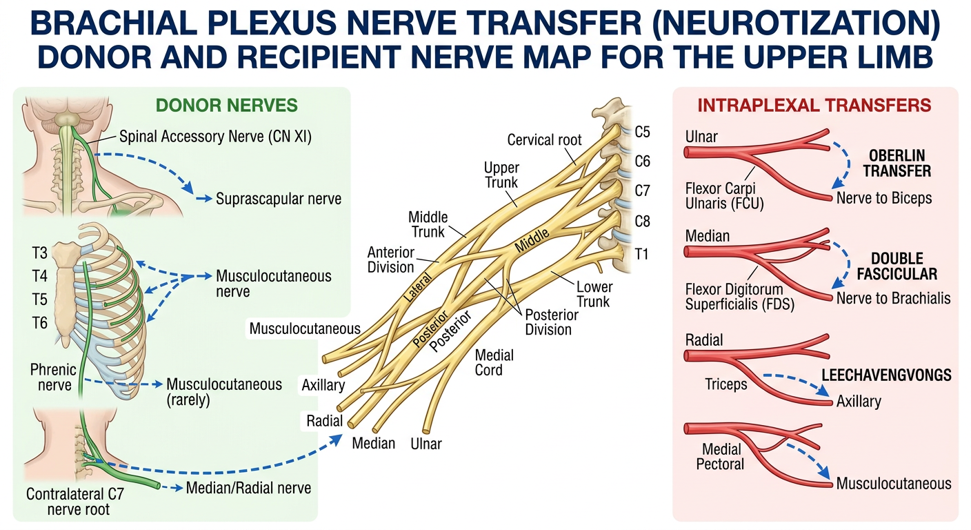
1. DEFINITION
| Feature | Nerve Grafting | Neurotization |
|---|---|---|
| Proximal stump | Available | Unavailable (avulsion) |
| What is transferred | Expendable sensory nerve | Functioning motor/mixed nerve |
| Mechanism | Bridges the gap | Creates entirely new nerve pathway |
| Donor sacrifice | Structural only | Functional (acceptable morbidity) |
2. INDICATIONS
- Brachial plexus root avulsion - the hallmark indication; roots torn from spinal cord (preganglionic injury), making any proximal repair impossible
- Irreparable long-gap proximal nerve injury where grafting distance is prohibitive
- Proximal nerve injury where regeneration distance to target muscle is so long that irreversible atrophy would occur before axons arrive
- Cervical spinal cord injury / tetraplegia - restoring elbow flexion, wrist extension, and hand grasp (expanding frontier)
- Brain injury with upper limb hemiplegia - contralateral C7 transfer (emerging)
- Obstetric brachial plexus palsy (OBPP) not recovering by 3-6 months
3. PRE-OPERATIVE ASSESSMENT
| Investigation | Finding and Purpose |
|---|---|
| Clinical exam + MRC grading | Baseline motor map; identify preserved function (guides donor selection) |
| EMG/NCS at 3-4 weeks | Confirms denervation in recipient muscles; fibrillations = denervation |
| MRI cervical spine (T2 coronal + axial) | Identifies absent rootlets, pseudomeningocele = root avulsion (sensitivity 81-93%) |
| CT myelography | Gold standard when MRI equivocal for avulsion |
| Intraoperative nerve stimulation | Real-time identification of pure motor fascicles in donor nerve |
| Horner's sign | Ptosis + miosis + anhidrosis = preganglionic lower plexus injury (C8/T1 avulsion) |
| Elevated hemidiaphragm on CXR | Phrenic nerve damage = C5 avulsion |
Left: Coronal T2 - rootlets visible on right, absent on left with meningoceles in foramina. Right: Axial T2 - asymmetric rootlet absence. These findings are pathognomonic of preganglionic avulsion, confirming neurotization as the only repair option.
4. CLASSIFICATION OF DONOR NERVES
A. EXTRAPLEXAL DONORS
| Donor Nerve | Recipient | Function Restored | Key Result |
|---|---|---|---|
| Spinal accessory (CN XI) | Suprascapular nerve | Shoulder abduction + external rotation | 95% improvement; most reliable extraplexal donor |
| Intercostal nerves T3-T6 | Musculocutaneous nerve | Elbow flexion | MRC ≥3 in 80% if done within 6 months |
| Phrenic nerve | Musculocutaneous nerve | Elbow flexion | Significant respiratory morbidity; rarely used now |
| Contralateral C7 | Median or radial nerve | Wrist / finger function | Used in total plexus; long regeneration, limited results |
B. INTRAPLEXAL DONORS
| Donor | Recipient | Eponym | Function Restored |
|---|---|---|---|
| Ulnar nerve fascicle (FCU branch) | Nerve to biceps | Oberlin transfer (1994) | Elbow flexion |
| Median nerve fascicle (FDS branch) | Nerve to brachialis | Double fascicular transfer | Elbow flexion (combined with Oberlin) |
| Radial nerve - long head triceps branch | Axillary nerve | Leechavengvongs transfer | Deltoid / shoulder abduction |
| Medial pectoral nerve | Musculocutaneous nerve | Pectoral transfer | Elbow flexion (85-90% MRC ≥3) |
| Thoracodorsal nerve | Musculocutaneous nerve | - | Elbow flexion (salvage) |
5. TECHNIQUES
THE OBERLIN TRANSFER (Campbell's Technique 67.4)
- Supine position; anterior arm incision 8-10 cm, 4 cm distal to pectoralis major insertion
- Incise biceps fascia; retract biceps laterally to expose musculocutaneous nerve and its branch to biceps muscle
- Identify ulnar nerve in the brachial canal medially
- Using intraoperative electrical stimulator, isolate the fascicle innervating flexor carpi ulnaris (FCU) - this is the sacrificed donor; FCU function is compensated by FCR and other wrist flexors
- Divide selected ulnar fascicle; coapt directly to nerve to biceps - no gap, no graft needed
- Secure with 9-0 nylon sutures + fibrin glue (shortened operative time, secure coaptation)
A: 8-10 cm incision on anterior arm below pectoralis major. B: Musculocutaneous nerve exposed; nerve to biceps identified; biceps retracted laterally. C: Ulnar nerve in brachial canal; electrical stimulator selects the FCU motor fascicle. D: Transfer completed - fascicle sutured with nylon and fibrin glue; distal end of ulnar fascicle coapted to nerve to biceps.
DOUBLE FASCICULAR TRANSFER
- Oberlin (ulnar fascicle → nerve to biceps) PLUS median nerve FDS branch → nerve to brachialis
- Provides dual innervation to both elbow flexors
- Superior, more reliable elbow flexion than single fascicular transfer
- Brachialis reinnervated separately = stronger combined flexion force
SPINAL ACCESSORY → SUPRASCAPULAR NERVE
- Accessory nerve divided at trapezius entry; coaptation to suprascapular nerve (which innervates supraspinatus + infraspinatus)
- Results: mean 115 degrees shoulder abduction after combined accessory to suprascapular + triceps branch to axillary nerve transfers (Leechavengvongs et al.)
- Caveat: A 2024 systematic review (Mendiratta et al., Front Pediatr) found that in brachial plexus birth injury, spinal accessory transfer showed no benefit over supraclavicular exploration + nerve grafting for shoulder abduction
INTERCOSTAL → MUSCULOCUTANEOUS
- T3-T6 intercostal nerves harvested at midaxillary line (to preserve length)
- 3-4 intercostals sutured directly to musculocutaneous nerve trunk
- MRC ≥3 elbow flexion in 80% if operated within 6 months (Rockwood & Green)
6. SURGICAL PRINCIPLES
- Timing: Operate within 3-6 months of injury - denervation changes irreversible by 12-18 months
- Tension-free coaptation: Direct end-to-end; no graft needed (donor brought to recipient)
- Donor morbidity: Must be minimal - any lost function must be compensated by adjacent muscles or overlapping territories
- Fascicular selectivity: Intraoperative electrical stimulation to identify pure motor fascicles - essential before sacrifice
- Magnification: Operating microscope mandatory; 9-0 or 10-0 monofilament nylon ± fibrin glue
- Distal preference: The closer to the target muscle, the shorter the regeneration distance - basis of distal nerve transfers
7. PRIORITIES BY INJURY PATTERN
| Injury Level | Priority 1 | Priority 2 | Priority 3 |
|---|---|---|---|
| Upper (C5-C6) | Elbow flexion (MCN) | Shoulder abduction (suprascapular) | External rotation (axillary) |
| Upper + C7 | Elbow flexion | Shoulder | Wrist/finger extension |
| Total (C5-T1) | Elbow flexion (lateral cord) | Hand sensation | Deltoid / axillary |
8. RESULTS SUMMARY
| Transfer | Target Function | % MRC ≥3 |
|---|---|---|
| Ulnar fascicle → Biceps (Oberlin) | Elbow flexion | 75-80% |
| Medial pectoral → MCN | Elbow flexion | 85-90% |
| Double fascicular (Oberlin + median) | Elbow flexion | ~90% |
| Spinal accessory → Suprascapular | Shoulder abduction | ~95% |
| Intercostal → MCN | Elbow flexion | 80% (within 6 months) |
| Triceps branch → Axillary | Deltoid | 70-80% |
9. RECENT ADVANCES (2023-2026)
A. DISTAL NERVE TRANSFERS - Dominant Paradigm Shift
- Dramatically reduces time to reinnervation (weeks to months vs. 12-18 months)
- Bertelli et al. J Hand Surg Eur 2024: "Recent years have witnessed notable progress in nerve transfer procedures, markedly enhancing outcomes of upper limb reconstruction for peripheral nerve, brachial plexus, and spinal cord injuries"
- Examples: PIN branch → AIN (wrist extension), radial sensory → median sensory, motor branches within forearm
B. NERVE TRANSFERS FOR CENTRAL NERVOUS SYSTEM INJURY
- Nerve transfer procedures can restore elbow flexion, wrist extension, and hand grasping functions in C5-C6 tetraplegics
- Efficacy depends on activation of plasticity in the spinal cord distal to the injury site
- Various intraplexal and extraplexal transfers have been adapted for SCI patients
- Xu et al. Chinese J Reconstr Surg 2026: "Nerve transfer is expanding from peripheral repair to induction of central plasticity - a new strategy for central upper limb paralysis"
- Contralateral C7 transfer: reduces muscle tone, improves partial motor function
- Limitations: improvement in fine motor skills and key muscle strength remains limited; maladaptive plasticity and co-activatory patterns persist
- Mechanisms involve interhemispheric reorganization and cortico-red nucleus-spinal pathway activation
C. BRAIN-COMPUTER INTERFACE (BCI) INTEGRATION
- BCI technology facilitates cortical remapping after nerve transfers by providing synchronized closed-loop feedback
- In BPI: BCI focuses on in-situ muscle activation via "neural bypass" to prevent disuse atrophy and restore agency
- BCI-mediated neuromodulation shows potential in alleviating chronic deafferentation pain by down-regulating pathological cortical hyperexcitability
- Song et al. Ann Med 2026: Emerging conductive hydrogels + hybrid BCI systems addressing signal stability
D. TARGETED MUSCLE REINNERVATION (TMR)
- Residual nerve stumps after amputation transferred to nearby motor nerve branches
- Dual purpose: eliminates neuroma pain AND creates additional EMG signal sources for myoelectric prosthetic control
- Greater degrees of freedom in prosthetic limb control
E. REGENERATIVE PERIPHERAL NERVE INTERFACE (RPNI)
- Residual nerve stumps embedded in free muscle grafts
- Amplifies EMG signal for high-fidelity prosthetic control
- Prevents painful neuroma formation
F. INTRAOPERATIVE ELECTRICAL STIMULATION PROTOCOL
- Brief ES (20 Hz, 1 hour) immediately post-coaptation accelerates axonal sprouting from donor stump
- Increases Schwann cell activity, BDNF release, and faster target muscle reinnervation
- Now being incorporated into brachial plexus surgical programs as standard adjunct
G. CELL / GENE THERAPY COMBINATION
- Integration of cell therapy (Schwann cells, ADSCs) + gene transfer (BDNF, GDNF) + electrical stimulation + systematic rehabilitation = next frontier to enhance central plasticity post-neurotization
- Currently translational / experimental stage
10. COMPLICATIONS
| Complication | Cause | Management |
|---|---|---|
| Donor function loss | Over-zealous fascicle harvest | Careful ES-guided selection |
| Co-contraction | Same brain area used for donor and recipient | Motor re-education, biofeedback |
| Failed reinnervation | Late surgery, poor coaptation | Re-explore if no Tinel at 3 months |
| Respiratory compromise | Bilateral phrenic nerve sacrifice | Avoid in bilateral use; spirometry pre-op |
| Neuroma at coaptation | Tension, technique | Tension-free repair, fibrin glue |
11. FACTORS AFFECTING OUTCOME
| Favorable | Unfavorable |
|---|---|
| Surgery within 3-6 months | Delay > 12 months |
| Young age (CNS plasticity) | Age > 40 years |
| Distal coaptation (near muscle) | Proximal coaptation (long distance) |
| Pure motor donor to motor recipient | Type mismatch |
| Partial plexus injury | Total plexus avulsion |
| Good post-op motor re-education | Poor rehabilitation compliance |
KEY EXAM POINTS (10-mark summary)
- Neurotization = nerve transfer: functioning expendable donor → denervated recipient; proximal stump not needed
- Oberlin transfer (1994) = ulnar FCU fascicle → nerve to biceps = prototype procedure; 75-80% MRC ≥3
- Double fascicular (Oberlin + median FDS branch) = superior to single transfer for elbow flexion
- Spinal accessory → suprascapular = most reliable extraplexal transfer (95%)
- Intercostals → MCN = 80% MRC ≥3 if done within 6 months (total plexus injuries)
- Timing: within 3-6 months; >12 months = poor prognosis due to irreversible muscle atrophy
- Distal nerve transfers = biggest recent advance; shorter distance = faster and better reinnervation
- Expanding frontier: nerve transfers now used in spinal cord injury (tetraplegia) and brain injury (hemiplegia), inducing central plasticity
- BCI integration = next-generation adjunct; closes the loop between cortical signals and reinnervated muscles
- TMR/RPNI = neurotization adapted for amputees; serves both pain prevention and prosthetic control
Schwann cell in orthopaedics 10 marks pg answer with flow chart and images
Schwann cell peripheral nerve regeneration orthopaedic repair
PMID: 42047941
Schwann cell orthopaedics nerve regeneration recent advances 2024 2025 stem cell exosome therapy
A comprehensive medical flowchart titled "SCHWANN CELLS IN ORTHOPAEDICS" with clean professional blue and white clinical design, showing the complete biology and clinical significance. TOP SECTION - "SCHWANN CELL PHENOTYPES" with three colored boxes side by side: LEFT (blue): "MYELINATING SCHWANN CELL - Surrounds single large-diameter axon - Produces myelin sheath (galactocerebroside) - Footprint ~100 µm - Enables saltatory conduction - Node of Ranvier between cells" CENTER (green): "NON-MYELINATING (REMAK) SCHWANN CELL - Surrounds multiple small-diameter unmyelinated axons - Forms Remak bundles - Covers neuromuscular junction terminals - Does NOT produce myelin" RIGHT (orange): "REPAIR SCHWANN CELL - Dedifferentiated phenotype post-injury - Activated by c-Jun transcription factor - Produces BDNF, NGF-β, GDNF, IGF-1 - Recruits macrophages - Forms Bands of Büngner" MIDDLE SECTION - Large central flowchart: "NERVE INJURY" → arrow to "WALLERIAN DEGENERATION" box listing: Axonal swelling 8-24h, Granular cytoskeletal disintegration, Myelin breakdown, Blood-nerve barrier disruption → arrow down to "SCHWANN CELL RESPONSE" box: Phase 1 DEDIFFERENTIATION (Days 1-3): Downregulate myelin genes (MBP, P0), Upregulate c-Jun, Activate EMT genes, Begin myelin autophagy (myelinophagy) Phase 2 PROLIFERATION (Days 3-7): SC divide rapidly, Secrete GGF/neuregulins, Recruit macrophages via cytokines, Clear 50% of myelin debris Phase 3 BANDS OF BÜNGNER (Week 1-2): Arrange in longitudinal columns inside endoneurial tubes, Form regeneration tracks, Express laminin and fibronectin on external lamina Phase 4 AXON GUIDANCE (Weeks 2 onward): Growth cone of axon sprout follows filopodia along bands, Axon grows at 1-3 mm/day, Neurotropism via NGF/BDNF gradients → arrow to "REMYELINATION" box: Axonal contact triggers SC redifferentiation, Upregulate myelin genes, Downregulate c-Jun, Form new myelin sheath proximal to distal BOTTOM SECTION - "CLINICAL / ORTHOPAEDIC SIGNIFICANCE" with boxes: • Nerve repair (neurorrhaphy): SCs guide axon across repair site • Nerve grafts: donor SC provide regeneration scaffold • Nerve conduits: SC-seeded conduits superior to empty tubes • Neuroma prevention: organized SC bands prevent chaotic sprouting • RECENT ADVANCES: SC transplantation, SC-seeded scaffolds, SC-derived exosomes, iPSC-to-SC differentiation, FK506 immunosuppression for SC allografts Color coded: injury phase red, SC response gold/orange, regeneration green, clinical significance blue. Professional medical infographic typography.
SCHWANN CELLS IN ORTHOPAEDICS
10-Mark PG Answer with Flowchart, Images & Recent Advances
COMPREHENSIVE FLOWCHART
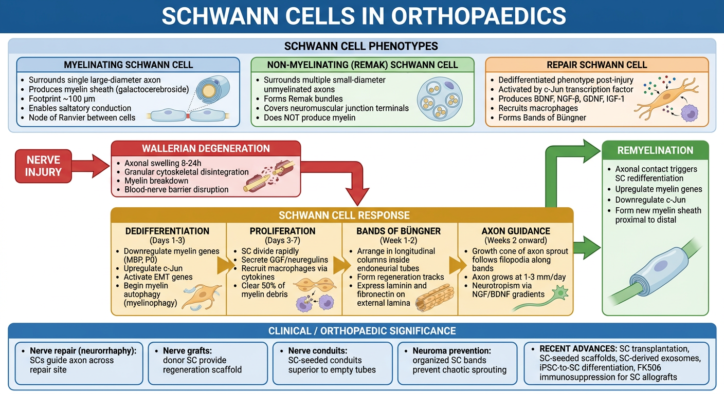
1. INTRODUCTION AND ORIGIN
Miller's Review: "Schwann cells originate in neural crest and are important in post-traumatic nerve regeneration. They produce NGF-β, BDNF, IGF-1, and erythropoietin."
2. SCHWANN CELL PHENOTYPES
A. MYELINATING SCHWANN CELLS
- Surround single large-diameter axons (>1 µm diameter) in a 1:1 ratio
- Produce the myelin sheath composed primarily of galactocerebroside
- Footprint approximately 100 µm per cell along the axon
- The gap between adjacent Schwann cells = Node of Ranvier (concentrated Na⁺ channels)
- Enable saltatory conduction between nodes - faster, energy-efficient impulse propagation
- Thicker myelin sheath = faster conduction velocity
B. NON-MYELINATING (REMAK) SCHWANN CELLS
- Surround multiple small-diameter unmyelinated axons simultaneously
- Form Remak bundles - multiple axons embedded in grooves of a single Schwann cell cytoplasm
- Most unmyelinated fibers = postganglionic sympathetic and parasympathetic axons
- Some migrate to neuromuscular junctions (perisynaptic/terminal Schwann cells / teloglia)
- Do NOT produce myelin but are essential for PNS development and function
C. REPAIR (DEDIFFERENTIATED) SCHWANN CELLS
- The injury-response phenotype - derived from conversion of both myelinating and Remak cells
- Activated by the c-Jun transcription factor - the master regulator of the repair program
- Key functions: myelin debris clearance, macrophage recruitment, axon guidance via Bands of Büngner
- This phenotype makes PNS regeneration possible (absent in CNS - where oligodendrocytes undergo apoptosis instead)
3. NEUROTROPHIC FACTORS SECRETED BY SCHWANN CELLS
| Factor | Full Name | Action |
|---|---|---|
| NGF-β | Nerve Growth Factor-β | Promotes sensory and sympathetic axon survival and growth |
| BDNF | Brain-Derived Neurotrophic Factor | Supports motor and sensory neuron survival; key after injury |
| GDNF | Glial Cell Line-Derived Neurotrophic Factor | Promotes motor neuron survival and axon growth |
| IGF-1 | Insulin-like Growth Factor-1 | Enhances axon regeneration and SC proliferation |
| GGF | Glial Growth Factors (neuregulins) | Potent SC proliferation stimulators |
| Erythropoietin | - | Neuroprotective; promotes regeneration |
| CNTF | Ciliary Neurotrophic Factor | Motor neuron survival |
4. WALLERIAN DEGENERATION AND SCHWANN CELL RESPONSE
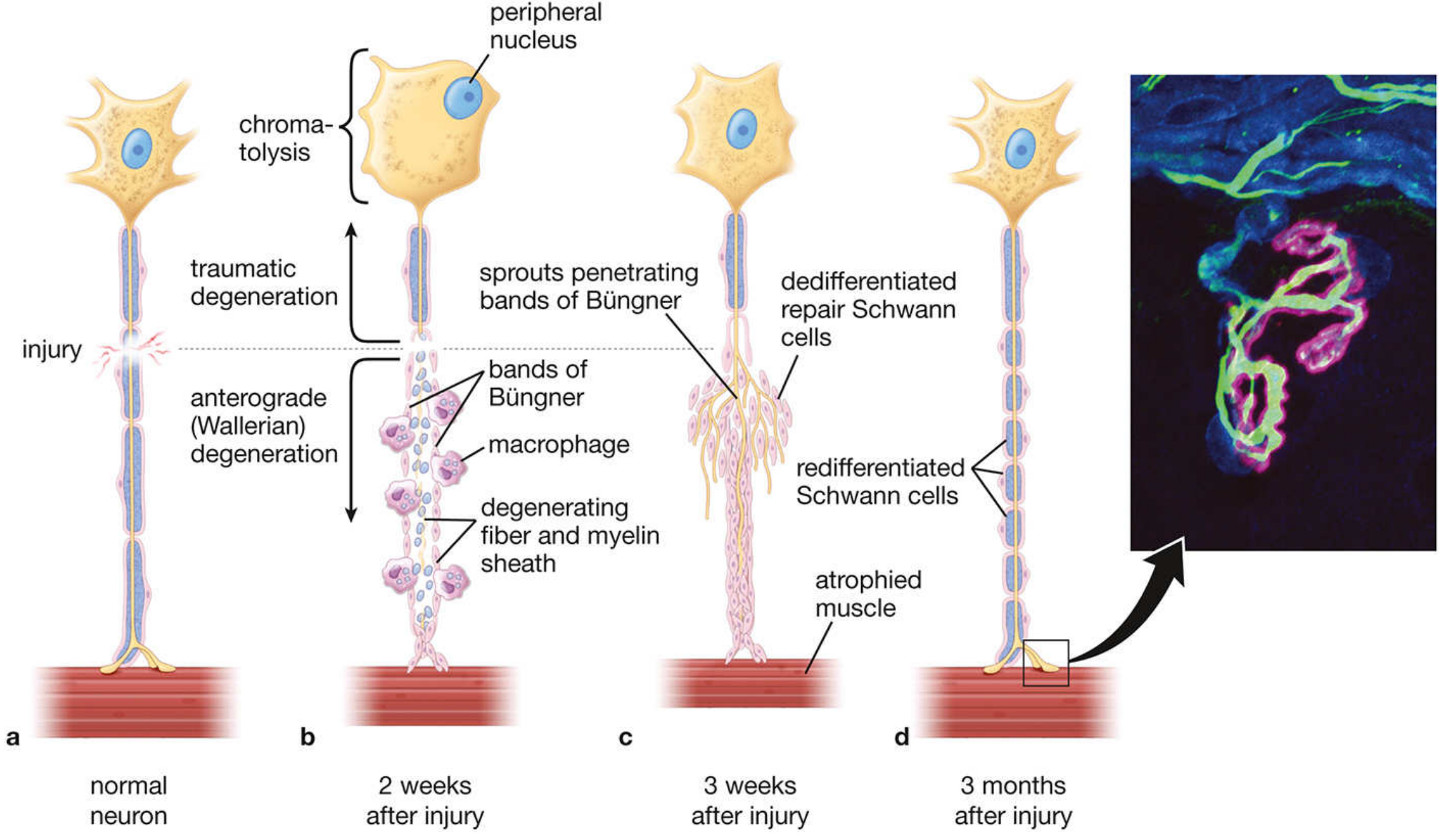
a Normal neuron. b 2 weeks post-injury: chromatolysis in cell body; anterograde Wallerian degeneration; bands of Büngner with macrophages clearing debris. c 3 weeks: multiple axon sprouts penetrating Büngner bands; pronounced muscle atrophy. d 3 months: successful reinnervation; redifferentiated Schwann cells remyelinating; new NMJ reformed. Inset: confocal image - motor axons (green), NMJ (pink), repair Schwann cells (blue).
5. SEQUENTIAL PHASES OF SCHWANN CELL RESPONSE TO INJURY
PHASE 1 - DEDIFFERENTIATION (Hours to Day 3)
- Loss of axonal contact triggers downregulation of myelin genes (MBP, P0, periaxin)
- c-Jun transcription factor is upregulated - the master switch for the repair program
- Genes associated with epithelial-to-mesenchymal transition (EMT) are activated
- Myelin autophagy (myelinophagy) begins - SC break down their own myelin internaly
- Blood-nerve barrier disrupted along entire length of the injured axon (unlike CNS)
PHASE 2 - PROLIFERATION AND MACROPHAGE RECRUITMENT (Days 3-7)
- Repair SCs divide rapidly under GGF/neuregulin stimulation
- Secrete cytokines (MCP-1, TNF-α, IL-1β) to recruit monocyte-derived macrophages
- Macrophages phagocytose myelin debris; SC autophagy accounts for ~50% of initial clearance
- Rapid clearance (completed in 2-3 weeks in PNS) is critical - allows axon regeneration
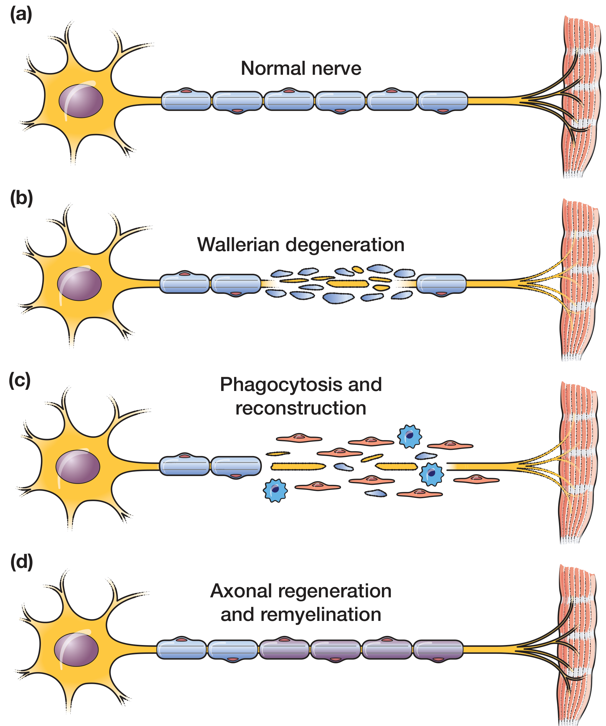
(a) Normal nerve with intact Schwann cell myelin. (b) Wallerian degeneration - myelin and axon fragment. (c) Schwann cells (red/pink) + macrophages (blue) clear debris. (d) Axonal regeneration and remyelination restores nerve-muscle connection.
PHASE 3 - BANDS OF BÜNGNER FORMATION (Week 1-2)
- Proliferating repair SCs elongate and arrange into longitudinal parallel columns
- Organized within the collapsed endoneurial tubes
- Form Bands of Büngner - "biological tunnels" that act as regeneration tracks
- Express laminin and fibronectin on their external laminae - key adhesion substrates for growth cones
- Secrete NGF/BDNF gradients creating a chemotactic gradient (neurotropism) toward target
PHASE 4 - AXON GUIDANCE (Weeks 2 onward)
- Growth cones develop at tips of proximal axon sprouts, rich in actin-filled filopodia
- Filopodia interact with laminin/fibronectin on SC external laminae → preferential adhesion
- Sprouts grow along Bands of Büngner at 1-3 mm/day (clinical rate ~1 mm/day)
- Many sprouts degenerate; large number increases chance of successful target reinnervation
- Neurotropism: regenerating axons preferentially directed to original target tissue
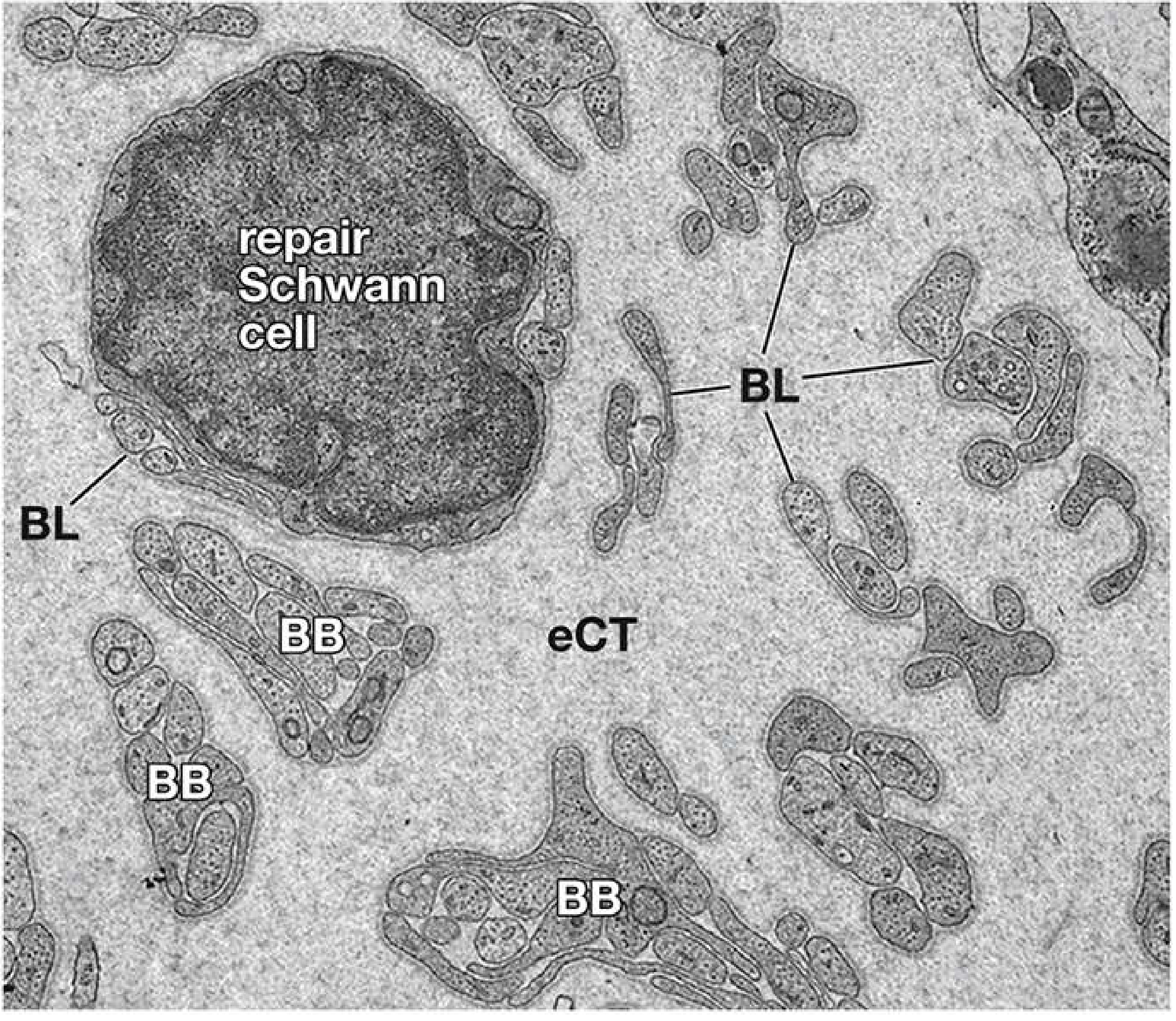
EM at ×65,000: Large repair Schwann cell (center) with basal lamina (BL). Multiple Büngner bands (BB) in endoneurial connective tissue (eCT) contain elongated SC processes - each surrounded by its own BL forming "tubes within tubes." These are the biological rails guiding axonal regeneration.
PHASE 5 - REMYELINATION
- Successful axonal contact triggers SC redifferentiation - reverse of the injury response
- Proceeds in a proximal-to-distal direction following axon growth
- Myelin genes re-expressed (MBP, P0, MAG); c-Jun downregulated
- New myelin sheath forms - initially thinner than original
- Regenerated axons remain smaller diameter with reduced conduction velocity (permanent)
6. KEY DIFFERENCES: PNS vs CNS REGENERATION
| Feature | PNS (Schwann cells) | CNS (Oligodendrocytes) |
|---|---|---|
| Response to injury | Dedifferentiation → repair SC | Apoptosis |
| Myelin clearance | Rapid (weeks) by SC + macrophages | Slow (months-years) |
| Blood barrier disruption | Along entire axon length | Only at injury site |
| Macrophage access | Massive infiltration → efficient clearance | Restricted; microglia insufficient |
| Scar | None | Astrocyte glial scar - inhibits regeneration |
| Regeneration | Possible | Usually fails |
| Reason for difference | SC: dedifferentiate and support | OL: apoptose; CNS inhibitory environment |
This fundamental difference is why peripheral nerve injuries can regenerate but spinal cord injuries do not recover spontaneously.
7. CLINICAL SIGNIFICANCE IN ORTHOPAEDICS
A. NERVE REPAIR (NEURORRHAPHY)
- Accurate Schwann cell Bands of Büngner guide axons across the repair site
- Quality of repair correlates with how well SC tracks are preserved and aligned
- Fascicular matching in perineurial repair = aligning SC tracks for motor-to-motor, sensory-to-sensory guidance
B. NERVE GRAFTING
- Sural nerve autograft works because donor Schwann cells survive and form new Büngner bands in the graft
- Cell labeling experiments confirm graft SCs actively remyelinate regenerating axons (not just a scaffold)
- Acellular nerve allografts work because recipient SCs migrate in from both stumps and repopulate the graft
C. NERVE CONDUITS
- Empty conduits allow neurotrophic factor accumulation (Lundborg principle) but are limited to gaps <3 cm
- SC-seeded conduits dramatically outperform empty conduits - SCs immediately establish Büngner-like bands inside the lumen
- SC provide trophic support, guide axons, and begin remyelination within the conduit
D. NEUROMA PREVENTION
- Organized SC activity in Büngner bands = directional, organized axon growth
- Disrupted SC organization = chaotic sprouting → painful neuroma
- Conduits and tension-free repair preserve SC track organization → reduces neuroma risk
E. TIMING OF NERVE REPAIR
- Repair SCs and their Büngner bands persist and remain functional for approximately 12-18 months after injury
- Beyond this, SCs gradually dedifferentiate further, lose their regeneration-supporting capacity, and endoneurial tubes collapse permanently
- This is the biological basis for the 12-18 month window for nerve repair/neurotization
8. RECENT ADVANCES IN SCHWANN CELL BIOLOGY (2023-2026)
A. AUTOLOGOUS SCHWANN CELL TRANSPLANTATION
- Harvest and ex vivo expansion of patient's own SCs from a peripheral nerve biopsy
- Transplanted into nerve conduits or at repair sites to supplement endogenous SCs
- Particularly useful for long-gap injuries where distal SC reserves are depleted
- Key challenge: current preparation times too slow (weeks) to intervene before irreversible muscle atrophy; accelerating autologous SC expansion is an active research priority (Nerve SPACE 2025 consensus)
B. iPSC-DERIVED SCHWANN CELLS
- Induced pluripotent stem cells (iPSCs) differentiated into Schwann cell-like cells
- Theoretically unlimited supply; patient-specific (no immunosuppression needed)
- iPSC-SCs loaded into conduits show superior nerve regeneration vs empty conduits in animal models
- Challenge: ensuring full SC maturation and myelination capacity before clinical use
C. SCHWANN CELL-DERIVED EXOSOMES
- SCs release exosomes containing miRNA, proteins, and neurotrophic factors
- Exosome therapy: cell-free approach preserving SC paracrine benefits without transplantation risks
- Promote Schwann cell proliferation, macrophage M2 polarization (anti-inflammatory), and axonal growth
- Ultrasound-targeted microbubble delivery enhances exosome targeting to injury site
- Liu et al. Neuromolecular Med 2026: SC and macrophages are key regulators of the injury repair environment; modulating the immune microenvironment via SC-exosomes is a frontier strategy
D. FK506 (TACROLIMUS) TO SUPPORT SC ALLOGRAFTS
- Local FK506 delivery allows allogeneic SC grafts to survive without systemic immunosuppression
- SC allografts seeded into acellular nerve conduits bridge long gaps without donor site morbidity
- FK506 also has direct neurotrophic effects independent of immunosuppression
- Optimal local delivery strategies and dosing schedules remain under investigation
E. ELECTRICAL STIMULATION TO ENHANCE SC ACTIVITY
- Intraoperative electrical stimulation (20 Hz, 1 hr) promotes SC proliferation and BDNF secretion
- Graphene-based and piezoelectric conductive scaffolds provide sustained local ES to SC-seeded conduits
- SC exposed to ES show enhanced laminin production → improved axon-SC adhesion and guidance
- Bordett et al. Biomed Mater 2024: Electrical, ultrasound, and magnetic stimulation all upregulate SC neurotrophic factor secretion
F. PLATELET-RICH PLASMA (PRP) + SCHWANN CELLS
- PRP provides growth factors (PDGF, TGF-β, VEGF) that synergize with SC activity
- Stimulates SC proliferation in vitro and in vivo
- Combined PRP + neurorrhaphy shows faster functional recovery (Yan et al. JPRAS Open 2026)
G. MODULATING c-JUN FOR PROLONGED SC REPAIR PHENOTYPE
- c-Jun is the master transcription factor for SC dedifferentiation into repair phenotype
- Sustained c-Jun activity maintains SC in the repair state for longer - fighting premature re-differentiation before axons arrive
- Experimental strategies to maintain c-Jun expression without pathological side effects are under active investigation
9. SUMMARY TABLE
| SC Function | Orthopaedic Relevance |
|---|---|
| Myelination | Enables normal nerve conduction; restored after repair |
| Neurotrophic factor production | Essential for axon survival and regeneration after injury |
| Dedifferentiation to repair SC | Without this, no peripheral nerve regeneration is possible |
| Bands of Büngner | Physical guide tracks for regenerating axons |
| Macrophage recruitment | Efficient debris clearance - enables regeneration (PNS advantage over CNS) |
| Remyelination | Restores conduction; proceeds proximal → distal |
| SC graft / conduit seeding | Active area of clinical translation |
| SC exosomes | Cell-free therapeutic frontier for nerve repair |
KEY EXAM POINTS
- Three SC phenotypes: Myelinating (1:1 axon), Remak/non-myelinating (many axons), Repair (injury-induced dedifferentiation)
- c-Jun = master transcription factor for repair SC activation; downregulated during remyelination
- Bands of Büngner = longitudinal SC columns inside endoneurial tubes = the biological rails guiding axon regeneration
- PNS regenerates; CNS does not = because SC dedifferentiate and support; oligodendrocytes apoptose and astrocytes form inhibitory scar
- Axon growth rate: 1-3 mm/day (histological); ~1 mm/day (clinical); track with Tinel's sign
- SC support window: 12-18 months after injury; beyond this, tracks collapse = basis for urgency in nerve repair
- Neurotrophins produced by SC: NGF-β, BDNF, GDNF, IGF-1, erythropoietin
- SC-seeded conduits outperform empty conduits - active SC immediately form Büngner-like tracks
- Recent advances: SC transplantation, iPSC-SCs, SC-derived exosomes, FK506 allografts, electrical stimulation
- The 50% of axons that fail to cross a repair site degenerate because they fail to contact SC Büngner bands - the regeneration-guiding function of SCs is irreplaceable