
Alkaptonuria. Orthopaedics context for 10 marks
alkaptonuria ochronosis spine disc calcification radiograph
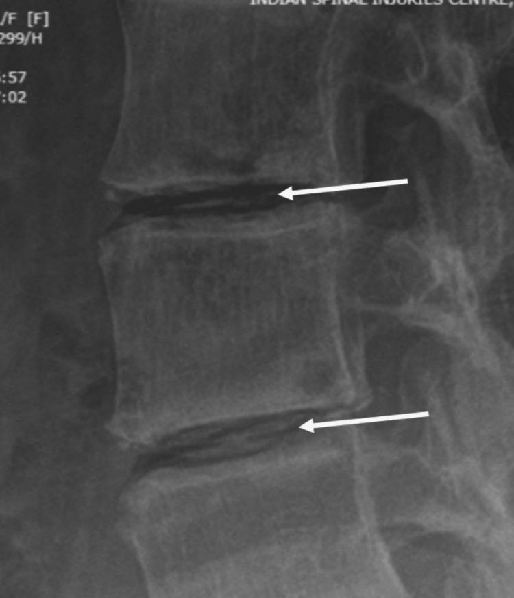
This lateral lumbar spine radiograph demonstrates significant intradiscal calcification, a hallmark finding in Alkaptonuria (ochronosis). The calcifications appear as opaque, oblong, wafer-like densities within the intervertebral disc spaces, specifically highlighted by white arrows. Accompanying these findings are generalized degenerative changes including narrowing of the disc spaces and vertebral endplate sclerosis, which appears as increased radiodensity at the superior and inferior margins of the vertebral bodies. There is also evidence of reduced bone density (osteopenia). These visual features are characteristic of ochronotic spondylosis, where homogentisic acid deposition leads to rapid disc degeneration and secondary calcification. This diagnostic image is intended for intermediate to advanced medical education, illustrating the differential diagnosis for spinal calcification beyond standard degenerative disc disease or ankylosing spondylitis.
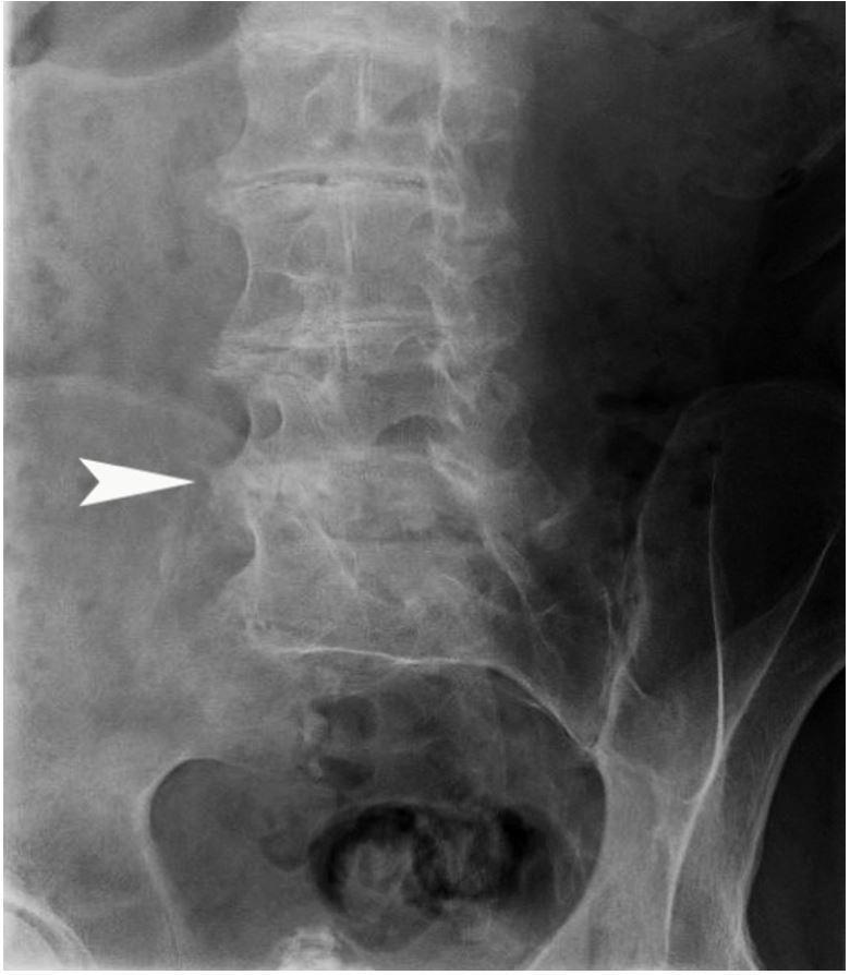
This diagnostic image is an anteroposterior (AP) X-ray radiograph of the lower lumbar spine and pelvis, demonstrating characteristic musculoskeletal manifestations of alkaptonuria (ochronosis). The image reveals severe, multi-level degenerative disc disease characterized by significant narrowing of the intervertebral disc spaces. A hallmark finding is the presence of dense, linear radiopaque calcifications within the intervertebral discs, indicated specifically by a white arrowhead at the L4-L5 level. Despite the advanced disc space narrowing and calcification, there is a notable absence of prominent marginal osteophytes, which is a classic radiographic sign distinguishing ochronotic arthropathy from standard age-related osteoarthritis. Additional findings include irregularity of the vertebral endplates and evidence of facet joint degeneration. The pelvic structures, including the sacroiliac joints and superior pubic rami, are partially visualized at the bottom of the frame. This radiograph illustrates the progressive spinal involvement typical in patients with endogenous ochronosis, where metabolic accumulation of homogentisic acid leads to premature and severe connective tissue degradation.
ochronosis knee joint pigmentation cartilage
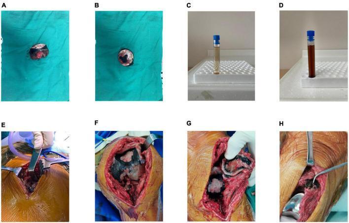
A multi-panel clinical photograph series documenting macroscopic findings of alkaptonuria (ochronosis). Panels A and B show a resected femoral head with severe cartilage destruction and characteristic black/dark brown pigmentation of the articular surface and subchondral bone. Panels C and D illustrate a diagnostic urine test: initial urine (C) appears normal light yellow, while the sample exposed to air for 24 hours (D) shows significant darkening to a chocolate-brown color due to homogentisic acid oxidation. Panels E, F, and G provide intraoperative views of the hip and knee joints, revealing extensive ochronotic pigmentation of the synovial tissues, tendons, and joint capsules, as well as the articular surfaces of the distal femur and patella. Panel H displays the joint following total knee arthroplasty (TKA) prosthesis placement. This collection demonstrates the classic multisystem pigmentary manifestations of ochronosis in orthopedic surgery and clinical biochemistry.
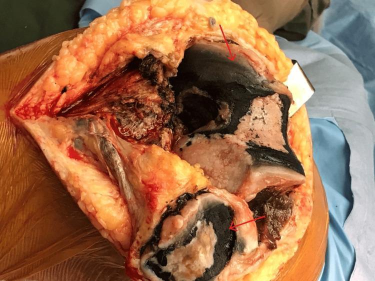
This clinical photograph captures an intraoperative view of an exposed left knee joint during a total knee arthroplasty. The image displays a significant and distinctive pathological finding: extensive blackish-to-inky pigmentation (ochronosis) covering the articular bone surfaces. Red arrows highlight the dense, charcoal-colored deposits concentrated on the femoral condyles and tibial plateau. The surrounding tissues include yellowish subcutaneous adipose tissue, reddish muscle fibers, and white-to-tan bone surfaces where the pigment is less dense. The pigmentation appears as irregular, granular patches and streaks that adhere closely to the articular cartilage and subchondral bone. This visual finding is characteristic of Alkaptonuria, a rare metabolic disorder where homogentisic acid accumulation leads to systemic connective tissue discoloration (ochronosis). The image serves as a clinical specimen example of advanced ochronotic arthropathy within an orthopedic surgical context.
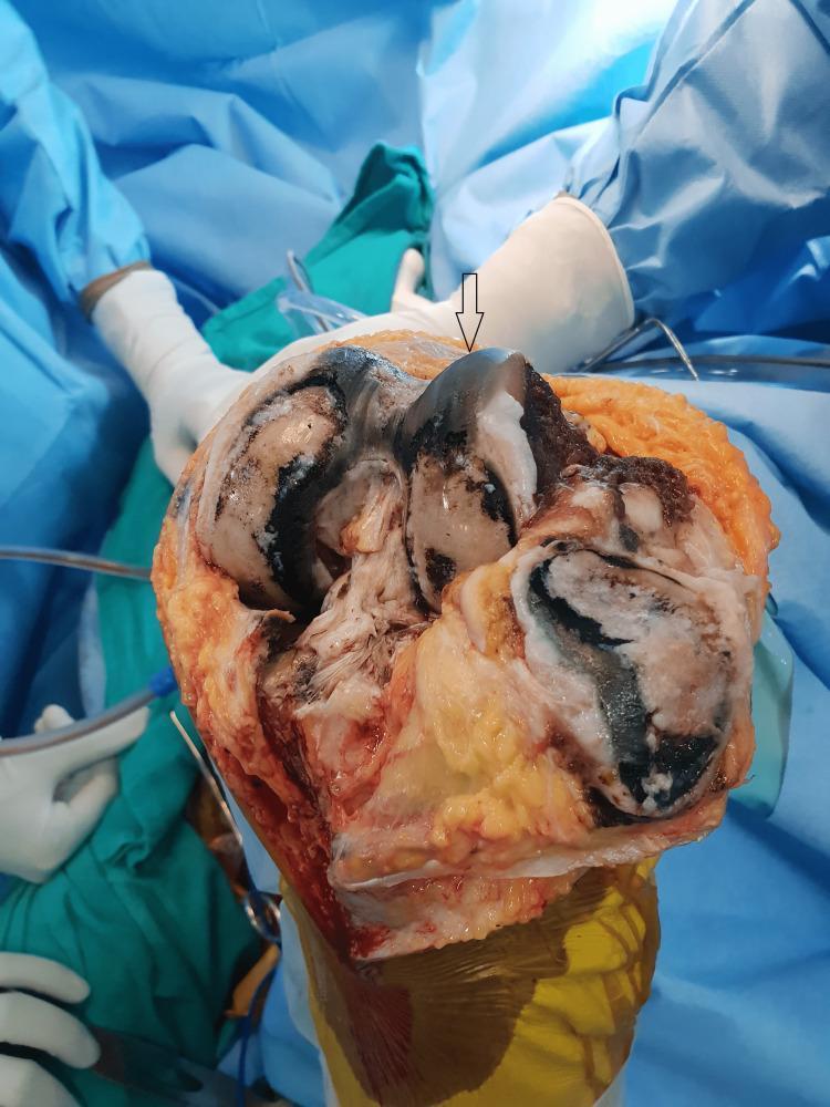
This intraoperative clinical photograph displays a human knee joint during a total knee arthroplasty (TKA). The image shows the distal femur and patellar articular surfaces heavily affected by alkaptonuric ochronosis. There is striking, diffuse dark blue to black pigmentation (ochronotic deposition) visible across the articular cartilage of the femoral condyles and the posterior aspect of the patella. The cartilage exhibits significant degenerative changes, including thinning and fragmentation, consistent with secondary osteoarthritis. The surrounding synovial tissues and soft tissues also demonstrate similar dark pigmentation. A surgical arrow highlights the most densely discolored region on the femoral articular surface. This visual evidence is a hallmark of ochronotic arthropathy, where the accumulation of homogentisic acid leads to the characteristic 'ink-black' appearance of connective tissues. The context suggests a patient with advanced joint destruction requiring prosthetic replacement.
Alkaptonuria (Ochronosis) - Orthopaedic Context
Definition and Genetics
Pathophysiology (Orthopaedic Relevance)
- HGA accumulates in cartilage, intervertebral discs, tendons, ligaments, and synovium
- Pigment deposition causes collagen cross-linking and brittleness of the matrix
- Local accumulation of pyrophosphate within joints contributes to the destructive arthropathy
- The term "ochronosis" refers to the ochre-like (yellow-brown to black) pigmentation seen macroscopically
Clinical Features
Age of Presentation
- Dark urine on standing or with alkali addition may be the only sign for years (detectable from birth)
- Low back pain typically begins at 30-40 years of age
- Peripheral arthropathy follows ~a decade after spinal involvement
Triad of Ochronosis
- Dark urine on addition of alkali (turns black on standing due to HGA oxidation; note: 25% of patients lack this)
- Ochronotic pigmentation (bluish-black discoloration of sclerae, ear cartilage/auricle, nasal cartilage, skin)
- Arthritis
Musculoskeletal Features
| Feature | Detail |
|---|---|
| Spine | Thoracolumbar involvement predominates (not lumbosacral) |
| Mimics | Resembles ankylosing spondylitis (thoracic kyphosis, stiffness) |
| Key difference | Sacroiliac joints are SPARED (unlike AS) |
| Peripheral joints | Hips, knees, shoulders - large joints; hands and feet typically spared |
| Symptoms | Pain, stiffness, crepitation, flexion contractures, limited ROM, joint-locking from loose osteochondral bodies |
| Tendons/Ligaments | Ruptures with minimal provocation (Achilles tendon, quadriceps tendon); symmetric tendon involvement |
| Bone | Increased bone resorption, osteoporosis even without immobility |
Radiological Features
- Calcification of intervertebral discs (wafer-like/linear opacities within disc spaces) - this is pathognomonic
- Disc space narrowing at multiple levels
- Vertebral endplate sclerosis/irregularity
- Absence of prominent marginal osteophytes (differentiates from OA)
- Thoracolumbar distribution
- Destruction and joint space narrowing
- Features resembling osteoarthritis but at an earlier age
Intraoperative Findings
- Articular cartilage
- Synovium, joint capsule
- Tendons and ligaments
- Periarticular bone
Differential Diagnosis
| Condition | Differentiating Point |
|---|---|
| Ankylosing spondylitis | AS involves sacroiliac joints; ochronosis spares SI joints; disc calcification is distinctive |
| Osteoarthritis | OA has osteophytes, lumbosacral > thoracolumbar; no disc calcification pattern |
| Rheumatoid arthritis | RA involves small joints symmetrically; positive RF/anti-CCP |
| CPPD arthropathy | No dark urine/pigmentation; meniscal calcification pattern |
Diagnosis
- Urine organic acid analysis: elevated HGA (turns black with alkali or on standing)
- Ferric chloride test: urine turns black
- Benedict's reagent: urine turns dark
- Serum/urine HGA levels
- Genetic testing: HGD gene mutations
- Intraoperative pigmentation (often first diagnosis)
Treatment
Conservative
- Standard osteoarthritis management (NSAIDs, physiotherapy, occupational therapy)
- Dietary restriction of phenylalanine and tyrosine (limited benefit)
- High-dose ascorbic acid (antioxidant - theoretically slows pigment deposition; unproven efficacy)
- Surveillance for aortic dilatation, valvular calcification, renal/prostate stones
Medical (Disease-Modifying)
- Nitisinone (Orfadin, 10 mg/day): Inhibits 4-hydroxyphenylpyruvate dioxygenase, an earlier enzyme in the tyrosine pathway, dramatically reducing urinary HGA to normal levels. Originally approved for hereditary tyrosinemia type 1. It delays disease progression and improves clinical signs of alkaptonuria. Side effect: elevates blood tyrosine (corneal deposits possible).
Surgical
- Joint replacement (arthroplasty): Average age at joint replacement in ochronosis is 53 years vs. national mean of 67 years for OA - patients require surgery significantly earlier
- Total hip arthroplasty (THA) and total knee arthroplasty (TKA) are the standard surgical interventions
- Spinal surgery for severe kyphosis or neurological compromise
- Tendon repair for spontaneous ruptures
Systemic Associations (Important for Preoperative Assessment)
- Cardiac: Pigmentation of aortic/mitral valves → aortic stenosis requiring valve replacement (especially after age 60); coronary artery calcification
- Urological: Pigmented renal and prostatic calculi
- Otolaryngological: Tympanic membrane pigmentation, laryngeal cartilage involvement
Summary Box (Examination Recall)
- Gene: HGD (Chromosome 3q21-q23)
- Enzyme deficient: Homogentisate 1,2-dioxygenase
- Metabolite accumulated: Homogentisic acid
- Inheritance: Autosomal recessive
- Pathognomonic X-ray: Calcification of intervertebral discs
- Pathognomonic intraoperative finding: Jet-black pigmented cartilage
- Sacroiliac joints: Spared (key differentiator from AS)
- Joint replacement age: ~53 years (14 years earlier than typical OA)
- Drug treatment: Nitisinone
- First described by: Sir Archibald Garrod (1896) - the first "inborn error of metabolism"