
H pylori associated lesion and their pathogenesis for md pathology exam
I'll read the medical library skill first, then research H. pylori comprehensively for your MD Pathology exam.
Reading File
Now let me search the library and PubMed in parallel for comprehensive H. pylori pathology content.
Finding Sources
Finding Sources
Searching PubMed
"Helicobacter pylori"[MeSH Terms] AND pathogenesis AND gastric cancer
Now let me read the most relevant textbook sections - the pathology-focused blocks and a key surgical reference in parallel.
Finding Sources
Finding Sources
Finding Sources
Now let me read the most pathology-rich textbook sections in parallel.
Reading File
Reading File
Reading File
Excellent content. Let me continue reading the Yamada gastroenterology section and the Jawetz pathogenesis section to complete the picture.
Reading File
Reading File
Now let me get the key pathogenesis section from the Sherris microbiology book and the Correa cascade/gastric carcinoma section:
Reading File
Finding Sources
Now let me read the Maingot's Correa cascade section and also fetch the key histology images:
Reading File
Loading Image
Loading Image
Loading Image
I now have comprehensive, authoritative content from multiple textbooks. Let me compile the full MD Pathology exam answer.
Helicobacter pylori - Associated Lesions and Pathogenesis
MD Pathology Exam - Comprehensive Notes
1. The Organism
H. pylori is a spiral-shaped, Gram-negative, microaerophilic rod with multiple sheathed polar flagella. It is catalase-, oxidase-, and urease-positive - urease positivity is its single most important virulence characteristic. Humans are the primary reservoir.
First bacterium declared a Class I (definite) human carcinogen by the WHO/IARC in 1994.
2. Virulence Factors and Mechanism of Survival
H. pylori has evolved multiple strategies to colonize the hostile acid environment of the gastric mucosa:
| Virulence Factor | Mechanism |
|---|---|
| Urease | Hydrolyses urea → NH₃ + CO₂; neutralises local pH, creating a protective ammonia "cloud" |
| Flagella | Corkscrew motility; allows the organism to swim beneath the mucus layer to the less-acidic epithelial surface |
| Outer membrane proteins (OMPs) | Adhesins (BabA, SabA) bind Lewis-b blood-group antigens on gastric epithelial cells |
| CagA (cytotoxin-associated gene A) | Injected into host cells via a Type IV Secretion System (T4SS); triggers actin cytoskeleton reorganisation, activates oncogenic signalling cascades (SHP-2, NF-κB, Wnt/β-catenin) |
| VacA (vacuolating cytotoxin A) | Also delivered by T4SS; causes vacuolization of endosomal compartment, mitochondrial damage, apoptosis, and T-cell suppression |
| CagPAI (pathogenicity island) | ~40-kb genomic segment encoding the T4SS apparatus; strains possessing it (Cag+) are significantly more virulent and carcinogenic |
| Urease + NH₃ | Direct cytotoxicity to mucosal cells; damages tight junctions |
The organisms colonise beneath the mucus overlying the antrum, remain extracellular, and do not invade tissue. They are found adherent to the apical surface of surface epithelium and gastric pit (foveolar) cells but do not colonise intestinal metaplastic foci.
Sherris & Ryan's Medical Microbiology, 8e; Jawetz Melnick & Adelberg's Medical Microbiology, 28e
3. H. pylori-Associated Lesions
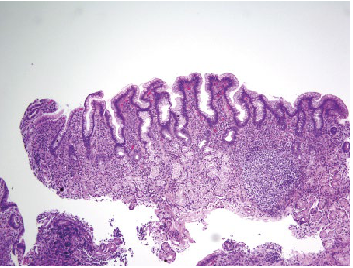
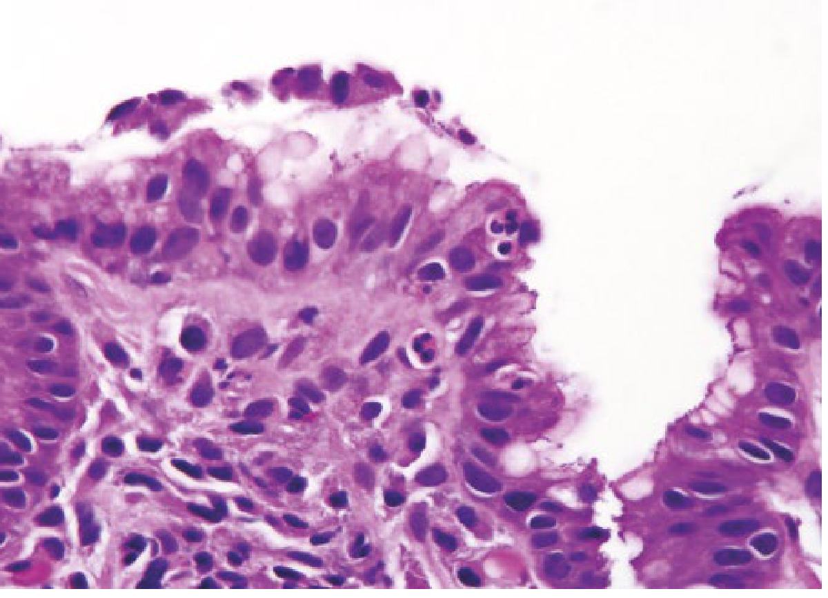
3.1 Chronic Active Gastritis (Most Common; Universal in Infection)
Formerly called: Diffuse antral gastritis, Type B gastritis, Superficial gastritis.
Gross: Antral-predominant with nodularity ("mamillated" corpus appearance endoscopically). Erythema, mucosal friability.
Histology (the cornerstone for exam):
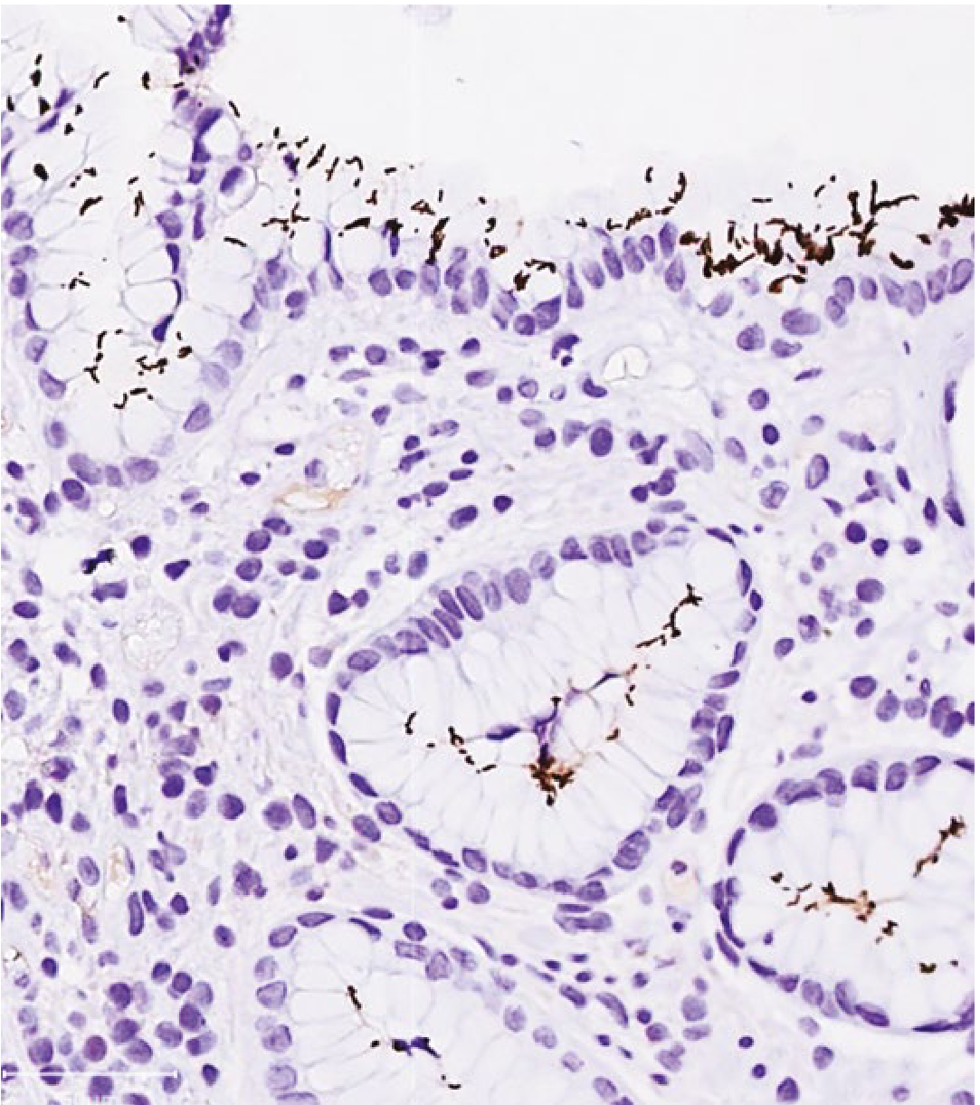
- Organisms visible in gastric mucus and adherent to the apical surface of foveolar cells on H&E; highlighted by Giemsa, Warthin-Starry silver stain, or immunohistochemistry (IHC) - the most sensitive method
- Superficial gastritis pattern - inflammation concentrated in the upper mucosa
- Lamina propria - heavy lymphoplasmacytic (chronic) infiltrate
- Active component - neutrophils in the epithelium ("cryptitis"); neutrophilic infiltration into gastric pits forms pit abscesses
- Lymphoid follicles (MALT - mucosa-associated lymphoid tissue) in the lamina propria - physiologically absent from normal gastric mucosa; their presence is pathognomonic of H. pylori infection
- Distribution: Antrum >> Body; if both affected = pangastritis
After eradication: Active (neutrophilic) component resolves within weeks; chronic inflammatory component persists for months to years.
3.2 Peptic Ulcer Disease (PUD)
H. pylori causes ~70% of gastric ulcers and ~90-95% of duodenal ulcers (excluding NSAID-related disease).
Pathogenesis of Duodenal Ulcer:
- H. pylori antral gastritis → ↓ somatostatin from D-cells → ↑ gastrin from G-cells → ↑ acid secretion (hypersecretion)
- Acid load in duodenum → gastric metaplasia in the duodenal cap (duodenal mucosa acquires gastric-type epithelium)
- H. pylori colonises this metaplastic epithelium → active duodenitis → ulceration
- CagA+ strains also express a duodenal ulcer-promoting gene (dupA)
Pathogenesis of Gastric Ulcer:
- H. pylori infection causes mucosal injury directly via ammonia, cytotoxins (VacA), and inflammatory mediators (IL-8, TNF-α)
- ↓ prostaglandin synthesis (disrupted by inflammation) → impaired mucus/bicarbonate barrier
- Normal or low acid (vs. duodenal ulcer which has high acid) - injury is primarily mucosal defence failure rather than acid excess
Key histological features of H. pylori-associated peptic ulcer:
- Punched-out ulcer with clean base (benign), heaped edges
- Base shows necrotic slough, granulation tissue, fibrosis, endarteritis obliterans of vessels at the ulcer margin
- Adjacent mucosa shows chronic active gastritis with H. pylori organisms
Sherris & Ryan's Medical Microbiology, 8e; Jawetz, 28e; Maingot's Abdominal Operations
3.3 Atrophic Gastritis and Intestinal Metaplasia
Chronic H. pylori gastritis can progress to multifocal atrophic gastritis (MAG):
- Loss of native gastric glands (oxyntic glands in the body, antral glands in the antrum)
- Replacement by intestinal metaplasia (IM) - goblet cells, absorptive cells, Paneth cells appear in the gastric mucosa
Types of intestinal metaplasia:
- Type I (complete/small-intestinal): Mucin-secreting goblet cells with brush border; sialomucins; lower malignant potential
- Type II/III (incomplete/colonic type): Goblet cells with sulphomucins; no brush border; higher malignant potential
This forms the basis of the Correa cascade (see below).
3.4 Gastric Adenocarcinoma - The Correa Cascade
H. pylori is a WHO Class I definite carcinogen. It is linked to distal (non-cardia) gastric adenocarcinoma; there is no proven link to cardia carcinoma.
Correa Cascade (sequential model of gastric carcinogenesis):
Normal Gastric Mucosa
        ↓  (H. pylori infection)
Chronic Active Gastritis
        ↓  (years–decades)
Multifocal Atrophic Gastritis
        ↓
Intestinal Metaplasia (complete → incomplete)
        ↓
Low-Grade Dysplasia
        ↓
High-Grade Dysplasia (Carcinoma in situ)
        ↓
INVASIVE INTESTINAL-TYPE GASTRIC ADENOCARCINOMA
Molecular mechanisms of H. pylori carcinogenesis:
- CagA oncoprotein - directly injected into epithelial cells via T4SS; activates SHP-2 tyrosine phosphatase, ERK/MAPK, Wnt/β-catenin pathways; promotes proliferation, inhibits apoptosis
- NF-κB activation - by CagA, VacA, and lipopolysaccharide → ↑ IL-8, IL-1β, TNF-α → sustained mutagenic inflammation
- Reactive oxygen species (ROS) - generated by activated neutrophils and macrophages → DNA strand breaks, TP53 mutations
- Epigenetic silencing - H. pylori-induced promoter methylation of tumour suppressor genes (CDH1/E-cadherin, RUNX3, MLH1)
- Microsatellite instability (MSI) - epigenetic silencing of MMR gene MLH1
- ↑ cell proliferation / ↓ apoptosis - VacA disrupts apoptosis pathways; CagA disrupts cell polarity
Relative risk: H. pylori seropositivity confers a 2.1 to 20-fold increased risk of gastric cancer. In a landmark Japanese prospective study, gastric cancer developed in 2.9% of seropositive patients and in none of seronegative patients.
Maingot's Abdominal Operations; Sherris & Ryan, 8e; Duan et al., J Hematol Oncol 2025
3.5 Gastric MALT Lymphoma (MALToma)
Mechanism:
- Normal gastric mucosa contains no lymphoid tissue
- H. pylori infection induces acquired MALT (lymphoid follicles in lamina propria - the reactive lymphocytes seen in gastritis)
- Chronic antigenic stimulation by H. pylori → T-cell-driven B-cell proliferation
- Initially polyclonal; with accumulating genetic mutations → monoclonal B-cell proliferation
- Monoclonal expansion = low-grade MALT lymphoma (also called marginal zone B-cell lymphoma of MALT type; formerly called "pseudolymphoma" in early descriptions)
- Further mutations (e.g., t(11;18)(q21;q21) - BIRC3-MALT1 translocation) → progression to high-grade diffuse large B-cell lymphoma (DLBCL)
Key pathological points for exam:
- Histology: lymphoepithelial lesions (infiltration of gastric gland epithelium by lymphoid cells, causing gland destruction) - this is the defining histological feature
- Cells are centrocyte-like (monocytoid) B-cells in the marginal zone
- Reactive germinal centres are invaded by the neoplastic B cells
- IHC: CD20+, CD79a+, CD5-, CD10- (marginal zone phenotype)
The unique biology - regression with antibiotics:
- Low-grade MALT lymphoma dependent on T-cell stimulation by H. pylori antigen
- H. pylori eradication → tumour regression in ~75-80% of cases (gastric MALToma is essentially the only cancer known to regress with antibiotic therapy)
- Cases with t(11;18) translocation are typically H. pylori-independent and do NOT regress with eradication
Sherris & Ryan, 8e; Comprehensive Clinical Nephrology, 7e; Yamada's Textbook of Gastroenterology, 7e
4. Summary Table - H. pylori Associated Lesions
| Lesion | Key Feature | Mechanism |
|---|---|---|
| Chronic active gastritis | Lymphoid follicles + neutrophilic epithelial infiltration + H. pylori on surface epithelium | Direct cytotoxicity (urease, VacA) + immune activation (CagA → IL-8, NF-κB) |
| Duodenal ulcer (90-95%) | Antral predominant; hypersecretory state | ↓ D-cells → ↑ gastrin → ↑ acid; dupA gene; gastric metaplasia in duodenum |
| Gastric ulcer (~70%) | Antrum/lesser curve; normal/low acid | Mucosal defence failure; ↓ prostaglandins; cytotoxin-mediated damage |
| Gastric atrophy + intestinal metaplasia | Loss of gastric glands; goblet cells | Chronic inflammation → gland destruction → metaplastic replacement |
| Gastric adenocarcinoma (intestinal type) | Distal stomach; non-cardia | Correa cascade; CagA oncogenic signalling; ROS-mediated DNA damage; epigenetic silencing |
| Gastric MALT lymphoma | Lymphoepithelial lesions; marginal zone B-cells | MALT acquisition → antigen-driven B-cell clonal expansion; regresses with eradication |
5. Histological Diagnosis
| Method | Notes |
|---|---|
| H&E stain | Curved/spiral organisms in gastric mucus, adherent to surface epithelium; reliable when organisms numerous |
| Giemsa stain | Good for H. pylori detection on histology sections |
| Warthin-Starry (silver) stain | Excellent sensitivity; organisms appear black |
| Immunohistochemistry (IHC) | Most sensitive; detects coccoid forms in partially treated patients; organisms highlighted in brown on surface epithelium |
| Modified Giemsa/Cresyl violet | Rapid, cheap, adequate sensitivity |
Rapid urease test (CLO test): Biopsy-based; urease-positive organisms produce NH₃ → colour change; results in <2 hours.
6. H. pylori and the Sydney Classification of Gastritis
The Updated Sydney System grades gastritis in biopsies from 5 sites (2 antrum, 2 corpus, 1 incisura angularis) and scores:
- H. pylori density (0-3+)
- Chronic inflammation (mononuclear cells)
- Activity (neutrophil infiltration)
- Glandular atrophy
- Intestinal metaplasia
7. Key Exam Points (Quick Recall)
- H. pylori is exclusively extracellular and does not invade mucosa
- It colonises the gastric-type epithelium only (not intestinal metaplastic foci, except with PPI use when deeper gland colonisation occurs)
- Lewis-b antigen is the main mucosal receptor (blood group O → slightly higher risk)
- CagA strains are more virulent and more strongly associated with carcinoma and ulcers
- The lymphoid follicle in the gastric mucosa is pathognomonic of H. pylori infection
- Correa cascade is the accepted model; it is potentially reversible with eradication at early stages
- MALT lymphoma is the only neoplasm that can regress with antibiotic therapy
- H. pylori is classified as a Group 1 carcinogen (IARC)
- Duodenal ulcer = high acid; Gastric ulcer = normal/low acid - this distinction traces back to H. pylori affecting gastrin/somatostatin balance
- VacA causes vacuolization; CagA causes cytoskeletal changes and oncogenic signalling - remember both are delivered by Type IV Secretion System
Sources: Yamada's Textbook of Gastroenterology 7e; Jawetz Melnick & Adelberg's Medical Microbiology 28e; Sherris & Ryan's Medical Microbiology 8e; Maingot's Abdominal Operations; Schwartz's Principles of Surgery 11e; Duan et al., J Hematol Oncol, 2025 - PMID 39849657
Please in corporate Robbins too
Finding Sources
Finding Sources
Both Robbins books are in the library. Let me search them directly now.
Finding Sources
Finding Sources
Found all the Robbins sections. Let me read them all in parallel now.
Reading File
Reading File
Reading File
Excellent Robbins content retrieved. Let me now get the MORPHOLOGY section from Robbins PBD and the gastric ulcer + MALT lymphoma / carcinoma Robbins sections.
Reading File
Reading File
Reading File
Now let me get the Robbins PBD sections on gastric carcinoma pathogenesis (H. pylori angle) and the MALToma section, plus fetch the key Robbins histology figure:
Reading File
Reading File
Loading Image
Loading Image
I now have everything I need from both Robbins books along with the classic pathogenesis diagram and histology panel. Let me fetch the MALToma histology image too:
Loading Image
I now have comprehensive Robbins content from both books (Robbins & Kumar Basic Pathology and Robbins, Cotran & Kumar Pathologic Basis of Disease) along with the key figures. Here is the fully updated, Robbins-integrated answer:
Helicobacter pylori - Associated Lesions and Pathogenesis
MD Pathology Exam - Comprehensive Notes (Robbins + Multitext)
1. The Organism
H. pylori is a spiral-shaped/curved, Gram-negative, microaerophilic bacillus with multiple sheathed polar flagella. It is catalase-, oxidase-, and urease-positive. Humans are the primary reservoir; transmission is mainly fecal-oral, typically acquired in childhood and persisting for life without treatment.
"First bacterium classified as a definite (Group I) carcinogen" - WHO/IARC 1994, and the first bacterium declared a carcinogen.
- Robbins, Cotran & Kumar Pathologic Basis of Disease
"H. pylori now has acquired the dubious distinction of being the first bacterium classified as a carcinogen... implicated in the genesis of both gastric adenocarcinoma and gastric lymphomas."
- Robbins & Kumar Basic Pathology
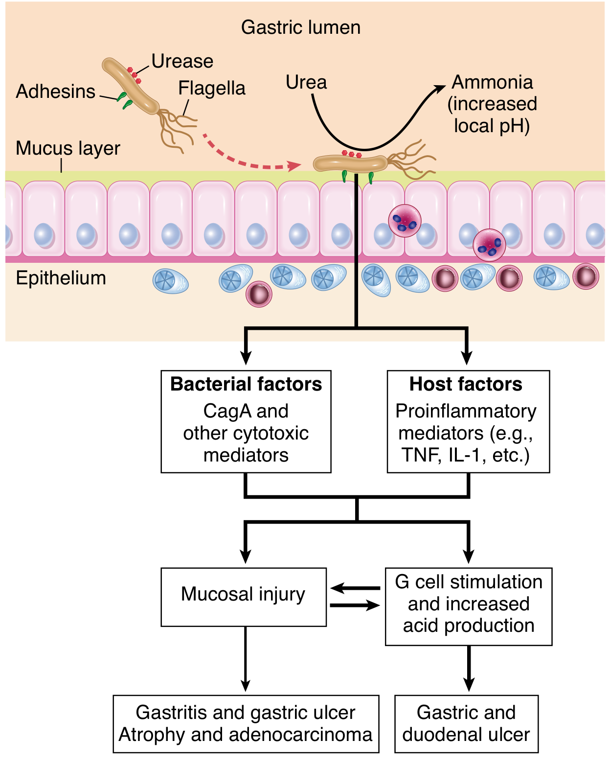
2. Virulence Factors - Robbins' Four Key Features
Robbins Basic Pathology identifies four main virulence factors (memorise these for exam):
| # | Factor | Mechanism |
|---|---|---|
| 1 | Flagella | Allow motility in viscous mucus; bacteria swim beneath the mucus layer to the less-acidic epithelial surface |
| 2 | Urease | Generates ammonia from endogenous urea → elevates local pH → protects bacteria from gastric acid |
| 3 | Adhesins | Outer membrane proteins (BabA, SabA) bind Lewis-b antigens on surface foveolar cells, enhancing adherence |
| 4 | Toxins (CagA, CagE) | Stimulate release of IL-8 (potent neutrophil chemotactic factor) → initiate and sustain innate and adaptive immune responses → mucosal damage |
(Robbins & Kumar Basic Pathology, 2022)
3. Pathogenesis - The Two Disease Patterns (Robbins PBD)
Robbins PBD highlights a critical dual-pathway model based on which part of the stomach is predominantly infected:
H. pylori infection
        |
        +---> Predominantly ANTRAL gastritis (most common)
        |         - Inflammation stimulates G cells → ↑ gastrin
        |         - ↑ parietal cell expansion in body → ↑ acid secretion
        |         - OUTCOME: Peptic ulcer disease (gastric or duodenal)
        |
        +---> PANGASTRITIS (body + antrum, long-standing infection)
                  - Progressive atrophy of parietal cell mass
                  - ↓ acid secretion
                  - Intestinal metaplasia
                  - OUTCOME: Gastric adenocarcinoma
                  - (Inverse relationship: atrophy/carcinoma vs. ulcer)
"This results in an inverse relationship between gastric adenocarcinoma, which is associated with atrophy and intestinal metaplasia, and peptic ulcer disease, which requires at least some residual acid secretion."
- Robbins, Cotran & Kumar Pathologic Basis of Disease
Host factors also matter: polymorphisms in cytokine gene promoters (especially IL-1β and TNF) influence whether infection remains antral or progresses to pangastritis.
4. H. pylori-Associated Lesions
4.1 Chronic Active Gastritis (Universal in Infection)
Formerly called: Type B gastritis, Diffuse antral gastritis, Superficial gastritis.
Pathogenesis (Robbins):
- H. pylori organisms colonise the antral mucosa; the bacteria and associated inflammation stimulate G cells → gastrin release → hyperacidity and increased risk of PUD
- Released CagA (and other cytotoxins) trigger IL-8 secretion from epithelial cells → neutrophil recruitment
- Sustained chronic inflammation produces the histological picture below
MORPHOLOGY - Robbins (verbatim, exam-ready):
"The organism is concentrated within the superficial mucus overlying epithelial cells in the surface and neck regions. The distribution can be irregular, with areas of heavy colonization adjacent to those with few organisms."
Histological features (Robbins PBD + Basic):
- H. pylori organisms visible in gastric mucus and on apical surface of foveolar cells on H&E or Giemsa; most reliably identified by IHC or Warthin-Starry silver stain
- Large numbers of plasma cells, often in clusters or sheets in the superficial lamina propria - this is a hallmark feature
- Increased lymphocytes, macrophages and neutrophils in lamina propria
- Neutrophils cross the basement membrane into the epithelium, and accumulate in gland lumens = pit abscesses
- Lymphoid aggregates with germinal centers (submucosal) = induced MALT - normally absent from the stomach; their presence = pathognomonic of H. pylori infection; potential for lymphomatous transformation
- Intestinal metaplasia (goblet cells + columnar absorptive cells) in longstanding disease = risk factor for adenocarcinoma
- Thickened rugal folds with intense inflammation (mimics infiltrative malignancy endoscopically)
- Hyperplastic/inflammatory polyps may develop due to regenerative changes from mucosal damage
"Submucosal lymphoid aggregates, some with germinal centers, are frequently present and represent an induced form of mucosa-associated lymphoid tissue (MALT) that has the potential to transform into lymphoma."
- Robbins & Kumar Basic Pathology
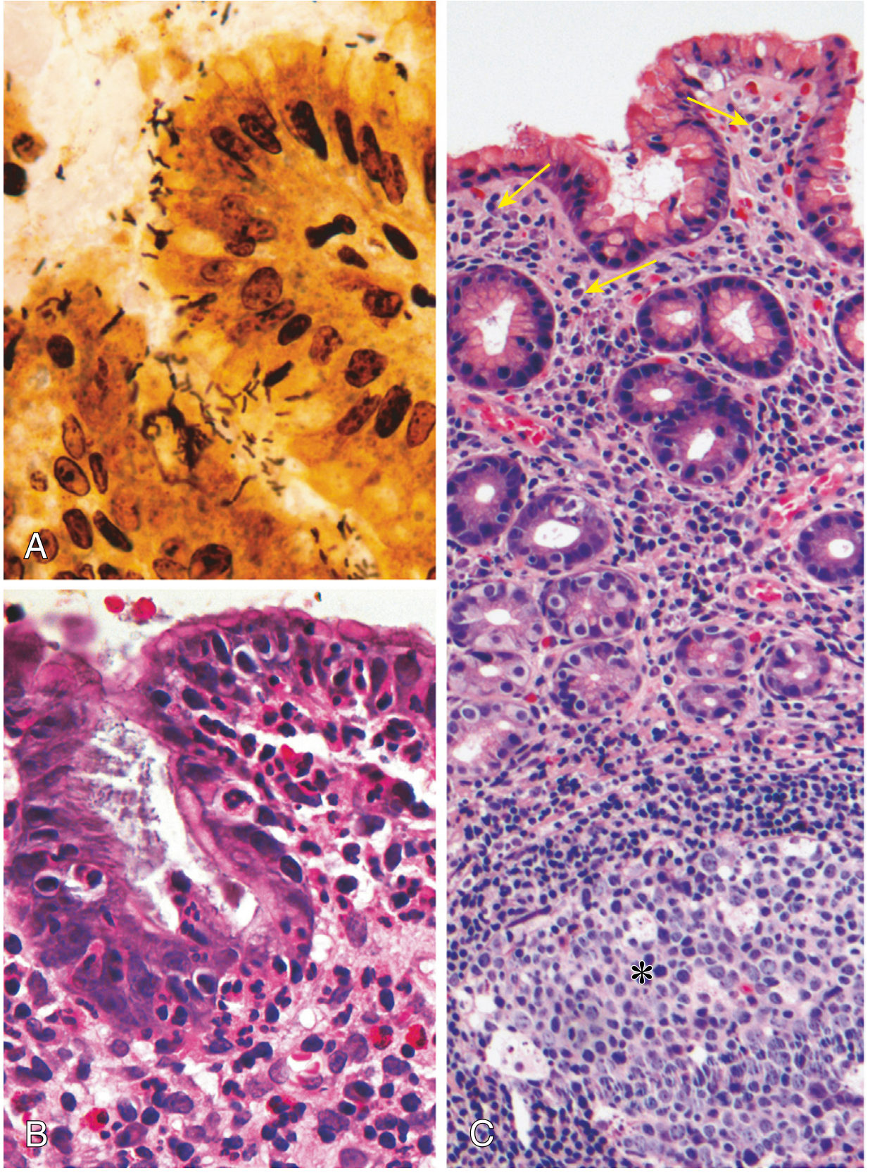
H. pylori tropism - important exam point (Robbins):
- The organism shows tropism for gastric foveolar epithelium
- Not found in areas of intestinal metaplasia, acid-producing oxyntic mucosa of the gastric body, or duodenal epithelium
- Therefore antral biopsy is the preferred site for H. pylori evaluation (unless on PPIs - then organisms may be seen deeper in body mucosa)
4.2 Peptic Ulcer Disease
Robbins Key Concepts summary: "Peptic ulcer disease is usually secondary to chronic H. pylori-induced gastritis and the resulting hyperchlorhydria. Ulcers can develop in the stomach or duodenum and usually heal after suppression of gastric acid production and H. pylori eradication."
Duodenal Ulcer Mechanism:
- Antral H. pylori gastritis → inflammation inhibits D-cells (↓ somatostatin) → G cells release ↑ gastrin
- ↑ Gastrin → parietal cell hyperplasia and expansion in the body → hypersecretion of acid
- Acid load delivered to duodenum → gastric metaplasia of duodenal mucosa
- H. pylori colonises this metaplastic epithelium → active duodenitis → ulceration
- CagA+ strains additionally express dupA (duodenal ulcer-promoting gene)
Gastric Ulcer Mechanism:
- Direct mucosal cytotoxicity (ammonia, VacA, CagA-mediated IL-8/cytokine release)
- ↓ Prostaglandin synthesis → impaired mucus-bicarbonate barrier
- Acid level normal or low (cf. duodenal ulcer - high acid)
- Primarily a failure of mucosal defence rather than acid excess
Histology of peptic ulcer (Robbins classic description):
- Active phase: base shows necrotic slough + granulation tissue
- Healing: fibrosis in base; endarteritis obliterans of vessels at the ulcer margin
- Adjacent mucosa invariably shows H. pylori chronic active gastritis
- Benign ulcer: punched-out, regular margins, perpendicular walls, smooth base
- Malignant ulcer (contrast): irregular heaped-up margins, irregular base
4.3 Atrophic Gastritis and Intestinal Metaplasia
Longstanding H. pylori pan-gastritis → multifocal atrophic gastritis (MAG):
- Loss of oxyntic glands in the body; loss of antral glands in the antrum
- Replacement by intestinal metaplasia (goblet cells, absorptive enterocytes, Paneth cells)
- Not associated with autoantibodies (unlike autoimmune/Type A gastritis) and is patchy
- Reduced acid secretion → mild ↑ gastrin (but not to the extreme degree of autoimmune gastritis where all parietal cells are lost)
Types of intestinal metaplasia:
- Complete (Type I): Sialomucins; brush border; lower risk
- Incomplete (Type II/III): Sulphomucins; no brush border; higher malignant risk
4.4 Gastric Adenocarcinoma - The Correa Cascade
Robbins' statement on carcinogenesis:
"The proposed scenario for the development of gastric adenocarcinoma in the setting of H. pylori infection involves increased epithelial cell proliferation in a background of chronic inflammation, which may generate genotoxic agents such as reactive oxygen species. The H. pylori genome also contains genes directly implicated in oncogenesis. Strains associated with gastric adenocarcinoma have a 'pathogenicity island' that contains the cytotoxin-associated gene A (CagA). Although H. pylori is noninvasive, released CagA can penetrate gastric epithelial cells, where it has a variety of effects including the initiation of a signaling cascade that mimics growth factor stimulation."
- Robbins & Kumar Basic Pathology
"Persistent infection first leads to development of chronic gastritis, followed by gastric atrophy, intestinal metaplasia of the lining cells, dysplasia, and cancer. This sequence takes decades to complete and occurs in less than 5% of infected individuals."
Correa Cascade (Robbins-integrated):
Normal Gastric Mucosa
     ↓ H. pylori infection (CagA+ strain)
Chronic Active Gastritis (antrum)
     ↓ progression to body/fundus (pangastritis)
Multifocal Atrophic Gastritis
     ↓ (years-decades)
Intestinal Metaplasia (incomplete/Type III is high risk)
     ↓
Low-grade Dysplasia
     ↓
High-grade Dysplasia
     ↓
INTESTINAL-TYPE GASTRIC ADENOCARCINOMA (distal/non-cardia)
Molecular mechanisms:
- CagA (injected by T4SS) → mimics growth factor stimulation → activates SHP-2, ERK/MAPK, Wnt/β-catenin → proliferation, inhibited apoptosis
- NF-κB activation → ↑ pro-inflammatory cytokines (IL-8, IL-1β, TNF-α) → sustained mutagenic inflammation
- Reactive oxygen species (ROS) generated by neutrophils/macrophages → DNA strand breaks, TP53 mutations
- Epigenetic silencing of tumour suppressor genes (CDH1/E-cadherin, RUNX3, MLH1) via CpG promoter methylation
- Host cytokine polymorphisms - IL-1β and TNF promoter polymorphisms (Robbins PBD emphasis) → amplified inflammatory response → greater mucosal injury and carcinogenesis risk
H. pylori is linked to distal gastric adenocarcinoma only; no proven association with cardia carcinoma. IARC classified it as a definite Group I carcinogen in 1994.
4.5 Gastric MALT Lymphoma (MALToma)
Robbins (PBD & Basic) explanation is the standard exam answer:
Pathogenesis (step by step from Robbins):
- Normal stomach has no MALT (lymphoid tissue is not present)
- H. pylori infection induces appearance of H. pylori-reactive T cells
- These T cells stimulate a polyclonal B-cell proliferation (the MALT seen in chronic gastritis)
- In chronic infections, mutations accumulate giving certain B-cell clones a growth advantage
- These cells grow out as a monoclonal lymphoma that still remains dependent on T-cell stimulation of NF-κB
- At this stage the tumour is localized to the stomach → H. pylori eradication "cures" the lymphoma by removing the antigenic stimulus
- With persistent disease, additional mutations cause constitutive NF-κB activation (e.g., t(11;18)(q21;q21) BIRC3-MALT1 translocation) → tumour no longer requires bacterial antigen → does not regress with eradication → spreads beyond the stomach
"At this stage, the B-cell proliferation remains localized to the stomach and eradication of H. pylori by antibiotic therapy 'cures' the lymphoma by removing the antigenic stimulus for T cells."
- Robbins & Kumar Basic Pathology
MORPHOLOGY (Robbins PBD):
- Dense lymphocytic infiltrate in the lamina propria
- Lymphoepithelial lesions = neoplastic lymphocytes infiltrating and destroying gastric glands - the defining histological feature
- Reactive-appearing B-cell follicles may be present
- ~40% of tumours show plasmacytic differentiation
IHC (Robbins PBD):
- CD19+, CD20+ (B-cell markers)
- CD5-, CD10- (marginal zone phenotype - not CLL or follicular lymphoma)
- CD43+ in ~25% (unusual feature, diagnostically helpful)
- Monoclonality confirmed by: restricted κ or λ light chain expression, or clonal IgH rearrangement by molecular analysis
Transformation to high-grade DLBCL:
- Associated with p53 and p16 tumour suppressor gene inactivation (Robbins PBD)
- Histology: sheets of large, transformed B cells - diffuse large B-cell lymphoma (DLBCL)
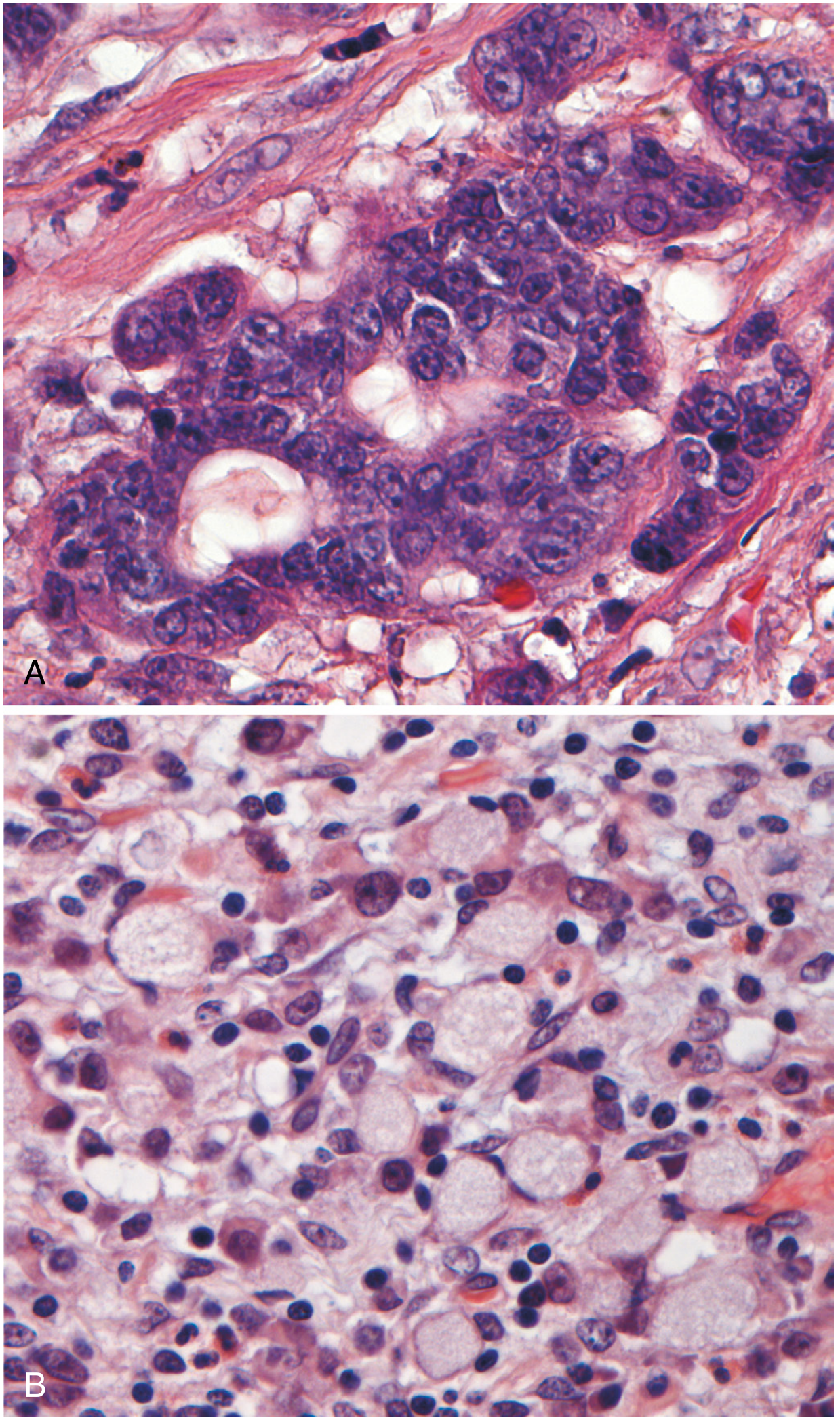
5. Robbins Key Concepts Summary (exam-ready, direct from text)
From Robbins PBD Key Concepts - Gastritis:
- Most common cause of chronic gastritis = H. pylori infection
- H. pylori gastritis: antrum predominantly, associated with increased gastric acid production
- Later involvement of body → atrophy → reduced acid → risk of adenocarcinoma
- H. pylori gastritis induces MALT that can give rise to B-cell lymphomas (MALTomas)
- Intestinal metaplasia develops in all forms of chronic gastritis and is a risk factor for gastric adenocarcinoma
- Peptic ulcer disease: secondary to H. pylori-induced gastritis and resulting hyperchlorhydria
6. Diagnostic Methods for Exam (Robbins + Jawetz)
| Test | Type | Notes |
|---|---|---|
| H&E stain | Invasive (biopsy) | Spiral organisms in mucus on surface epithelium; demonstrates gastritis pattern |
| Giemsa stain | Invasive | Reliable, cheap; good sensitivity |
| Warthin-Starry (silver) stain | Invasive | Organisms appear black; high sensitivity |
| IHC | Invasive | Most sensitive; detects coccoid forms in partially treated patients |
| Rapid urease test (CLO) | Invasive | Colour change within 2 hours; false-negative if on PPI |
| Urea breath test | Non-invasive | Detects ammonia from urease; best test for confirming eradication |
| Stool antigen test | Non-invasive | Good for initial diagnosis and post-eradication confirmation |
| Serology (IgG) | Non-invasive | Cannot distinguish active from past infection; useful for epidemiology |
| PCR | Invasive | Detects H. pylori DNA from biopsy; can detect antibiotic resistance mutations |
7. Master Summary Table
| Lesion | Robbins Key Point | Mechanism | Exam Highlight |
|---|---|---|---|
| Chronic active gastritis | Antral-predominant; organisms in surface mucus; plasma cells + neutrophils + MALT | CagA/VacA → IL-8 → neutrophil recruitment; T-cell driven chronic inflammation | Lymphoid follicles in gastric mucosa = PATHOGNOMONIC |
| Duodenal ulcer | 90-95% H. pylori-related | Antral gastritis → ↑ gastrin → ↑ acid → gastric metaplasia of duodenum → colonisation → ulcer | High acid state |
| Gastric ulcer | ~70% H. pylori-related | Mucosal defence failure; cytotoxins; ↓ prostaglandins | Normal/low acid state |
| Atrophic gastritis + intestinal metaplasia | Patchy; no autoantibodies | Longstanding inflammation → gland destruction → metaplastic replacement | Precursor to carcinoma; not autoimmune |
| Gastric adenocarcinoma | CagA PAI; Correa cascade; <5% infected individuals; decades | CagA oncogenic signalling + ROS + epigenetic silencing + host cytokine polymorphisms | Distal/non-cardia; Group I IARC carcinogen |
| MALT lymphoma (MALToma) | NF-κB dependent; regresses with antibiotics if no t(11;18) | H. pylori T cells → polyclonal B-cell → monoclonal lymphoma (NF-κB driven) | Lymphoepithelial lesions; CD20+, CD5-, CD10- |
Sources: Robbins, Cotran & Kumar Pathologic Basis of Disease (9e); Robbins & Kumar Basic Pathology (10e); Yamada's Textbook of Gastroenterology, 7e; Jawetz Melnick & Adelberg's Medical Microbiology, 28e; Sherris & Ryan's Medical Microbiology, 8e; Maingot's Abdominal Operations
Now mix both of the above results than give better answer
Here is the fully synthesised, single best answer combining all sources - Robbins PBD, Robbins Basic Pathology, Yamada's Gastroenterology, Jawetz, Sherris & Ryan, and Maingot's:
Helicobacter pylori - Associated Lesions and Pathogenesis
MD Pathology Exam - Master Answer
1. The Organism
H. pylori is a spiral-shaped/curved, Gram-negative, microaerophilic bacillus with multiple sheathed polar flagella. It is catalase-, oxidase-, and urease-positive. Humans are its only reservoir; transmission is by the fecal-oral route, typically acquired in childhood and persisting lifelong without treatment.
"H. pylori has acquired the dubious distinction of being the first bacterium classified as a carcinogen - implicated in the genesis of both gastric adenocarcinoma and gastric lymphomas."
- Robbins & Kumar Basic Pathology
It was also called an "accidental tourist" - established in human stomachs thousands of years ago and remaining bound to migrating human populations ever since. (Sherris & Ryan)
2. Virulence Factors
Robbins Basic Pathology identifies four core virulence factors - the most exam-tested framework:
| # | Factor | Mechanism |
|---|---|---|
| 1 | Flagella | Corkscrew motility; allows bacteria to swim beneath viscous mucus to the less-acidic epithelial surface |
| 2 | Urease | Hydrolyses urea → NH₃ + CO₂; neutralises local pH, creating a protective ammonia microenvironment |
| 3 | Adhesins (OMPs) | BabA, SabA bind Lewis-b blood-group antigens on surface foveolar cells; anchor the organism to epithelium |
| 4 | Toxins - CagA and CagE | Stimulate IL-8 (potent neutrophil chemotactic factor) → initiate and sustain innate + adaptive immune damage |
Additional detail (Sherris, Jawetz):
- CagA is injected directly into gastric epithelial cells via a Type IV Secretion System (T4SS) encoded by the cag Pathogenicity Island (CagPAI) - a ~40 kb genomic insert. Inside the cell, CagA reorganises the actin cytoskeleton, activates SHP-2 phosphatase, ERK/MAPK, Wnt/β-catenin, and NF-κB, mimicking growth-factor stimulation and promoting malignant transformation
- VacA (vacuolating cytotoxin A) - also delivered by the T4SS; causes vacuolization of the endosomal compartment, mitochondrial damage, apoptosis, and T-cell suppression
- NH₃ from urease is directly cytotoxic to the mucosa and disrupts tight junctions
- Urease expression is pH-regulated; it is upregulated when gastric pH rises (e.g., after a meal), allowing the organism to fine-tune its acid protection
Key biology: H. pylori is extracellular. It does not invade tissue. Colonisation of the surface is sufficient to produce disease. (Robbins Basic Pathology)
3. Pathogenesis - The Dual-Pathway Model (Robbins PBD)
This is the most exam-important conceptual framework:
H. pylori infection acquired in childhood
              |
    ┌─────────┴──────────┐
    ↓                    ↓
ANTRAL-PREDOMINANT    PANGASTRITIS
   GASTRITIS         (body + antrum)
(most common)      (longstanding disease)
    |                    |
↑ G-cell gastrin    Parietal cell
↑ Acid secretion      atrophy
    |                ↓ Acid secretion
    ↓                    |
 PEPTIC ULCER       INTESTINAL
  DISEASE           METAPLASIA
(gastric +              |
duodenal)               ↓
                GASTRIC ADENOCARCINOMA
"This results in an inverse relationship between gastric adenocarcinoma (associated with atrophy) and peptic ulcer disease (which requires at least some residual acid secretion)." - Robbins PBD
Host factors also govern this divergence: polymorphisms in IL-1β and TNF promoters amplify the inflammatory response and tip the balance toward pangastritis, atrophy, and carcinoma. (Robbins PBD)
4. Associated Lesions - Individual Discussion
4.1 Chronic Active Gastritis
Formerly called: Type B gastritis / Diffuse antral gastritis / Superficial gastritis.
Pathogenesis:
- H. pylori colonises antral mucosa → organisms + inflammation stimulate G cells → ↑ gastrin → ↑ acid → increased PUD risk
- CagA/VacA/urease → IL-8 secretion by epithelial cells → neutrophil infiltration
- T-cell-driven B-cell accumulation → lymphoid follicle formation (MALT)
- With time, infection may spread to involve body and fundus → atrophic change
MORPHOLOGY (Robbins - verbatim framework):
The organism is concentrated in the superficial mucus overlying foveolar epithelial cells in the surface and neck regions of glands. Distribution can be irregular, with areas of heavy colonisation adjacent to those with few organisms. Findings include:
- H. pylori organisms in surface mucus and on foveolar cell apices - visible on H&E/Giemsa; best shown by IHC or Warthin-Starry silver stain
- Large numbers of plasma cells, often in clusters or sheets in the superficial lamina propria (hallmark)
- Increased lymphocytes and macrophages in the lamina propria
- Neutrophils crossing the basement membrane into the epithelium
- Neutrophils accumulating in gland lumens = pit abscesses (pathognomonic of active gastritis)
- Submucosal lymphoid aggregates with germinal centres = induced MALT; absent in normal stomach; potential to transform into lymphoma (Robbins Basic + PBD)
- Intestinal metaplasia (goblet cells + columnar absorptive cells) in longstanding disease - risk factor for adenocarcinoma
- Thickened rugal folds with intense inflammation - can mimic infiltrative malignancy endoscopically
- Hyperplastic polyps (elongated foveolar glands with inflamed stroma) from regenerative repair
"Submucosal lymphoid aggregates, some with germinal centers, are frequently present and represent an induced form of MALT that has the potential to transform into lymphoma. Thus, risk of lymphoma, as well as adenocarcinoma, is increased in chronic H. pylori gastritis." - Robbins & Kumar Basic Pathology
H. pylori tropism (critical exam point):
- Shows tropism for gastric-type foveolar epithelium only
- Not found in areas of intestinal metaplasia, oxyntic (acid-secreting) mucosa of the body (unless patient is on PPIs), or duodenal epithelium
- Therefore antral biopsy is the preferred site for H. pylori evaluation
After eradication: Active (neutrophilic) component resolves within weeks. Chronic inflammatory component (lymphoplasmacytic) persists for months to years. (Yamada)
4.2 Peptic Ulcer Disease
"Peptic ulcer disease is usually secondary to chronic H. pylori-induced gastritis and the resulting hyperchlorhydria." - Robbins PBD Key Concepts
H. pylori causes ~90-95% of duodenal ulcers and ~70% of gastric ulcers (non-NSAID related).
Duodenal Ulcer - Pathogenesis:
H. pylori antral gastritis
    ↓
Inflammation → ↓ D-cell somatostatin
    ↓
Disinhibition of G cells → ↑ Gastrin secretion
    ↓
↑ Parietal cell mass (body) → HYPERSECRETION OF ACID
    ↓
Excess acid delivered to duodenal bulb
    ↓
Gastric metaplasia of duodenal mucosa
(duodenum acquires gastric-type foveolar epithelium)
    ↓
H. pylori colonises this metaplastic epithelium
    ↓
Active duodenitis → Duodenal ulceration
CagA+ strains also harbour a duodenal ulcer-promoting gene (dupA). (Sherris)
Gastric Ulcer - Pathogenesis:
- Direct mucosal cytotoxicity: ammonia (urease), VacA, CagA-mediated IL-8/cytokine release
- ↓ Prostaglandin E₂ synthesis (disrupted by inflammation) → impaired mucus/bicarbonate barrier
- Acid level is normal or low (cf. duodenal ulcer with high acid) - primary mechanism is mucosal defence failure
- Sites: Lesser curve of antrum/incisura most common (watershed area with poor blood supply)
Histology of peptic ulcer:
- Active phase: necrotic slough (zone 1) + granulation tissue (zone 2) + fibrosis (zone 3) + endarteritis obliterans (zone 4) at ulcer margin
- Adjacent mucosa: invariably shows H. pylori chronic active gastritis with organisms
- Benign features: punched-out margins, perpendicular walls, smooth base (vs. malignant ulcer with heaped-up irregular margins)
4.3 Atrophic Gastritis and Intestinal Metaplasia
Longstanding H. pylori pangastritis → Multifocal Atrophic Gastritis (MAG) / Environmental Metaplastic Atrophic Gastritis (EMAG):
"Changes seen in atrophic H. pylori gastritis are sometimes referred to as environmental metaplastic atrophic gastritis, to distinguish it from autoimmune metaplastic atrophic gastritis." - Robbins PBD
Features:
- Loss of oxyntic glands (body) and antral glands → glandular atrophy (patchy, unlike autoimmune gastritis which is diffuse and body-only)
- No autoantibodies against parietal cells or intrinsic factor (distinguishes from autoimmune/Type A)
- Replaced by intestinal metaplasia: goblet cells, absorptive enterocytes, Paneth cells appear in gastric mucosa
Types of intestinal metaplasia:
- Type I (Complete/small intestinal): Sialomucins, brush border, complete absorptive cell differentiation; lower risk
- Type II/III (Incomplete/colonic): Sulphomucins, no brush border; higher malignant risk
"Intestinal metaplasia develops in all forms of chronic gastritis and is a risk factor for gastric adenocarcinoma." - Robbins PBD Key Concepts
4.4 Gastric Adenocarcinoma - The Correa Cascade
H. pylori was classified as a WHO/IARC Group I (definite) human carcinogen in 1994 - the first bacterium to receive this designation.
Correa Cascade (the accepted stepwise model):
Normal Gastric Mucosa
    ↓  H. pylori infection (especially CagA+ strain)
Chronic Active Gastritis (antral-predominant)
    ↓  spread to body, decades of inflammation
Multifocal Atrophic Gastritis
    ↓
Intestinal Metaplasia
(Complete → Incomplete/Type III - higher risk)
    ↓
Low-Grade Dysplasia
    ↓
High-Grade Dysplasia (Carcinoma in situ)
    ↓
INVASIVE INTESTINAL-TYPE GASTRIC ADENOCARCINOMA
(distal/non-cardia stomach)
"This sequence takes decades to complete and occurs in less than 5% of infected individuals." - Robbins Basic Pathology
Molecular Mechanisms of Carcinogenesis (Robbins + Sherris synthesis):
| Mechanism | Detail |
|---|---|
| CagA oncogenic signalling | CagA injected by T4SS → activates SHP-2, ERK/MAPK, Wnt/β-catenin → mimics growth factor stimulation → ↑ proliferation, ↓ apoptosis (Robbins) |
| NF-κB activation | CagA, VacA, LPS → ↑ IL-8, IL-1β, TNF-α → sustained mutagenic inflammation |
| ROS-mediated DNA damage | Activated neutrophils/macrophages → reactive oxygen species → DNA strand breaks, TP53 mutations |
| Epigenetic silencing | Promoter methylation of CDH1/E-cadherin, RUNX3, MLH1 → TSG silencing; MSI via MLH1 methylation |
| Host cytokine polymorphisms | IL-1β and TNF promoter polymorphisms → amplified inflammation → pangastritis → atrophy (Robbins PBD specific emphasis) |
| Increased cell proliferation | Chronic mucosal injury + H. pylori-driven proliferative signals → accumulation of somatic mutations |
Evidence: In a landmark Japanese prospective study, gastric cancer developed in 2.9% of H. pylori seropositive patients and in none of seronegative patients. Relative risk conferred: 2.1 to 20-fold across studies. (Maingot's)
Link is to distal (non-cardia) gastric cancer only. No proven association with cardia/GEJ carcinoma.
4.5 Gastric MALT Lymphoma (MALToma)
"H. pylori gastritis induces MALT that can give rise to B-cell lymphomas (MALTomas)." - Robbins PBD Key Concepts
The Normal Stomach has NO MALT - its development is entirely a consequence of H. pylori infection.
Pathogenesis - Step by Step (Robbins):
H. pylori chronic gastritis
    ↓
H. pylori-reactive T cells appear
    ↓
T cells stimulate POLYCLONAL B-cell proliferation
(the lymphoid follicles/MALT of chronic gastritis)
    ↓
Accumulation of mutations → individual B-cell clones
gain growth advantage
    ↓
MONOCLONAL B-cell lymphoma
(still NF-κB dependent; still T-cell stimulated)
    ↓ ERADICATION WORKS HERE (75-80% regression)
    ↓ further mutations (e.g., t(11;18)(q21;q21) BIRC3-MALT1)
CONSTITUTIVE NF-κB ACTIVATION
(tumour is now antigen-independent)
    ↓ eradication no longer effective
Spread beyond stomach
    ↓
Additional mutations (p53, p16 inactivation)
HIGH-GRADE DLBCL TRANSFORMATION
MORPHOLOGY (Robbins PBD):
- Dense lymphocytic infiltrate in the lamina propria
- Lymphoepithelial lesions = neoplastic centrocyte-like B-cells infiltrating and destroying gastric gland epithelium - THE defining histological feature
- Reactive B-cell follicles may be present
- ~40% show plasmacytic differentiation
IHC Profile:
- CD19+, CD20+, CD79a+ (B-cell)
- CD5-, CD10- (marginal zone phenotype; not CLL, not follicular lymphoma)
- CD43+ in ~25% (diagnostically helpful unusual feature - Robbins PBD)
- Monoclonality confirmed by: restricted κ or λ light chain expression OR clonal IgH rearrangement by molecular methods
The unique biology - Robbins:
"At this stage, the B-cell proliferation remains localized to the stomach and eradication of H. pylori by antibiotic therapy 'cures' the lymphoma by removing the antigenic stimulus for T cells."
MALToma is the only neoplasm known to regress with antibiotic therapy. Cases with t(11;18) are H. pylori-independent and do NOT regress with eradication. (Sherris; Robbins)
5. Histological Identification Methods
| Method | Notes |
|---|---|
| H&E | Curved/spiral organisms on surface epithelium; reliable when numerous |
| Giemsa | Good sensitivity; organisms stain dark purple/blue |
| Warthin-Starry (silver) | Organisms appear black against yellow background; excellent sensitivity |
| IHC | Most sensitive; detects coccoid forms in partially treated patients; gold standard |
| Modified Giemsa/Cresyl violet | Rapid, inexpensive |
| Rapid urease test (CLO) | Biopsy-based; colour change in <2 hours; false-negative on PPIs |
| Urea breath test | Non-invasive; best for confirming eradication post-treatment |
| Stool antigen | Non-invasive; useful for initial diagnosis and eradication confirmation |
| Serology (IgG) | Cannot distinguish active from past infection; epidemiological use |
6. Master Comparison: Type A vs Type B Gastritis
| Feature | Type A (Autoimmune) | Type B (H. pylori) |
|---|---|---|
| Site | Body/fundus | Antrum (initially) |
| Spread | Diffuse | Patchy; may become pangastritis |
| Acid | Low/absent (achlorhydria) | Initially high; low with atrophy |
| Gastrin | Very high (G-cell hyperplasia) | Mildly elevated with atrophy |
| Autoantibodies | Anti-parietal cell, anti-IF | Absent |
| Atrophy | Extensive, body-predominant | Multifocal, patchy |
| Complications | Pernicious anaemia; neuroendocrine tumours | PUD; adenocarcinoma; MALToma |
| Cause | Autoimmune | H. pylori |
7. All H. pylori-Associated Lesions - Summary Table
| Lesion | Key Robbins Statement | Pathogenesis | Distinguishing Feature |
|---|---|---|---|
| Chronic active gastritis | "Most common cause of chronic gastritis" | CagA/VacA → IL-8 → neutrophils; MALT formation | Pit abscesses + lymphoid follicles + plasma cell sheets |
| Duodenal ulcer | "Hyperchlorhydria"; 90-95% H. pylori | ↑ Gastrin → ↑ acid → gastric metaplasia of duodenum | High acid; antral disease |
| Gastric ulcer | "Mucosal defence failure" | ↓ Prostaglandins; cytotoxin-mediated injury | Normal/low acid |
| Atrophic gastritis | "Patchy; no autoantibodies" | Longstanding inflammation → gland destruction | Environmental/EMAG; distinguishable from Type A |
| Intestinal metaplasia | "Risk factor for adenocarcinoma in ALL forms of gastritis" | Chronic injury → metaplastic replacement | Type III (incomplete) = highest risk |
| Gastric adenocarcinoma | "First bacterium classified as carcinogen; <5% of infected" | Correa cascade; CagA oncogenic signalling; ROS; epigenetics | Distal/non-cardia; intestinal type |
| MALT lymphoma | "Eradication cures the lymphoma" by removing T-cell stimulus | MALT induction → polyclonal → monoclonal (NF-κB) | Lymphoepithelial lesions; CD20+, CD5-, CD10- |
8. High-Yield Quick Recall for Exam
- H. pylori = extracellular only; does not invade tissue
- Four virulence factors (Robbins): Flagella, Urease, Adhesins, Toxins (CagA/CagE)
- CagA is delivered by Type IV Secretion System encoded by cag Pathogenicity Island
- Lewis-b antigen is the main mucosal receptor; blood group O individuals have higher risk
- Organisms show tropism for gastric foveolar epithelium only - not intestinal metaplastic foci (unless on PPIs)
- Lymphoid follicle in gastric mucosa = pathognomonic of H. pylori (normally absent)
- Pit abscesses = neutrophils in gastric pit lumens = hallmark of ACTIVE gastritis
- Plasma cells in sheets in superficial lamina propria = characteristic of H. pylori gastritis
- Duodenal ulcer = high acid (↑ gastrin pathway); Gastric ulcer = normal/low acid (defence failure)
- Correa Cascade = normal → gastritis → atrophy → intestinal metaplasia → dysplasia → intestinal-type carcinoma
- Sequence takes decades; occurs in <5% of infected individuals (Robbins Basic)
- MALToma = only neoplasm that regresses with antibiotics (if NF-κB not constitutively active)
- t(11;18) = BIRC3-MALT1 translocation = H. pylori-independent; does NOT regress with eradication
- IL-1β and TNF polymorphisms (host genetics) determine whether antral-only disease or pangastritis/carcinoma develops (Robbins PBD)
- H. pylori = WHO/IARC Group I carcinogen since 1994 = first bacterium so classified
Sources: Robbins, Cotran & Kumar Pathologic Basis of Disease (9/10e) | Robbins & Kumar Basic Pathology (10e) | Yamada's Textbook of Gastroenterology (7e) | Jawetz Melnick & Adelberg's Medical Microbiology (28e) | Sherris & Ryan's Medical Microbiology (8e) | Maingot's Abdominal Operations
This is a shared conversation. Sign in to Orris to start your own chat.