
Surfactant, hb oxygen dissociation curve , functional residual volume, vital capacity, transport of gases, neural regulation of respiration,
Reading File
Finding Sources
Finding Sources
Finding Sources
Finding Sources
Finding Sources
Finding Sources
Finding Sources
Reading File
Reading File
Reading File
Reading File
Reading File
Reading File
Reading File
I have the offset wrong for FRC and vital capacity — those were from the wrong block. Let me read the correct sections.
Reading File
Reading File
Reading File
I now have rich content from multiple authoritative textbooks. Let me also read the CO2 transport section and the Guyton neural regulation section (pneumotaxic center and beyond).
Reading File
Reading File
Here is a comprehensive review of all six topics, drawn from authoritative physiology textbooks.
Respiratory Physiology: Core Topics
1. Surfactant
Definition: Surfactant is a mixture of phospholipids (primarily dipalmitoyl phosphatidylcholine, DPPC) lining the inner surface of alveoli, secreted by type II pneumocytes (type II alveolar cells).
Mechanism of action:
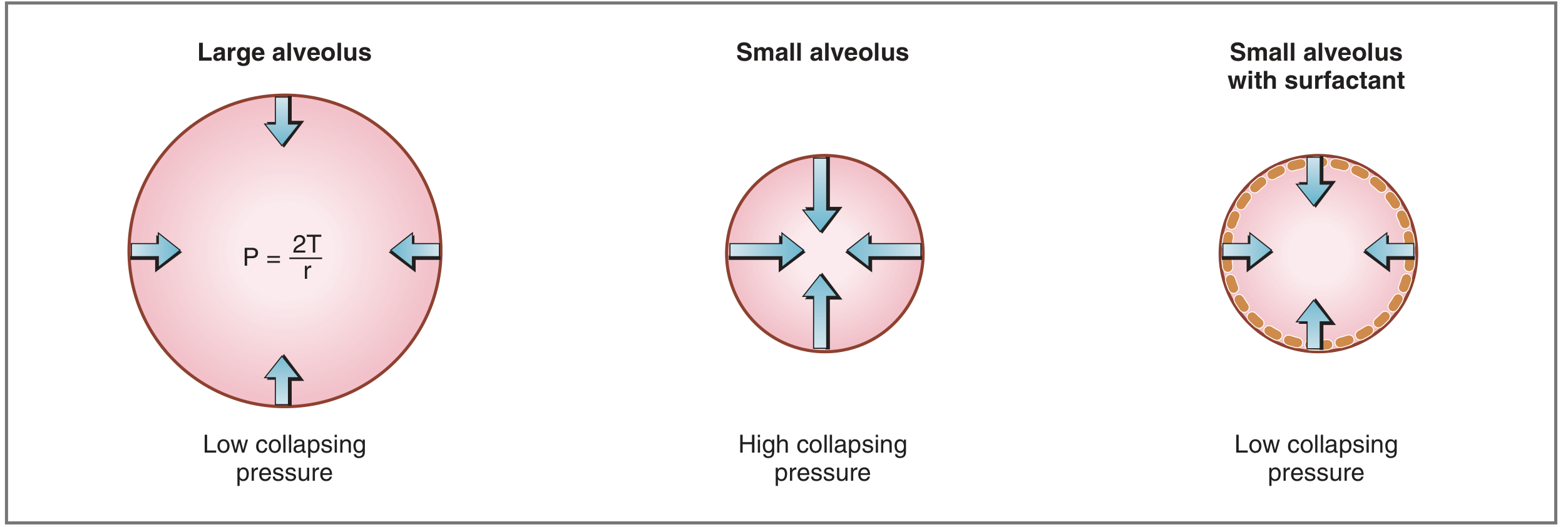
DPPC molecules are amphipathic — hydrophobic on one end, hydrophilic on the other. They align at the air-liquid interface of alveoli with hydrophobic ends facing inward and hydrophilic ends facing the aqueous layer. This arrangement disrupts the intermolecular attracting forces between water molecules that produce high surface tension, thereby reducing surface tension.
Why it matters — Law of Laplace:
$$P = \frac{2T}{r}$$
Where P = collapsing pressure, T = surface tension, r = radius. Without surfactant, small alveoli (small r) would have very high collapsing pressures and collapse (atelectasis). Surfactant lowers T, reducing P even for small alveoli, keeping them patent.
Functions:
- Prevents alveolar collapse — reduces collapsing pressure in small alveoli
- Increases lung compliance — less work required during inspiration
- Keeps small and large alveoli at equal pressure — stabilizes alveoli of different sizes
Clinical correlation — Neonatal Respiratory Distress Syndrome (NRDS):
- Surfactant synthesis begins at ~gestational week 24; reliably present by week 35
- Premature infants lack surfactant → increased surface tension → alveolar collapse → hypoxemia + increased work of breathing
- Treatment: exogenous surfactant therapy; antenatal corticosteroids to accelerate lung maturation
2. Hemoglobin–Oxygen Dissociation Curve
The curve represents the relationship between hemoglobin saturation (or O₂ content) and the partial pressure of oxygen (PO₂). It is sigmoidal (S-shaped) due to cooperative binding — binding of O₂ to one heme group changes the quaternary structure of hemoglobin and increases the affinity of the remaining heme groups.
Key values (normal adult):
| Parameter | Value |
|---|---|
| P50 (PO₂ at 50% saturation) | 26.6 mm Hg |
| Arterial PO₂ (SaO₂ ~97%) | ~100 mm Hg |
| Mixed venous PO₂ (SvO₂ ~75%) | ~40 mm Hg |
| O₂ extraction fraction at rest | ~25% |
Shape significance:
- Flat upper portion (PO₂ 70–100 mm Hg): Even if arterial PO₂ falls (e.g., lung disease), SaO₂ stays relatively normal as long as PO₂ ≥ 60 mm Hg — a safety margin for oxygen loading in the lungs.
- Steep middle portion (PO₂ 20–60 mm Hg): Large amounts of O₂ are released with modest drops in PO₂ — facilitates O₂ unloading at tissues.
Shifts of the curve:
| Direction | Causes | Effect |
|---|---|---|
| Right shift (↑P50) | ↑PCO₂, ↑H⁺ (↓pH), ↑temperature, ↑2,3-DPG | ↓O₂ affinity → more O₂ released to tissues |
| Left shift (↓P50) | ↓PCO₂, ↓H⁺ (↑pH), ↓temperature, ↓2,3-DPG, fetal Hb (HbF), CO poisoning | ↑O₂ affinity → less O₂ released |
The Bohr effect: ↑PCO₂ or ↑H⁺ in metabolically active tissues shifts the curve right, enhancing O₂ delivery precisely where it is most needed.

3. Functional Residual Capacity (FRC)
Definition: FRC is the lung volume at the end of a normal, passive exhalation. It is the sum of:
$$\text{FRC} = \text{Expiratory Reserve Volume (ERV)} + \text{Residual Volume (RV)}$$
Normal adult FRC ≈ 2300–2500 mL
Physiology: At FRC, the inward elastic recoil of the lung exactly balances the outward elastic recoil of the chest wall. This is the resting equilibrium point of the respiratory system — no muscular effort is needed to maintain this volume.
Measurement methods:
- Nitrogen washout — breathe 100% O₂ until all N₂ is washed out; measure total N₂ exhaled
- Helium dilution — inhale a known concentration of He; measure dilution at equilibrium
- Body plethysmography — most accurate (includes trapped gas)
Factors that decrease FRC:
| Factor | Mechanism |
|---|---|
| Obesity | ↓ chest wall compliance; ↑ abdominal pressure on diaphragm |
| Supine posture | Abdominal contents push diaphragm cephalad |
| Female sex | ~10% less than males |
| Pregnancy / ascites | ↑ intra-abdominal pressure |
| Restrictive lung disease | ↓ lung/chest compliance |
| Neuromuscular disease | ↓ diaphragmatic tone |
| Laparoscopic surgery / pneumoperitoneum | ↑ intra-abdominal pressure |
Factors that increase FRC:
- Tall stature (FRC directly proportional to height)
- COPD / emphysema (loss of elastic recoil → air trapping)
Clinical significance: FRC represents the body's main O₂ store during apnea. With FiO₂ = 0.21, FRC contains ~480 mL O₂. Pre-oxygenation with 100% O₂ before induction of anesthesia fills the FRC with ~2300 mL O₂, delaying desaturation for 4–5 minutes.
4. Vital Capacity (VC)
Definition: Vital capacity is the maximum volume of air that can be exhaled after a maximal inspiration.
$$\text{VC} = \text{IRV} + \text{TV} + \text{ERV}$$
$$\text{VC} = \text{TLC} - \text{RV}$$
Normal VC ≈ 4600–5000 mL (varies with age, sex, height)
Types:
- Slow VC (SVC): Exhaled slowly from TLC to RV
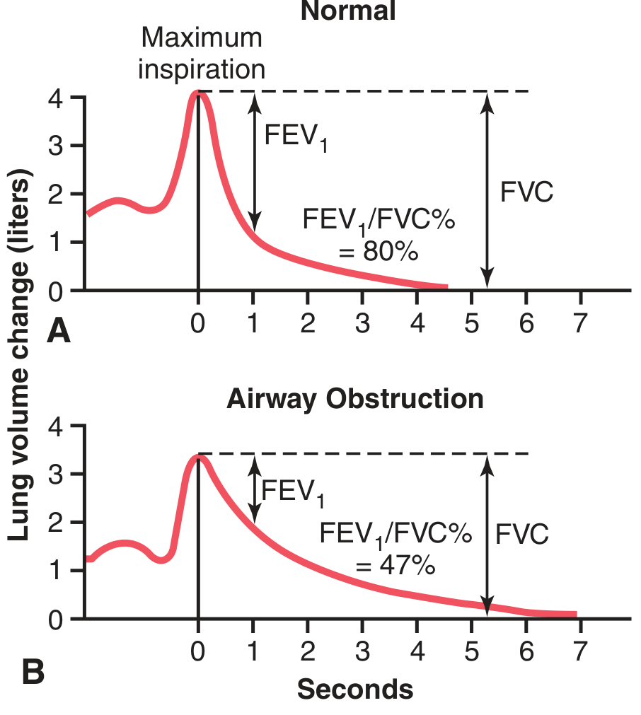
- Forced Vital Capacity (FVC): Exhaled with maximum effort as fast as possible — used in spirometry
FVC and FEV₁:
- FEV₁ = volume exhaled in the first second of FVC maneuver
- FEV₁/FVC ratio (Tiffeneau index):
- Normal: ≥ 80%
- Obstructive pattern (e.g., asthma, COPD): < 70% (FEV₁ ↓↓, FVC relatively preserved)
- Restrictive pattern (e.g., fibrosis): FEV₁/FVC normal or ↑ (both FEV₁ and FVC ↓ proportionally)
Conditions reducing VC:
- Restrictive: pulmonary fibrosis, pleural effusion, chest wall deformity, neuromuscular disease
- Obstructive: severe asthma, COPD (air trapping increases RV, reducing VC)
- VC < 15 mL/kg is an indication for mechanical ventilation
5. Transport of Gases
Oxygen Transport
O₂ is carried in two forms:
a) Dissolved O₂ (minor)
- By Henry's law: concentration = α × PO₂; α = 0.003 mL/dL/mm Hg
- At PaO₂ = 100 mm Hg → only 0.3 mL O₂/dL blood — insufficient alone
b) Bound to hemoglobin (major — ~98.5% of total)
- Each gram of Hb can carry 1.34–1.39 mL O₂
- Arterial O₂ content: CaO₂ = (Hb × 1.34 × SaO₂) + (0.003 × PaO₂) ≈ 20 mL/dL
O₂ delivery (DO₂):
$$\text{DO}_2 = \text{CaO}_2 \times \text{Cardiac Output} = 20 \text{ mL/dL} \times 50 \text{ dL/min} = \textbf{1000 mL O}_2\textbf{/min}$$
Fick equation:
$$\text{VO}_2 = \text{Q}_T \times (\text{CaO}_2 - \text{CvO}_2) \approx 250 \text{ mL/min at rest}$$
Normal extraction fraction = 25% (the body consumes only 1 in 4 O₂ molecules delivered).
CO₂ Transport
CO₂ is carried in three forms:
| Form | % of total | Notes |
|---|---|---|
| Bicarbonate (HCO₃⁻) | ~87% | Formed inside RBCs via carbonic anhydrase; HCO₃⁻ exits RBC in exchange for Cl⁻ (chloride shift) |
| Carbamino compounds | ~7–8% | CO₂ binds to terminal amino groups of proteins (esp. deoxyHb) |
| Dissolved CO₂ | ~5–6% | Solubility = 0.031 mmol/L/mm Hg — 20× more soluble than O₂ |
Chloride shift (Hamburger phenomenon): In systemic capillaries, CO₂ enters RBCs → carbonic anhydrase converts it to H₂CO₃ → H⁺ + HCO₃⁻. HCO₃⁻ is transported out of the RBC in exchange for Cl⁻. In pulmonary capillaries, this reverses.
Haldane effect: Deoxygenated Hb (deoxyHb) carries more CO₂ as carbamino compounds and buffers more H⁺ than oxyHb. This means that in metabolically active tissues, O₂ unloading automatically facilitates CO₂ loading — a complementary mechanism to the Bohr effect.
6. Neural Regulation of Respiration
The respiratory centers are distributed across the medulla oblongata and pons.
Medullary Centers
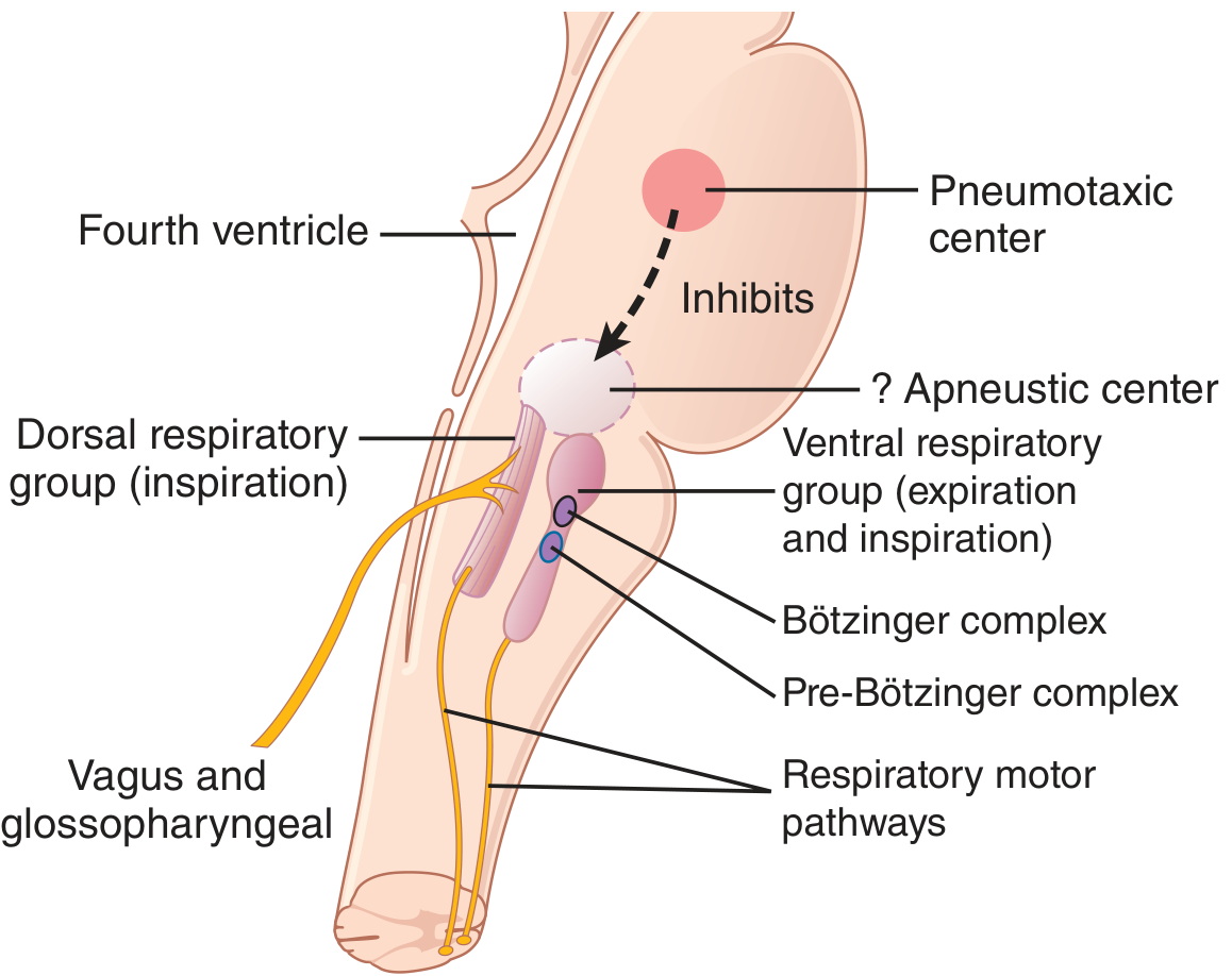
a) Dorsal Respiratory Group (DRG)
- Located in the nucleus tractus solitarius (NTS)
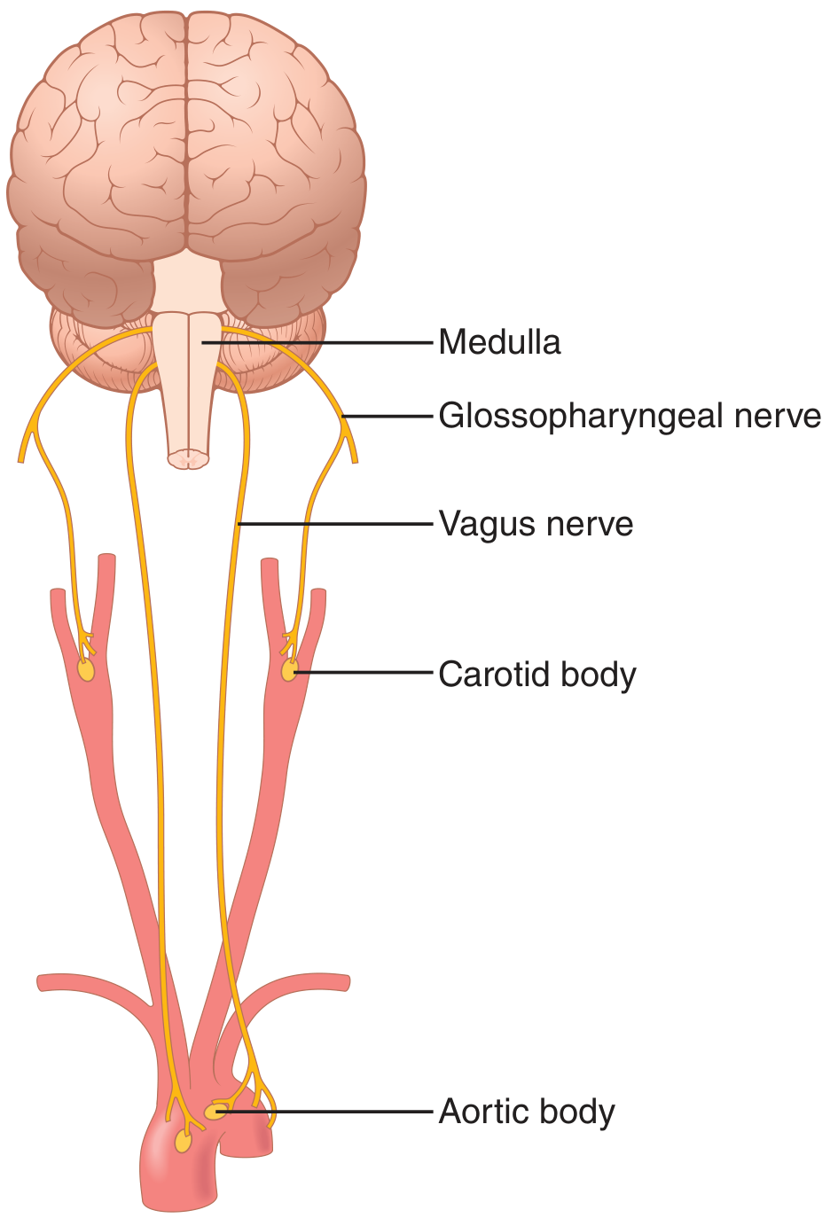
- Receives afferent inputs from: peripheral chemoreceptors (via CN IX/X), baroreceptors, lung stretch receptors
- Primarily drives inspiration; generates the basic rhythmic respiratory drive
- Produces an inspiratory "ramp" signal — a progressively increasing signal to the diaphragm over ~2 seconds, then abruptly ceases for ~3 seconds (passive expiration occurs)
b) Ventral Respiratory Group (VRG)
- Located in the nucleus ambiguus (rostral) and nucleus retroambiguus (caudal)
- Inactive during quiet breathing (eupnea)
- Activated during forced/heavy breathing — drives both inspiratory and expiratory muscles
- Pre-Bötzinger complex (small region in rostral VRG): contains pacemaker neurons; considered the primary respiratory rhythm generator
- Bötzinger complex: inhibitory neurons that switch off inspiration to initiate expiration
Pontine Centers
c) Pneumotaxic Center
- Located in the nucleus parabrachialis of the upper pons
- Sends signals to DRG to switch off the inspiratory ramp — limits duration of inspiration
- Strong signal → short inspiration → ↑ respiratory rate (up to 30–40 breaths/min)
- Weak signal → prolonged inspiration → ↓ rate (3–5 breaths/min)
d) Apneustic Center (lower pons)
- Tends to prolong inspiration (apneusis = sustained inspiratory gasp)
- Normally held in check by the pneumotaxic center
Reflex Controls
Hering-Breuer Inflation Reflex:
- Lung stretch receptors (in bronchial/bronchiolar walls) → vagus nerve → DRG
- Activated when tidal volume > ~1.5 L — inhibits further inspiration (protective reflex against over-inflation)
- More important in newborns; plays a minor role in normal adult tidal breathing
Chemical Control
Central chemoreceptors:
- Located on the ventral surface of the medulla
- Respond primarily to ↑PCO₂ / ↑H⁺ in CSF (CO₂ readily crosses blood-brain barrier; H⁺ does not)
- CO₂ is the dominant controller of ventilation — even small rises in PaCO₂ cause dramatic increases in ventilation
Peripheral chemoreceptors:
- Carotid bodies (CN IX — Hering's nerve) and aortic bodies (CN X — vagus)
- Primarily respond to ↓PaO₂ (below ~70 mm Hg), and to lesser extent ↑PCO₂ and ↑H⁺
- O₂ changes have little direct effect on the central respiratory center — this is entirely mediated peripherally
Quick Summary Table
| Topic | Key Point |
|---|---|
| Surfactant | DPPC from type II cells; reduces surface tension; prevents alveolar collapse; absent in prematurity → NRDS |
| Hb-O₂ curve | Sigmoidal; P50 = 26.6 mmHg; right shift = ↑CO₂, ↑H⁺, ↑temp, ↑2,3-DPG; Bohr effect |
| FRC | ERV + RV; ~2300 mL; lung elastic recoil = chest wall recoil; ↓ in obesity, supine, pregnancy |
| Vital Capacity | IRV + TV + ERV; FEV₁/FVC ≥ 80% normal; ↓ in restrictive; FEV₁/FVC ↓ in obstructive |
| Gas transport | O₂: 98.5% on Hb; CO₂: 87% as HCO₃⁻ via carbonic anhydrase (chloride shift) |
| Neural regulation | DRG (NTS) = rhythm; pre-Bötzinger = pacemaker; pneumotaxic = limits inspiration; CO₂ = dominant chemical controller |
Sources: Costanzo Physiology 7th Ed. | Guyton & Hall Textbook of Medical Physiology | Morgan & Mikhail's Clinical Anesthesiology 7th Ed. | Fishman's Pulmonary Diseases and DisordersYou've run out of free credits. Sign up for a free account to continue using Orris.
This is a shared conversation. Sign in to Orris to start your own chat.